

The Volar Cortical Hinge: An Independent Risk Factor for Distal Radius Fracture Displacement
- PMID: 38808183
- PMCID: PMC11129888
- DOI: 10.1055/s-0043-1771376
The Volar Cortical Hinge: An Independent Risk Factor for Distal Radius Fracture Displacement
Abstract
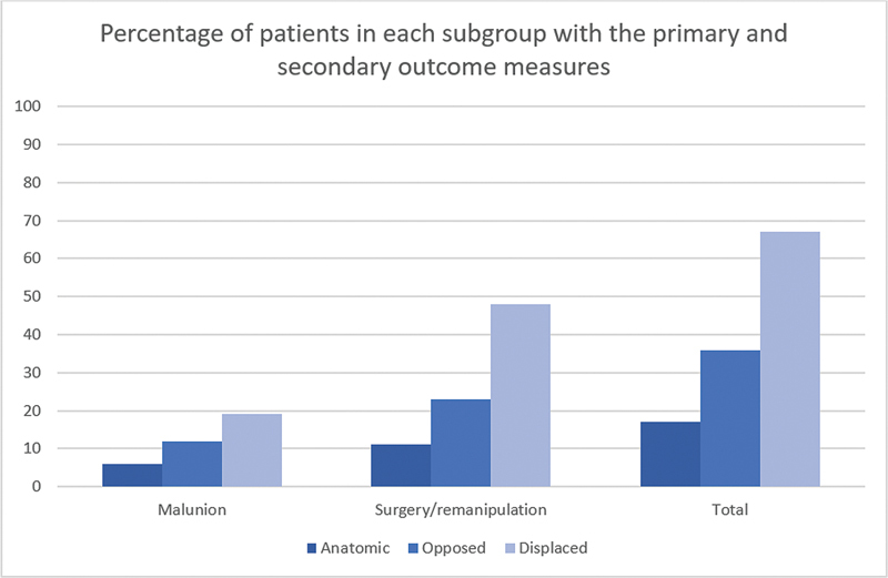
Background Displaced distal radius fractures are prone to redisplacement after manipulation. This can result in the need for delayed surgery. Several criteria have been studied to predict the likelihood of redisplacement. We hypothesized that reduction in the volar cortex would be an additional predictive factor. Purpose The aim of this study was to assess whether the quality of the volar cortex reduction predicts the subsequent need for further intervention (surgery or remanipulation). As a secondary outcome, we assessed whether the quality of the reduction predicts the rate of malunion. Methods A retrospective review was performed of displaced adult distal radius fractures over a 2-year period that had undergone closed reduction at presentation. We identified 105 patients and a review of their electronic notes and radiographs was then performed. The volar cortex reduction was defined as "anatomical," "opposed," or "displaced." We assessed the radial height, radial inclination, radial/ulnar translation, volar/dorsal angulation, teardrop angle, presence of dorsal comminution, quality of the cast (molding, cast index), and volar cortex reduction. These measurements were taken at five time points (prereduction, postreduction, 1 week, 2 weeks, and 6 weeks). All patients that subsequently required surgical fixation or repeat reduction were identified as the primary outcome measure. The 6-week radiographs were assessed for radiographic malunion as our secondary outcome measure. A statistical analysis was then performed to assess the factors that influenced a loss of position and the need for delayed surgical intervention. Results Of the 105 patients, 22 patients required delayed surgery, 3 patients underwent a repeat manipulation, and 12 patients had a radiographic malunion at 6 weeks. During the study period, the proportion of patients requiring surgery or repeat manipulation in the displaced group was 10/21 (47.6%), in the opposed group it was 11/50 (23.4%), and in the anatomic group it was 4/36 (11.1%; p = 0.008). We then included the patients with a radiographic malunion and found the proportion of patients with an adverse outcome in the displaced group was 14/21 (66.7%), in the opposed group it was 17/47 (36.2%), and in the anatomic group it was 6/36 (16.7%; p = 0.001). At the 1-week time point, this association was equally significant, as the proportion in the displaced group was 17/33 (51.5%), in the opposed group it was 15/45 (33.3%) and in the anatomic group it was 1/22 (4.5%; p = 0.001). The patients' age, quality of cast, presence of dorsal comminution, and degree of initial displacement did not predict the subsequent need for surgery or remanipulation. Conclusion The most important factor in our study for significant redisplacement of an initially dorsally displaced distal radius fracture is the association of the volar cortex. This parameter maintains significance at the 1-week time point. This data shows that volar cortex reduction is a useful clinical measurement in assessing which distal radius fractures will undergo delayed displacement requiring intervention. Level of evidence Level 3-Retrospective comparative study.
Keywords: displaced; distal radius; fracture; remanipulation; volar cortex; volar cortical hinge.
Thieme. All rights reserved.
Conflict of interest statement
Conflict of Interest None declared.
Figures


References
-
- Alemdaroğlu K B, İltar S, Aydoğan N H, Say F, Kilinç C Y, Tiftikçi U. Three-point index in predicting redisplacement of extra-articular distal radial fractures in adults. Injury. 2010;41(02):197–203. - PubMed
-
- LaMartina J, Jawa A, Stucken C, Merlin G, Tornetta P., III Predicting alignment after closed reduction and casting of distal radius fractures. J Hand Surg Am. 2015;40(05):934–939. - PubMed
-
- Mackenney P J, McQueen M M, Elton R. Prediction of instability in distal radial fractures. J Bone Joint Surg Am. 2006;88(09):1944–1951. - PubMed
-
- Walenkamp M MJ, Aydin S, Mulders M AM, Goslings J C, Schep N WL. Predictors of unstable distal radius fractures: a systematic review and meta-analysis. J Hand Surg Eur Vol. 2016;41(05):501–515. - PubMed
LinkOut - more resources
Full Text Sources
Research Materials
Miscellaneous
