. 2024 Sep 3;147(9):3083-3098.
doi: 10.1093/brain/awae173.

Establishing connectivity through microdissections of midbrain stimulation-related neural circuits

Affiliations

Establishing connectivity through microdissections of midbrain stimulation-related neural circuits

Georgios P Skandalakis et al. Brain. .

Abstract

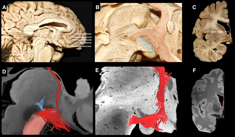
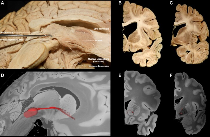
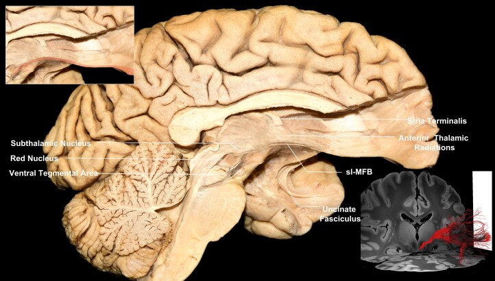
Comprehensive understanding of the neural circuits involving the ventral tegmental area is essential for elucidating the anatomofunctional mechanisms governing human behaviour, in addition to the therapeutic and adverse effects of deep brain stimulation for neuropsychiatric diseases. Although the ventral tegmental area has been targeted successfully with deep brain stimulation for different neuropsychiatric diseases, the axonal connectivity of the region is not fully understood. Here, using fibre microdissections in human cadaveric hemispheres, population-based high-definition fibre tractography and previously reported deep brain stimulation hotspots, we find that the ventral tegmental area participates in an intricate network involving the serotonergic pontine nuclei, basal ganglia, limbic system, basal forebrain and prefrontal cortex, which is implicated in the treatment of obsessive-compulsive disorder, major depressive disorder, Alzheimer's disease, cluster headaches and aggressive behaviours.

Keywords: deep brain stimulation; fibre tractography; neuropsychiatric neural circuits; ventral tegmental area.


Conflict of interest statement

V.A.C., as an employee of University of Freiburg, listed by the institution as inventor, has filed a US provisional patent application generally related to highly focused DBS in the treatment of obsessive–compulsive disorder (US patent application number 63/253,740). All other authors report no biomedical financial interests or potential competing interests. C.G.H. is a paid consultant for Hemerion Therapeutics, Synaptive Medical, Stryker Corp. and Integra. A.M.L. is a consultant to Abbott, Boston Scientific, Insightec and Medtronic and is Scientific Director at Functional Neuromodulation. V.A.C. receives a collaborative grant from BrainLab (Munich, Germany); he is a consultant for Ceregate (Munich, Germany), Cortec (Freiburg, Germany), ALEVA (Lausanne, Switzerland) and Inbrain (Barcelona, Spain); and he has ongoing investigator initiated trials (IITs) with Boston Scientific (USA) and has received personal honoraria and travel support for lecture work from Boston Scientific (USA), UNEEG and PRECISIS.

Figures

Figure 1
Figure 1
Illustration of the methods followed in this study. Initial characterization of ventral tegmental area (VTA)-related fibre tracts and their termination points was achieved through stepwise microdissections in cadaveric hemispheres. Regions of intetest (ROIs) were reconstructed manually for the VTA and recorded termination points. Reconstruction of the VTA tract was achieved through microdissection-guided fibre tractography in multiple databases. A connectivity-driven parcellation of the VTA was performed in an averaged template generated from data of 1065 healthy adults. Lead-DBS was used to study the relationship between the tracts we characterized anatomically and the previously used deep brain stimulation (DBS) targets.
Figure 2
Figure 2
VTA fibres running medial to the mammillothalamic tract. (A) Medial view of a left hemisphere. The ependymal/subependymal layer has been removed to expose fibres running between the VTA, medial hypothalamus, fornix and septal region. (B) Magnified view of the area depicting the trajectory of the fibres highlighted in red and mammillothalamic fibres highlighted in pale blue. (C) Coronal section at the level of the termination points depicting septal nuclei highlighted in red. (D) Tractography depicting the VTA fibres in red and mammillothalamic tract in pale blue. (E) Brainstem ex vivo tractography depicting VTA fibres. (F) Coronal section depicting fibres of the VTA within the septal nuclei. VTA = ventral tegmental area.
Figure 3
Figure 3
VTA fibres implicating raphe nuclei, NBM and BNST. (A) Medial view of a left hemisphere. The ventral mammillotegmental tract and mammillary body have been removed, exposing fibres running between the VTA, raphe nuclei, NBM and BNST. (B) Magnified view of the area, depicting the fibres highlighted in red. (C) Coronal section at the level of the termination point of VTA and stria terminalis fibres, depicting the BNST bounded by the anterior commissure inferiorly, the globus pallidus internus laterally, lateral ventricle medially and caudate nucleus superiorly. (D) Tractography showing VTA fibres in red. (E) Brainstem ex vivo tractography. (F) Coronal section, revealing tractography fibres within the BNST. BNST = bed nucleus of stria terminalis; NBM = nucleus basalis of Meynert; VTA = ventral tegmental area.
Figure 4
Figure 4
Temporo-insular fibres. (A) Medial view of a left hemisphere, showing fibres running between the VTA and insula highlighted in red. (B) Tractography, showing fibres running between the VTA and insula. (C) Coronal section at the level of the termination points, depicting the anterior insula. (D) Tractography, coronal section, revealing fibres within the insula. (E) Medial view of a left hemisphere following removal of insular fibres. Fibres running between the VTA and hippocampal region can be visualized arching laterally and posteriorly. (F) Tractography, showing fibres running between VTA and hippocampal area in red. (G) Coronal section at the level of the termination points, depicting the hippocampal area. (H) Tractography; coronal section, revealing fibres within the hippocampal area. (I) Medial view of a left hemisphere, showing fibres running between the VTA and amygdala/entorhinal cortex. (J) Tractography, showing the trajectory of the fibres between the amygdala/entorhinal cortex and VTA. (K) Coronal section of the contralateral hemisphere at the level of the termination points, depicting amygdala and entorhinal cortex. (L) Coronal section tractography, revealing VTA fibres within the amygdala. VTA = ventral tegmental area.
Figure 5
Figure 5
GP and NAc. (A) Medial view of a left hemisphere, showing fibres running between the VTA and basal ganglia region. (B) Coronal section at the level of the anterior termination points, showing the NAc. (C) Coronal section at the level of the posterior termination points, showing the GP. (D) Tractography, showing the trajectory of the fibres running between the VTA and NAc. (E) Tractography, coronal section, depicting VTA fibres within the NAc. (F) Tractography, coronal section, depicting VTA fibres within the GP. GP = globus pallidus; NAc = nucleus accumbens; slMFB = superolateral medial forebrain bundle; VTA = ventral tegmental area.
Figure 6
Figure 6
VTA PFC fibres (BA10, BA11 and BA47/12). Medial view of a left hemisphere, depicting fibres running within the ALIC between the VTA and BA10. Top left inset: magnification of the fibres implicating the VTA highlighted in red. Bottom right inset: tractography, showing fibres between the VTA and Brodmann areas 10, 11 and 47/12. ALIC = anterior limb of the internal capsule; BA = Brodmann area; PFC = prefrontal cortex; sl-MFB = superolateral medial forebrain bundle; VTA = ventral tegmental area.
Figure 7
Figure 7
Overview of VTA-related fibre tracts and DBS targets analysed. Overview of DBS targets, indications and electrode placements relative to VTA tracts that we characterized. Fibre tracts are denoted as follows: VTA temp, fibre tracts interconnecting the VTA with the insula, hippocampus, dorsal dentate gyrus, amygdala and entorhinal cortex; VTA-BF, fibre tracts interconnecting VTA with hypothalamus, fornix (fx), septal region, nucleus basalis of Meynert, mammillary body, raphe nuclei and bed nucleus of stria terminalis; VTA-PFC, VTA-PFC fibres; VTA-ExAmg, fibre tracts interconnecting the VTA with the GP and extended amygdala; and VTA-NAc, fibre tracts interconnecting the VTA with the nucleus accumbens (NAc). DBS = deep brain stimulation; ithp = inferior thalamic peduncle; MDD = major depressive disorder; OCD = obsessive–compulsive disorder; PHA = posterior hypothalamus; VC/VS = ventral capsule/ventral striatum; VTA = ventral tegmental area.
Figure 8
Figure 8
Relationship between VTA tracts and established DBS targets. (A) Fibre tracts interconnecting the VTA with the insula, hippocampus, dorsal dentate gyrus, amygdala and entorhinal cortex (VTA-temp) exhibited the highest ratio of percentage tract overlap to stimulation amplitude with ithp, slMFB, fornix and GPi DBS targets., (B) Fibre tracts interconnecting the VTA with the hypothalamus, fornix, septal region, nucleus basalis of Meynert, mammillary body, raphe nuclei and bed nucleus of stria terminalis (VTA-BF) exhibited the highest ratio of percentage tract overlap to stimulation amplitude with ithp, slMFB, fornix and NAc DBS targets.,,, (C) Fibre tracts interconnecting the VTA with the BA10, BA11 and BA47/12 (VTA-PFC) exhibited the highest ratio of percentage tract overlap to stimulation amplitude with VTA, slMFB and posterior hypothalamus DBS targets., (D) Fibre tracts interconnecting the VTA with the GP and extended amygdala (VTA-ExAmg) exhibited the highest ratio of percentage tract overlap to stimulation amplitude with fornix, GPi and NAc DBS targets.,,, (E) Fibre tracts interconnecting the VTA with the NAc (VTA-NAc) exhibited the highest ratio of percentage tract overlap to stimulation amplitude with BNST, NAc, ithp and slMFB DBS targets.,,, BNST = bed nucleus of stria terminalis; DBS = deep brain stimulation; GPi = globus pallidus internus; ithp = inferior thalamic peduncle; NAc = nucleus accumbens; slMFB = superolateral medial forebrain bundle; VTA = ventral tegmental area.

References

    1. Morales M, Margolis EB. Ventral tegmental area: Cellular heterogeneity, connectivity and behaviour. Nat Rev Neurosci. 2017;18:73–85. - PubMed
    1. Martinez Damonte V, Pomrenze MB, Manning CE, et al. Somatodendritic release of cholecystokinin potentiates GABAergic synapses onto ventral tegmental area dopamine cells. Biol Psychiatry. 2023;93:197–208. - PMC - PubMed
    1. Takahashi YK, Stalnaker TA, Mueller LE, et al. Dopaminergic prediction errors in the ventral tegmental area reflect a multithreaded predictive model. Nat Neurosci. 2023;26:830–839. - PMC - PubMed
    1. Ishino S, Kamada T, Sarpong GA, et al. Dopamine error signal to actively cope with lack of expected reward. Sci Adv. 2023;9:eade5420. - PMC - PubMed
    1. Grove JCR, Gray LA, La Santa Medina N, et al. Dopamine subsystems that track internal states. Nature. 2022;608:374–380. - PMC - PubMed