. 2024 Dec;13(1):2362392.
doi: 10.1080/22221751.2024.2362392. Epub 2024 Jun 10.

Characterization of genotype V Japanese encephalitis virus isolates from Republic of Korea

Affiliations

Characterization of genotype V Japanese encephalitis virus isolates from Republic of Korea

Ah-Ra Lee et al. Emerg Microbes Infect. 2024 Dec.

Abstract

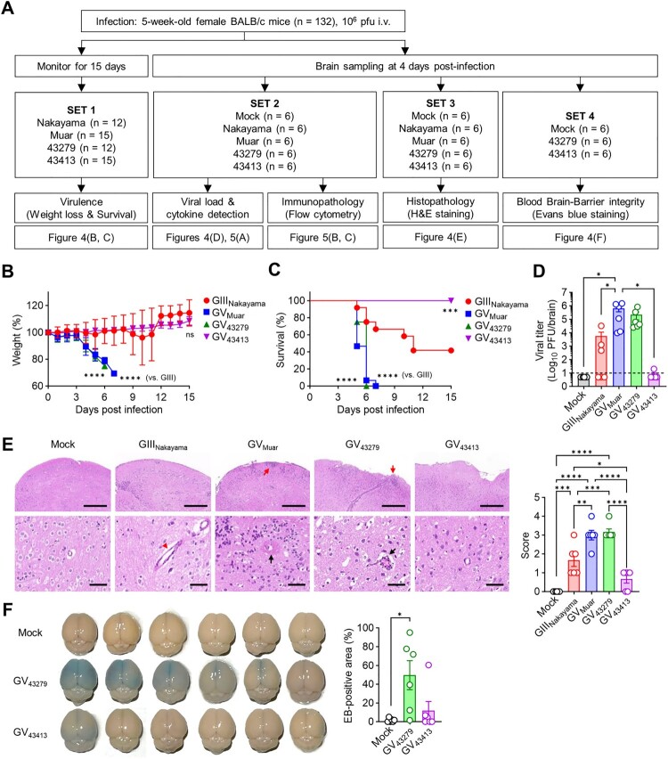
Japanese encephalitis (JE), caused by the Japanese encephalitis virus (JEV) infection, continues to pose significant public health challenges worldwide despite efficient vaccines. The virus is classified into five genotypes, among which genotype V (GV) was not detected for a long period after its initial isolation in 1952, until reports emerged from China and the Republic of Korea (ROK) since 2009. The characteristics of the virus are crucial in estimating its potential epidemiological impact. However, characterization of GV JEVs has so far been limited to two strains: Muar, the original isolate, and XZ0934, isolated in China. Two additional ROK GV JEV isolates, NCCP 43279 and NCCP 43413, are currently available, but their characteristics have not been explored. Our phylogenetic analysis revealed that GV virus sequences from the ROK segregate into two clades. NCCP 43279 and NCCP 43413 belong to different clades and exhibit distinct in vitro phenotypes. NCCP 43279 forms larger plaques but demonstrates inefficient propagation in cell culture compared to NCCP 43413. In vivo, NCCP 43279 induces higher morbidity and mortality in mice than NCCP 43413. Notably, NCCP 43279 shows more severe blood-brain barrier damage, suggesting superior brain invasion capabilities. Consistent with its higher virulence, NCCP 43279 displays more pronounced histopathological and immunopathological outcomes. In conclusion, our study confirms that the two ROK isolates are not only classified into different clades but also exhibit distinct in vitro and in vivo characteristics.

Keywords: Japanese encephalitis virus; characterization; genotype V; phylogenetic analysis; virulence.


Conflict of interest statement

No potential conflict of interest was reported by the author(s).

Figures

Figure 1.
Figure 1.
Nucleotide and amino acid sequence comparison of 43279 with K15P38 and 43413. (A) Viral genome sequences (upper panel) and amino acid sequences of polypeptide (lower panel) were compared between strains 43279 and K15P38. Mutations involving silent mutations and the corresponding amino acid positions are highlighted in red. Position numbers are calculated using only the open reading frame (ORF), omitting the untranslated regions. (B) Amino acid sequences of polypeptides were compared between strains 43279 and 43413. The amino acid positions related to gene mutations are indicated by dots at the top of the diagram, and positions with amino acid substitutions are highlighted in red.
Figure 2.
Figure 2.
Phylogenetic analysis of 43279 and 43413. Phylogenetic analysis was conducted using maximum likelihood (Tamura-Nei model) analysis. (A) The analysis utilized the whole open reading frame (ORF) sequences of all JEV genotypes, incorporating a total of 31 whole ORF sequences. (B) A phylogenetic analysis on the whole ORF sequences of GV JEVs and representative strains from genotypes GI to GIV, with 43279 and 43413 marked with closed circles. (C) A cluster analysis of GV JEVs from the ROK was performed based on 15 Env gene sequences. Sequences from distinct clades are highlighted in different colours. The scale bar represents estimated evolutionary distances with 0.01 (A, B) or 0.002 (C) changes per nucleotide position.
Figure 3.
Figure 3.
In vitro characterization of 43279 and 43413. (A) BHK-21 cells were cultured in a 6-well plate and infected with the indicated JEVs. Plaque sizes were measured at 70 hours post-infection (n = 23). Every dot represents the mean plaque size in each well. (B, C) BHK-21 cells were infected with JEVs at an MOI of 0.01 (n = 6). Virus titre (B) and the number of attached cells (C) were measured at indicated time points. Data were pooled from three independent experiments.
Figure 4.
Figure 4.
In vivo characterization of 43279 and 43413 in mice. (A) Flowchart illustrating the animal experiments conducted in this study. BALB/c mice were intravenously infected with 106 pfu of each JEV strain. (B, C) Weigh changes (B) and survival rates (C) were monitored for 15 days (n = 12-15). Data were pooled from three independent experiments. (D-F) Sets of BALB/c mice were infected with 106 pfu of each JEV strain and sacrificed for virus titration (D), evaluation of histological pathology (E), and BBB integrity test (F) in brain tissue (n = 6, each set). Data were pooled from two independent experiments. (E) Invasion of abnormal granular cells into outer molecular cortex layers (red arrow), extravasation of blood (red arrow head), and perivascular hemorrhage with massive cell infiltration (black arrow) are indicated. Scale bars = 500 µm (upper panel), 50 µm (lower panel).
Figure 5.
Figure 5.
Immunopathology of mice infected with 43279 and 43413. BALB/c mice were intravenously infected with 106 pfu of each JEV strain (n = 6). (A) TNF-α, IL-6, and MCP-1 levels were measured in brain homogenates (n = 6). (B) Representative flow cytometry analysis of brain myeloid cells (CD45hiCD11b+). Myeloid cells were further divided into neutrophils (Ly6CintLy6G+) and monocytes (Ly6ChiLy6G-). (C) Absolute number and proportion of myeloid cells in the brain (n = 6). Data were pooled from two independent experiments.

Similar articles

Cited by

References

    1. Quan TM, Thao TTN, Duy NM, et al. . Estimates of the global burden of Japanese encephalitis and the impact of vaccination from 2000-2015. Elife. 2020;9:e51027. doi:10.7554/eLife.51027 - DOI - PMC - PubMed
    1. Campbell GL, Hills SL, Fischer M, et al. . Estimated global incidence of Japanese encephalitis: a systematic review. Bull World Health Organ. 2011;89(10):774A–774E. - PMC - PubMed
    1. Zheng Y, Li M, Wang H, et al. . Japanese encephalitis and Japanese encephalitis virus in mainland China. Rev Med Virol. 2012;22(5):301–322. doi:10.1002/rmv.1710 - DOI - PubMed
    1. Pan XL, Liu H, Wang HY, et al. . Emergence of genotype I of Japanese encephalitis virus as the dominant genotype in Asia. J Virol. 2011;85(19):9847–9853. doi:10.1128/JVI.00825-11 - DOI - PMC - PubMed
    1. Han N, Adams J, Chen P, et al. . Comparison of genotypes I and III in Japanese encephalitis virus reveals distinct differences in their genetic and host diversity. J Virol. 2014;88(19):11469–11479. doi:10.1128/JVI.02050-14 - DOI - PMC - PubMed