. 2024 Jul 15;213(2):226-234.
doi: 10.4049/jimmunol.2300717.

Structural and Biochemical Requirements for Secretory Component Interactions with Dimeric IgA

Affiliations

Structural and Biochemical Requirements for Secretory Component Interactions with Dimeric IgA

Sonya Kumar Bharathkar et al. J Immunol. .

Abstract

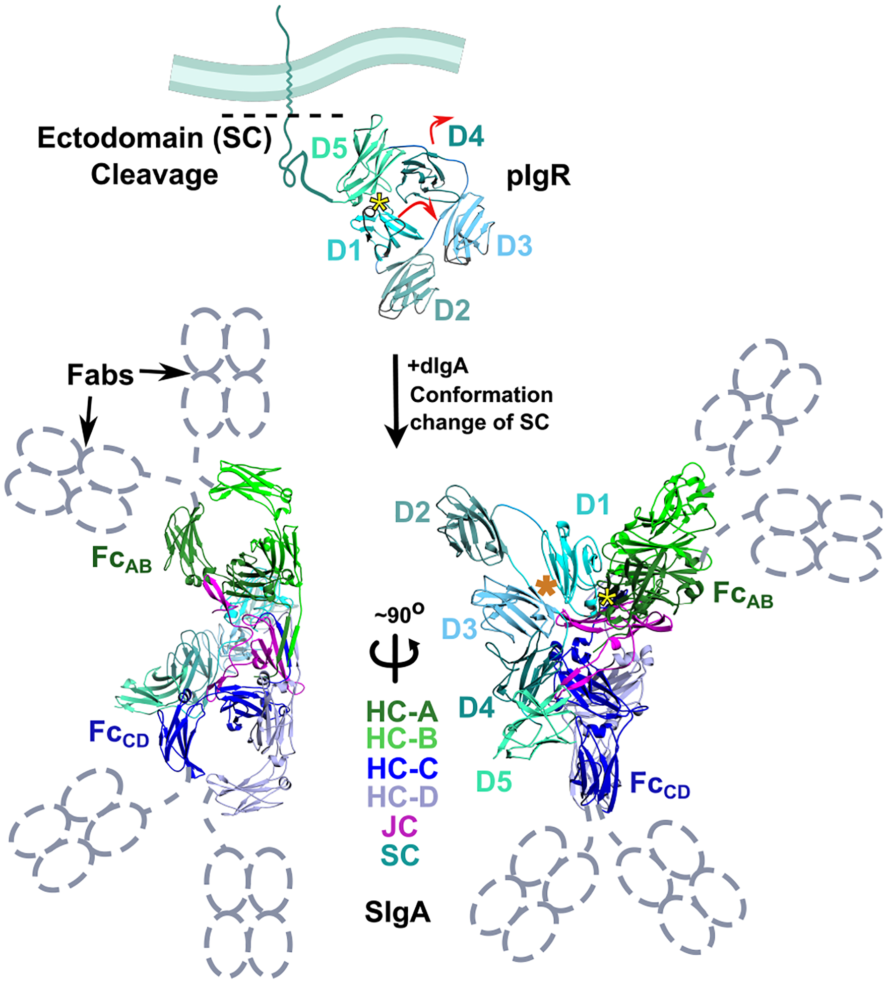
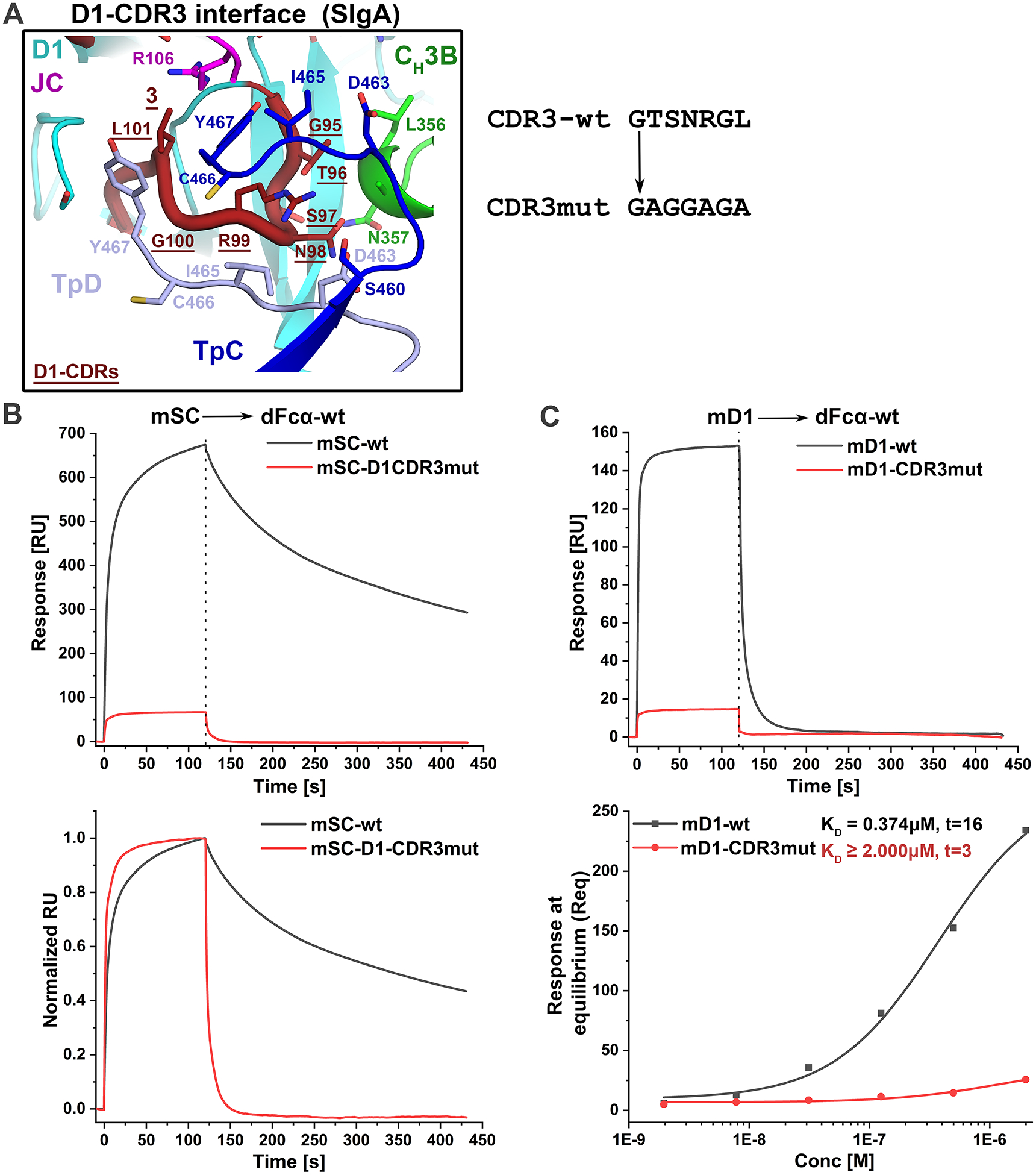
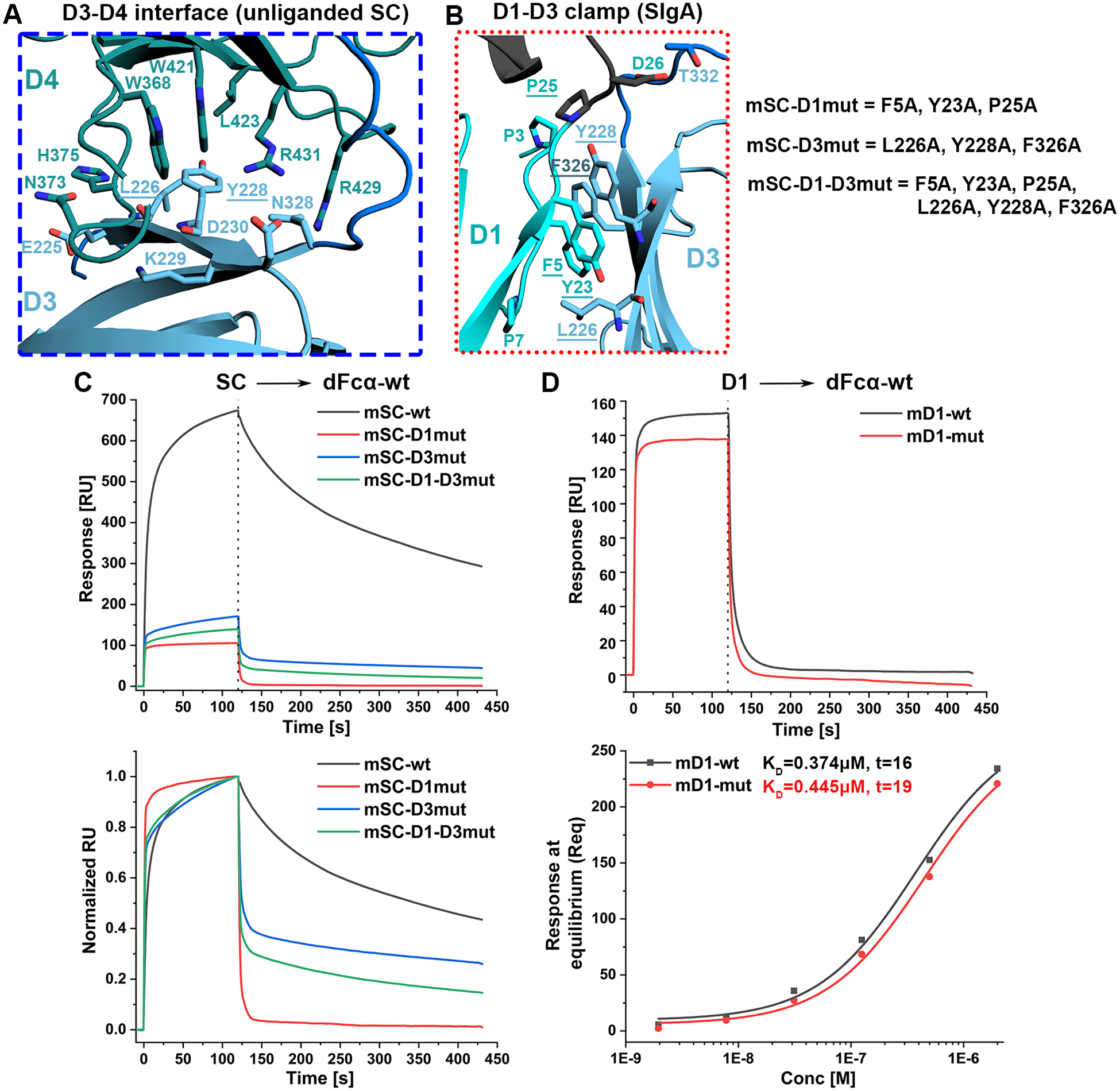
Secretory (S) IgA is the predominant mucosal Ab that protects host epithelial barriers and promotes microbial homeostasis. SIgA production occurs when plasma cells assemble two copies of monomeric IgA and one joining chain (JC) to form dimeric (d) IgA, which is bound by the polymeric Ig receptor (pIgR) on the basolateral surface of epithelial cells and transcytosed to the apical surface. There, pIgR is proteolytically cleaved, releasing SIgA, a complex of the dIgA and the pIgR ectodomain, called the secretory component (SC). The pIgR's five Ig-like domains (D1-D5) undergo a conformational change upon binding dIgA, ultimately contacting four IgA H chains and the JC in SIgA. In this study, we report structure-based mutational analysis combined with surface plasmon resonance binding assays that identify key residues in mouse SC D1 and D3 that mediate SC binding to dIgA. Residues in D1 CDR3 are likely to initiate binding, whereas residues that stabilize the D1-D3 interface are likely to promote the conformational change and stabilize the final SIgA structure. Additionally, we find that the JC's three C-terminal residues play a limited role in dIgA assembly but a significant role in pIgR/SC binding to dIgA. Together, these results inform models for the intricate mechanisms underlying IgA transport across epithelia and functions in the mucosa.


Figures

Figure-1:
Figure-1:. The structure of liganded and unliganded SC.
(Top) Cartoon representation of a mouse pIgR ectodomain homology model based on the human SC crystal structure (PDB:5D4K). The pIgR is depicted bound to an epithelial cell membrane with its five constitutive immunoglobin-like domains (D1-D5) adopting a closed, unliganded conformation. Upon encountering dIgA, D1-D5 undergoes conformational change (indicated by red arrows) to form SIgA (PDB:7JG2), which is shown as a cartoon representation in two orientations (Fabs are disordered and represented with dotted schematics in grey). The dimeric SIgA contains four heavy chains (HC-A to D), J-chain (JC), and one Secretory Component (SC). All chains are colored according to the key. The Fc formed by HC-A and HC-B is indicated as FcAB (green) and the Fc formed by HC-C and HC-D is indicated as FcCD (of blue). The JC occupies the center of the SIgA complex with FcAB and FcCD on opposite sides. The SC has five immunoglobin domains (D1-D5), and each domain (D1-D5) are colored with different shades of cyan. The yellow asterisk marks the location of D1 CDR loops in unliganded and liganded structures and the orange asterisk indicated the location of the D1-D3 interface in the liganded (SIgA) structure. Color scheme and labeling approach are adapted from S. Kumar Bharathkar et al., “The structures of secretory and dimeric immunoglobulin A,” eLife, vol. 9, p. e56098, Oct. 2020 (11).
Figure-2:
Figure-2:. Contribution of mSC-D1-CDR3 towards binding dIgA.
(A) Detailed view of mSC-D1-CDR3 and dIgA interactions found in mouse SIgA (PDB: 7JG2) (Fig.1, yellow asterisk); the structure is shown as a cartoon representation with interacting residues shown as sticks and labeled. D1-CDR3 residues that were mutated are underlined, and the unmutated and mutated sequences are shown on the right. (B) A representative SPR sensor-grams showing the response (top) and normalized response (bottom) of 0.5μM mSC-wt (black) and mSC-D1-CDR3mut (red) binding to dFcα-wt. (C) A representative SPR sensor-gram showing the response of 0.5μM of mD1-wt (black) and mD1-CDR3mut (red) binding to dFcα-wt (top) and the concentration dependent binding and steady state fit (bottom). The calculated KD value for mD1-wt is indicated; the t-value associated with mD1-CDR3mut binding is t<10 indicating a poor fit and uncertainty in the calculated KD value, which is estimated to be greater than 2 μM. Data from this figure are indicated by green inverted triangles in Fig.S5A. Results are consistent with 5 replicate experiments.
Figure-3:
Figure-3:. Role of SC-D1-D3 Clamp for the stabilization of SC and dIgA binding.
(A) Detailed view of the unliganded D3-D4 interface from the homology model of mSC and (B) the D1-D3 interface from the SIgA cryo-EM structure (Fig.1, orange asterisk) (PDB code 7JG2). In (A) and (B) structures are shown as a cartoon representation with interacting residues, shown as sticks and labeled. SC D1 and D3 residues that were mutated are underlined, and the unmutated and mutated sequences are shown on the right. (C) SPR sensor-grams showing the response (top) and normalized response (bottom) of 0.5μM mSC-wt (black) mSC-D1mut (red), mSC-D3mut (blue) and mSC-D1-D3mut (green) binding to dFcα-wt. (D) SPR sensor-grams showing the response of 0.5μM of mD1-wt (black) and mD1-mut binding to dFcα-wt (top) and the concentration dependent binding and steady state fit (bottom). The calculated KD value for each analyte is given to the right of the name along with the t-value. Data from this figure are indicated by green inverted triangles in Fig.S5A. Results are consistent with 5 replicate experiments.
Figure-4:
Figure-4:. Role of the JC YPD motif in binding mSC-wt or mD1-wt.
(A) The interaction of JC with SC-D1, and specifically D1-CDR1 shown as red loop with larger radius. The interacting residues are shown as sticks at this interface. Y135, P136 and D137 are underlined in JC, indicating that these residues were subjected to mutations, such as alanine substitutions or truncations. (B) The SEC chromatograms of the indicated dFcα-wt and dFcα mutant variants resulting from 25ml co-transfections and following Nickel-NTA purification; fractions corresponding to dimeric forms were used for SPR experiments. (C) SPR sensor-grams of 0.5μM mD1-wt binding to the indicated dFcαs with JC- YPD alanine mutations or truncations. (D) The steady state affinity analysis of mD1-wt binding to the indicated wt or mutant dFcα variants and (E) plot showing the resulting KD values for the indicated variants. The * represents the data points for which the steady state fitting t-value was <10 and/or KD≥2μM indicating a poor fit; the affinity for data meeting these criteria are estimated KD≥ 2μM. ** represents a KD value calculated from steady state analysis with t=11 and low maximal response indicating low confidence in the calculated value and estimated KD≥2μM. KD values calculated from replicate experiments are summarized in FigS5B. (F) SPR sensor-grams of 0.5μM mSC-wt binding to different dFcα with the indicated JC- YPD alanine mutations or truncations. (G) SPR sensor-grams showing the normalized response of 0.5μM mSC-wt analytes binding to the indicated dFcα variants. Results are consistent with 3 or more replicate experiments.

Update of

Similar articles

References

    1. Corthesy B 2013. Multi-Faceted Functions of Secretory IgA at Mucosal Surfaces. Frontiers in Immunology 4. - PMC - PubMed
    1. Mantis NJ, Rol N, and Corthésy B. 2011. Secretory IgA’s complex roles in immunity and mucosal homeostasis in the gut. Mucosal Immunol 4: 603–611. - PMC - PubMed
    1. de Sousa-Pereira P, and Woof JM. 2019. IgA: Structure, Function, and Developability. Antibodies 8: 57. - PMC - PubMed
    1. Zikan J, Mestecky J, Kulhavy R, and Bennett JC. 1986. The stoichiometry of J chain in human secretory dimeric IgA. Molecular Immunology 23: 541–544. - PubMed
    1. Lombana TN, Rajan S, Zorn JA, Mandikian D, Chen EC, Estevez A, Yip V, Bravo DD, Phung W, Farahi F, Viajar S, Lee S, Gill A, Sandoval W, Wang J, Ciferri C, Boswell CA, Matsumoto ML, and Spiess C. 2019. Production, characterization, and in vivo half-life extension of polymeric IgA molecules in mice. mAbs 11: 1122–1138. - PMC - PubMed

Publication types

MeSH terms

Substances

LinkOut - more resources