. 2024 May 27;16(10):9251-9263.
doi: 10.18632/aging.205869. Epub 2024 May 27.

Trimethylamine-N-oxide promotes osteoclast differentiation and oxidative stress by activating NF-κB pathway

Affiliations

Trimethylamine-N-oxide promotes osteoclast differentiation and oxidative stress by activating NF-κB pathway

Yangyang Zhao et al. Aging (Albany NY). .

Abstract

Background: Senile osteoporosis may be caused by an imbalance in intestinal flora and oxidative stress. Trimethylamine-N-oxide (TMAO), a metabolite of dietary choline dependent on gut microbes, has been found to be significantly increased in osteoporosis. However, the role of TMAO in bone loss during osteoporosis remains poorly understood. In this study, we examined the impact of TMAO on osteoclast differentiation and bone resorption in an in vitro setting.

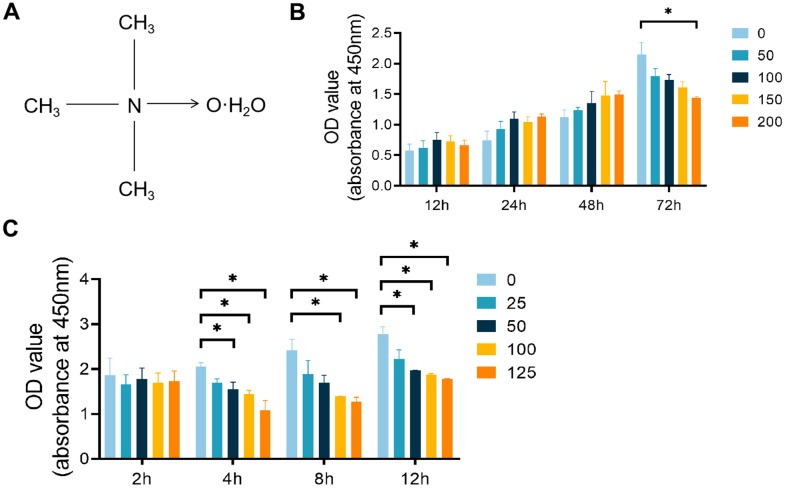
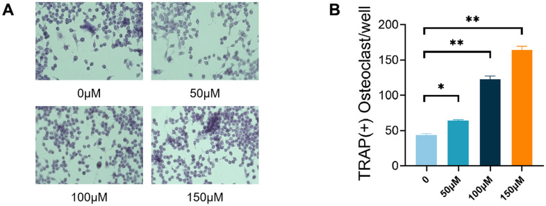
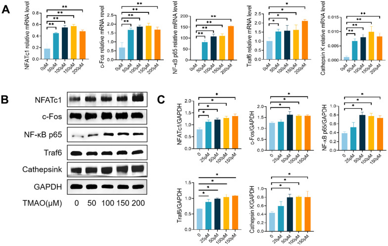
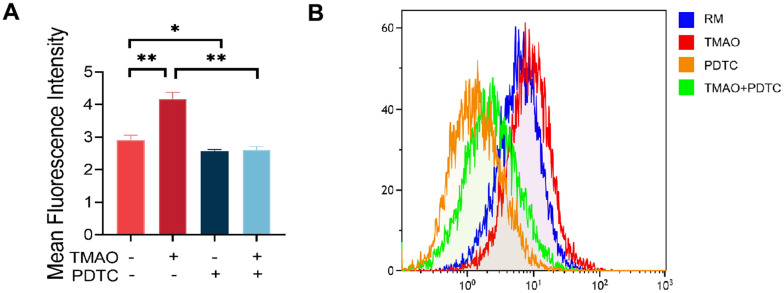
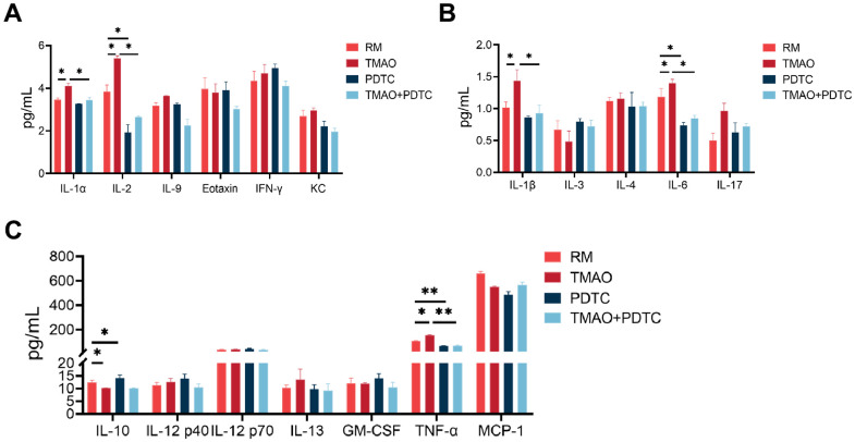
Methods: Osteoclast differentiation was induced by incubating RAW 264.7 cells in the presence of Receptor Activator for Nuclear Factor-κB Ligand (RANKL) and macrophage-stimulating factor (M-CSF). Flow cytometry, TRAP staining assay, CCK-8, and ELISA were employed to investigate the impact of TMAO on osteoclast differentiation and bone resorption activity in vitro. For mechanistic exploration, RT-PCR and Western blotting were utilized to assess the activation of the NF-κB pathway. Additionally, protein levels of secreted cytokines and growth factors were determined using suspension array technology.

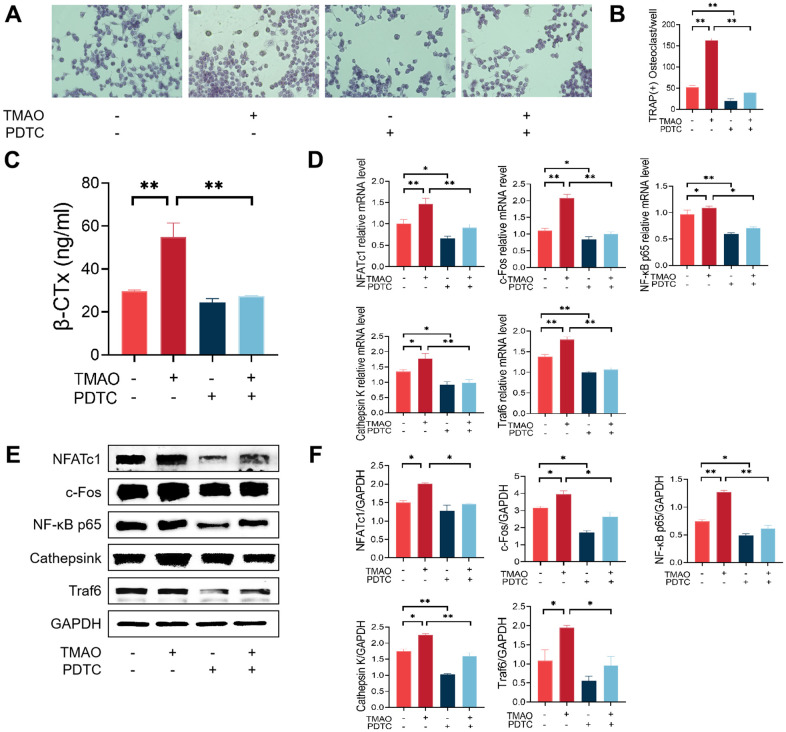
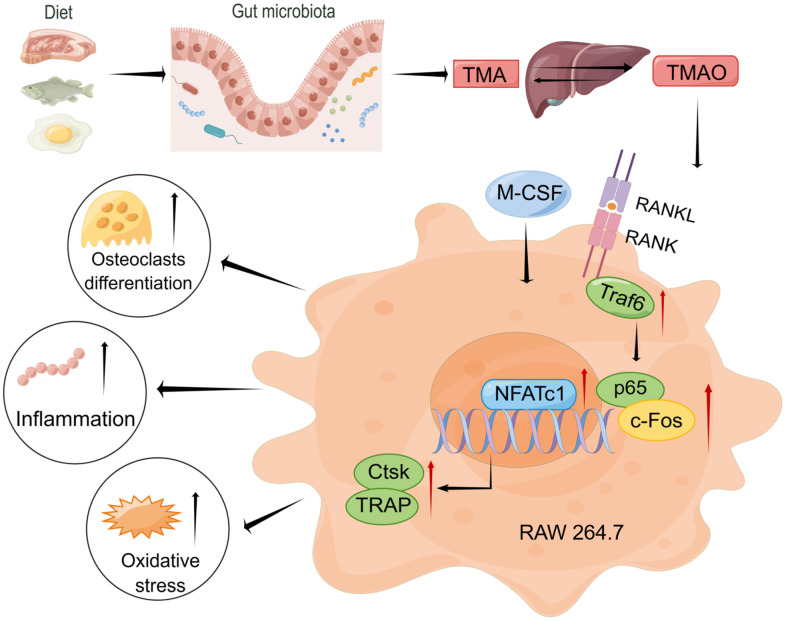
Results: Our findings demonstrate that TMAO enhances RANKL and M-CSF-induced osteoclast formation and bone resorption in a dose-dependent manner. Mechanistically, TMAO triggers the upregulation of the NF-κB pathway and osteoclast-related genes (NFATc1, c-Fos, NF-κB p65, Traf6, and Cathepsin K). Furthermore, TMAO markedly elevated the levels of oxidative stress and inflammatory factors.

Conclusions: In conclusion, TMAO enhances RANKL and M-CSF-induced osteoclast differentiation and inflammation in RAW 264.7 cells by activating the NF-κB signaling pathway. These findings offer a new rationale for further academic and clinical research on osteoporosis treatment.

Keywords: NF-κB; inflammation; osteoclast; reactive oxygen species; trimethylamine-N-oxide.


Conflict of interest statement

CONFLICTS OF INTEREST: The authors declare that they have no conflicts of interest.

Figures

Figure 1
Figure 1
Effects of TMAO and PDTC on the viability of RAW 264.7 cells. (A) Molecular structure of TMAO. (B) The cell viability of RAW 264.7 cells after treatment with different concentrations of TMAO for 12, 24, 48, and 72 h was evaluated by CCK-8. The cell viability of RAW 264.7 cells decreased significantly at 72 h with 200 μM TMAO treatment. (C) The cell viability of RAW 264.7 cells after treatment with different concentrations of PDTC for 2, 4, 8, and 12 h was evaluated by CCK-8. The cell viability of RAW 264.7 cells decreased significantly at 4 h with 50 μM PDTC treatment. * p < 0.05, ** p < 0.01.
Figure 2
Figure 2
TMAO promoted RANKL-induced osteoclast differentiation. (A) RAW 264.7 cells were cultured for 4 d with RANKL (50 ng/mL) and M-CSF (10 ng/mL) in the presence of varying concentrations of TMAO and then stained for TRAP activity. Representative photomicrographs were taken under a light microscope (magnification ×40). (B) TRAP-positive cells containing more than three nuclei were counted as osteoclasts; ** p < 0.01.
Figure 3
Figure 3
TMAO enhanced RANKL-induced osteoclast-associated gene expression. (A) TMAO enhanced osteoclast gene expression (n = 4 per group) as examined by real-time PCR. (B, C) TMAO enhanced NFATc1, c-Fos, Cathepsin K, NF-κB p65, and Traf6 protein expression as evaluated by Western blot (n = 3 per group). * p < 0.05, ** p < 0.01.
Figure 4
Figure 4
Inhibition of the NF-κB signaling pathway with PDTC reversed the effect of TMAO on osteoclast differentiation. (A, B) RAW 264.7 cells were pretreated with 25 μM PDTC for 2 h before being cultured with or without 100 μM TMAO. The number of TRAP-positive osteoclasts increased under 100 μM TMAO and decreased with PDTC pretreatment with or without TMAO. (magnification ×40). (C) The expression of β-CTx was up-regulated under 100 μM TMAO and suppressed by PDTC with or without TMAO treatment evaluated by ELISA (n = 3 per group). (D) The expression of osteoclast-specific genes was up-regulated under 100 μM TMAO and suppressed by PDTC with or without TMAO treatment evaluated by real-time PCR (n = 4 per group). (E, F) The protein expression levels of NFATc1, c-Fos, Cathepsin K, NF-κB p65, and Traf6 were promoted under 100 μM TMAO and suppressed by PDTC with or without TMAO treatment as examined by Western blot (n = 3 per group). * p < 0.05, ** p < 0.01.
Figure 5
Figure 5
TMAO increased ROS levels in RAW 264.7 cells during osteoclast differentiation. (A) RAW 264.7 cells were stimulated with RANKL and M-CSF for 4 days and then probed with 10 μM DCFH-DA for 30 min after being treated with TMAO or/and PDTC. TMAO treatment led to increased fluorescence intensity of ROS in RAW 264.7 cells, and PDTC treatment decreased TMAO-induced ROS generation (n = 5 per group). * p < 0.05, ** p < 0.01. (B) The ROS production in RAW 264.7 cells after being treated with TMAO or/and PDTC was detected by FCM. RM: RANKL and M-CSF.
Figure 6
Figure 6
TMAO enhanced inflammation level during osteoclast differentiation, analyzed by the Bio-Plex murine 23-Plex Panel Kit (Bio-Rad Laboratories). (A) IL-1α and IL-2 were elevated in the culture in the existence of TMAO stimulation. (B, C) IL-1β, IL-6, and TNF-α were also elevated, whereas IL-10 were significantly reduced in the TMAO cultures. RM: RANKL and M-CSF. * p < 0.05, ** p < 0.01.
Figure 7
Figure 7
Working model drawn by Figdraw illustrating the promotion mechanism of TMAO on RANKL-induced osteoclast differentiation.

Similar articles

Cited by

References

    1. Ensrud KE, Crandall CJ. Osteoporosis. Ann Intern Med. 2017; 167:ITC17–32. 10.7326/AITC201708010 - DOI - PubMed
    1. Ling Z, Yang C, Tan J, Dou C, Chen Y. Beyond immunosuppressive effects: dual roles of myeloid-derived suppressor cells in bone-related diseases. Cell Mol Life Sci. 2021; 78:7161–83. 10.1007/s00018-021-03966-9 - DOI - PMC - PubMed
    1. Hamilton JA. CSF-1 signal transduction. J Leukoc Biol. 1997; 62:145–55. 10.1002/jlb.62.2.145 - DOI - PubMed
    1. Suda T, Takahashi N, Udagawa N, Jimi E, Gillespie MT, Martin TJ. Modulation of osteoclast differentiation and function by the new members of the tumor necrosis factor receptor and ligand families. Endocr Rev. 1999; 20:345–57. 10.1210/edrv.20.3.0367 - DOI - PubMed
    1. Kong YY, Feige U, Sarosi I, Bolon B, Tafuri A, Morony S, Capparelli C, Li J, Elliott R, McCabe S, Wong T, Campagnuolo G, Moran E, et al.. Activated T cells regulate bone loss and joint destruction in adjuvant arthritis through osteoprotegerin ligand. Nature. 1999; 402:304–9. 10.1038/46303 - DOI - PubMed

Publication types

MeSH terms