. 2024 May 29;19(5):e0303643.
doi: 10.1371/journal.pone.0303643. eCollection 2024.

In vivo cisplatin-resistant neuroblastoma metastatic model reveals tumour necrosis factor receptor superfamily member 4 (TNFRSF4) as an independent prognostic factor of survival in neuroblastoma

Affiliations

In vivo cisplatin-resistant neuroblastoma metastatic model reveals tumour necrosis factor receptor superfamily member 4 (TNFRSF4) as an independent prognostic factor of survival in neuroblastoma

Catherine Murphy et al. PLoS One. .

Abstract

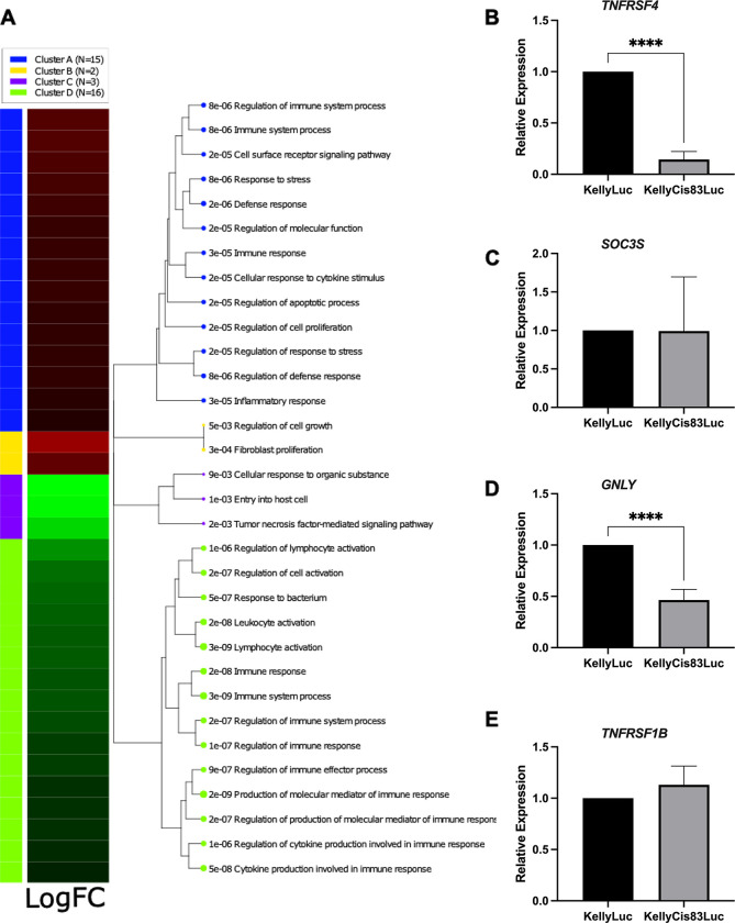
Neuroblastoma is the most common solid extracranial tumour in children. Despite major advances in available therapies, children with drug-resistant and/or recurrent neuroblastoma have a dismal outlook with 5-year survival rates of less than 20%. Therefore, tackling relapsed tumour biology by developing and characterising clinically relevant models is a priority in finding targetable vulnerability in neuroblastoma. Using matched cisplatin-sensitive KellyLuc and resistant KellyCis83Luc cell lines, we developed a cisplatin-resistant metastatic MYCN-amplified neuroblastoma model. The average number of metastases per mouse was significantly higher in the KellyCis83Luc group than in the KellyLuc group. The vast majority of sites were confirmed as having lymph node metastasis. Their stiffness characteristics of lymph node metastasis values were within the range reported for the patient samples. Targeted transcriptomic profiling of immuno-oncology genes identified tumour necrosis factor receptor superfamily member 4 (TNFRSF4) as a significantly dysregulated MYCN-independent gene. Importantly, differential TNFRSF4 expression was identified in tumour cells rather than lymphocytes. Low TNFRSF4 expression correlated with poor prognostic indicators in neuroblastoma, such as age at diagnosis, stage, and risk stratification and significantly associated with reduced probability of both event-free and overall survival in neuroblastoma. Therefore, TNFRSF4 Low expression is an independent prognostic factor of survival in neuroblastoma.


Conflict of interest statement

The authors declare that they have no competing interests. The funders had no role in the design of the study; in the collection, analyses, or interpretation of data; in the writing of the manuscript, or in the decision to publish the results.

Figures

Fig 1
Fig 1. Cisplatin-resistant KellyCis83Luc tail-vein injections cause more metastases in murine xenografts than drug-sensitive KellyLuc injections.
(A) Mean number of metastases per mouse for both cell lines. (B) Percentage of mice with more than 10 metastatic foci per cell line. (C) Metastatic foci grown in immunodeficient mice injected with the KellyLuc and KellyCis83Luc cell lines by tail vein. The graph represents the number and location of metastases in the indicated cell lines. (D) Schematic of major murine lymph nodes. (E) Representative histology of metastatic foci in each location by cell line. Scale bars: 1000 μm for whole slide images and 100 μm for magnifications. (F) Stiffness of lymph node metastasis tissues of the two cell lines KellyLuc (n = 2) and KellyCis83Luc (n = 5). Mets–metastases, Kelly–KellyLuc, KellyCis–KellyCis83Luc.
Fig 2
Fig 2. Establishment and analysis of a drug-resistant neuroblastoma murine xenograft model.
A) Fox Chase SCID Beige mice were injected with either cisplatin-sensitive KellyLuc or cisplatin-resistant KellyCis83Luc cells. B) After 40 days, metastases were resected, formalin-fixed paraffin embedded (FFPE) and stained with hematoxylin and eosin (H&E) to identify tumour cell-enriched regions. C) These positive regions were macrodissected for RNA sequencing via the HTG EdgeSeq Immuno-Oncology assay. D) The log fold-change (logFC) in the expression of each gene in cisplatin-sensitive vs cisplatin-resistant tumours was assessed. Using cut-off values of LogFC >±0.6 and p≤0.05, 36 candidate genes were identified. E) Pathway analysis software was used to gain insights into the molecular pathways underlying the candidate gene expression patterns. F) Kaplan‒Meier survival analysis was conducted to determine the clinical relevance of candidate genes in neuroblastoma. G) Expression trends of shortlisted genes were validated by RT‒qPCR on fresh-frozen xenograft tissue. H) The correlation between shortlisted genes and MYCN was calculated, and MYCN dependence was assessed via an inducible system. Created with biorender.com.
Fig 3
Fig 3. Functional enrichment analysis identifies pathways of immune system-related processes.
A) iDEP software analyses. N refers to the number of enriched GO biological processes for each cluster. For each enriched process, a p value is given. Gene sets closer on the tree share more genes. Dot sizes correspond to adjusted p values, the higher p value the bigger the dot. (B-E) Validation of some differentially expressed genes by RT‒qPCR.
Fig 4
Fig 4. Clinical relevance of TNFRSF4 in neuroblastoma.
Kaplan-Meier analysis ofTNFRSF4 expression for OS (A), EFS (B), TNFRSF4 mRNA expression correlations with clinical variables in neuroblastoma (N = 498) (C) EFS: Event-Free Survival; FAV: unfavorable/favorable (class label for extreme disease course); HR: High-Risk patients; OS: Overall Survival. High-Risk: patients with stage 4 disease >18 months at diagnosis and patients of any age and stage with MYCN-amplified tumours. The data generated using the IBM SPSS 21 software.
Fig 5
Fig 5. Univariate Cox regression analysis.
The SEQC neuroblastoma cohort was used for univariate Cox regression analysis for multiple prognostic variables in neuroblastoma, including the expression of TNFRSF4. Forest plots were generated based on the hazard ratios, 95% confidence intervals (CIs) and significance values (p). The data generated using the IBM SPSS 21 software.
Fig 6
Fig 6. Multivariate Cox regression analysis.
The SEQC neuroblastoma cohort was used for multivariate Cox regression analysis to assess whether the hazard ratios for INSS stage 4, MYCN amplification, age at diagnosis ≥ 18 months and low expression of TNFRSF4 were significant independent of each other. Forest plots were generated based on the hazard ratios, 95% confidence intervals (CI) and significance values (p). The data generated using the IBM SPSS 21 software.
Fig 7
Fig 7. Assessment of TNFRSF4 regulation by the MYCN oncogene and impact on cellular proliferation.
A) Correlation between MYCN and TNFRSF4 expression in a cohort of 498 neuroblastomas generated in R2. B) SHEP-Tet-21N cells were grown in the presence (MYCN-off) and absence (MYCN-on) of doxycycline, and RNA was extracted for RT‒qPCR to assess whether the expression of TNFRSF4 is MYCN dependent. C) Expression of MYCN and TNFRSF4 in SHEP-Tet21N cells grown in the presence and absence of doxycycline. D) Kelly and SK-N-AS cells were transfected with either a scrambled negative control (NC), siTNFRSF4 or siKIF11. Cell viability was assessed using the CytoTox-Glo™ Assay. The values were converted to a percentage of the viable cells compared to the negative control. The results of 2 independent experiments run in 3 biological replicates were combined. These data were statistically analysed using one-way ANOVA to determine whether cell viability was significantly reduced as a result of transfection. E) RT‒qPCR validation of downregulation of TNFRSF4 expression in Kelly and SK-N-AS cell transfectants. Two-tailed unpaired T tests were performed in GraphPad Prism to detect significant differences in expression (*p ≤ 0.05; **p ≤ 0.01; ***p≤ 0.001; ****p ≤ 0.0001). Analysis confirmed significant downregulation of TNFRSF4 in cells transfected with siTNFRSF4 compared to NC. Image created with Biorender.com.

References

    1. Lundberg K.I.; Treis D.; Johnsen J.I. Neuroblastoma Heterogeneity, Plasticity, and Emerging Therapies. Curr. Oncol. Rep. 2022, 24, 1053–1062, doi: 10.1007/s11912-022-01270-8 - DOI - PMC - PubMed
    1. Smith V.; Foster J. High-Risk Neuroblastoma Treatment Review. Children 2018, 5, 114, doi: 10.3390/children5090114 - DOI - PMC - PubMed
    1. Tolbert V.P.; Matthay K.K. Neuroblastoma: clinical and biological approach to risk stratification and treatment. Cell Tissue Res. 2018, 372, 195–209. doi: 10.1007/s00441-018-2821-2 - DOI - PMC - PubMed
    1. DuBois S.G.; Kalika Y.; Lukens J.N.; Brodeur G.M.; Seeger R.C.; Atkinson J.B.; et al.. Metastatic sites in stage IV and IVS neuroblastoma correlate with age, tumor biology, and survival. J. Pediatr. Hematol. Oncol. 1999, 21, 181–189, doi: 10.1097/00043426-199905000-00005 - DOI - PubMed
    1. Anderson J.; Majzner R.G.; Sondel P.M. Immunotherapy of Neuroblastoma: Facts and Hopes. Clin. Cancer Res. 2022, 28, 3196–3206, doi: 10.1158/1078-0432.CCR-21-1356 - DOI - PMC - PubMed