. 2024 May 30;390(20):1873-1884.
doi: 10.1056/NEJMoa2312665.

The Role of Interferon-γ in Autoimmune Polyendocrine Syndrome Type 1

Vasileios Oikonomou  1 Grace Smith  1 Gregory M Constantine  1 Monica M Schmitt  1 Elise M N Ferré  1 Julie C Alejo  1 Deanna Riley  1 Dhaneshwar Kumar  1 Lucas Dos Santos Dias  1 Joseph Pechacek  1 Yannis Hadjiyannis  1 Taura Webb  1 Bryce A Seifert  1 Rajarshi Ghosh  1 Magdalena Walkiewicz  1 Daniel Martin  1 Marine Besnard  1 Brendan D Snarr  1 Shiva Deljookorani  1 Chyi-Chia R Lee  1 Tom DiMaggio  1 Princess Barber  1 Lindsey B Rosen  1 Aristine Cheng  1 Andre Rastegar  1 Adriana A de Jesus  1 Jennifer Stoddard  1 Hye Sun Kuehn  1 Timothy J Break  1 Heidi H Kong  1 Leslie Castelo-Soccio  1 Ben Colton  1 Blake M Warner  1 David E Kleiner  1 Martha M Quezado  1 Jeremy L Davis  1 Kevin P Fennelly  1 Kenneth N Olivier  1 Sergio D Rosenzweig  1 Anthony F Suffredini  1 Mark S Anderson  1 Marc Swidergall  1 Carole Guillonneau  1 Luigi D Notarangelo  1 Raphaela Goldbach-Mansky  1 Olaf Neth  1 Maria Teresa Monserrat-Garcia  1 Justo Valverde-Fernandez  1 Jose Manuel Lucena  1 Ana Lucia Gomez-Gila  1 Angela Garcia Rojas  1 Mikko R J Seppänen  1 Jouko Lohi  1 Matti Hero  1 Saila Laakso  1 Paula Klemetti  1 Vanja Lundberg  1 Olov Ekwall  1 Peter Olbrich  1 Karen K Winer  1 Behdad Afzali  1 Niki M Moutsopoulos  1 Steven M Holland  1 Theo Heller  1 Stefania Pittaluga  1 Michail S Lionakis  1
Affiliations

The Role of Interferon-γ in Autoimmune Polyendocrine Syndrome Type 1

Vasileios Oikonomou et al. N Engl J Med. .

Abstract

Background: Autoimmune polyendocrine syndrome type 1 (APS-1) is a life-threatening, autosomal recessive syndrome caused by autoimmune regulator (AIRE) deficiency. In APS-1, self-reactive T cells escape thymic negative selection, infiltrate organs, and drive autoimmune injury. The effector mechanisms governing T-cell-mediated damage in APS-1 remain poorly understood.

Methods: We examined whether APS-1 could be classified as a disease mediated by interferon-γ. We first assessed patients with APS-1 who were participating in a prospective natural history study and evaluated mRNA and protein expression in blood and tissues. We then examined the pathogenic role of interferon-γ using Aire-/-Ifng-/- mice and Aire-/- mice treated with the Janus kinase (JAK) inhibitor ruxolitinib. On the basis of our findings, we used ruxolitinib to treat five patients with APS-1 and assessed clinical, immunologic, histologic, transcriptional, and autoantibody responses.

Results: Patients with APS-1 had enhanced interferon-γ responses in blood and in all examined autoimmunity-affected tissues. Aire-/- mice had selectively increased interferon-γ production by T cells and enhanced interferon-γ, phosphorylated signal transducer and activator of transcription 1 (pSTAT1), and CXCL9 signals in multiple organs. Ifng ablation or ruxolitinib-induced JAK-STAT blockade in Aire-/- mice normalized interferon-γ responses and averted T-cell infiltration and damage in organs. Ruxolitinib treatment of five patients with APS-1 led to decreased levels of T-cell-derived interferon-γ, normalized interferon-γ and CXCL9 levels, and remission of alopecia, oral candidiasis, nail dystrophy, gastritis, enteritis, arthritis, Sjögren's-like syndrome, urticaria, and thyroiditis. No serious adverse effects from ruxolitinib were identified in these patients.

Conclusions: Our findings indicate that APS-1, which is caused by AIRE deficiency, is characterized by excessive, multiorgan interferon-γ-mediated responses. JAK inhibition with ruxolitinib in five patients showed promising results. (Funded by the National Institute of Allergy and Infectious Diseases and others.).


Figures

Figure 1 (facing page).
Figure 1 (facing page).. Interferon-γ–Mediated Responses in Blood from Patients with APS-1.
Panels A through C show proximity extension assay–based surveys of 180 proteins, performed with serum from 35 patients with autoimmune polyendocrine syndrome type 1 (APS-1) and 40 healthy donors. Panel A is a heat map showing expression of each of the 58 differentially expressed proteins (DEPs) in each person. Red and blue indicate high and low expression, respectively; expression (expressed as the z score) ranges from −4 to 4. Panel B is a volcano plot comparing the serum proteins detected in patients with APS-1 as compared with healthy donors. Red data points are DEPs — that is, proteins with a log2 ratio of expression (patients:healthy donors) of greater than 0.4 or less than −0.4 (i.e., falling outside the area of the graph delineated by the two gray vertical lines) at a false discovery rate (FDR) of less than 0.05 (i.e., falling above the gray horizontal line). Select interferon-γ–regulated genes are labeled. Panel C shows the top five enriched Molecular Signatures Database “hallmark” gene sets in DEPs from Panel A. Colors indicate the –log10 FDR value, and the size of the data point indicates the gene count. The x axis shows the factor by which expression was higher in patients (e.g., 30 indicates 30 times as high in patients as in healthy donors). NF-κB denotes nuclear factor κB, and TNF-α tumor necrosis factor α. Panel D shows levels of interferon-γ and interferon-γ–inducible chemokines CXCL9 and CXCL10 in the serum of 28 to 86 patients with APS-1 and 20 to 47 healthy donors. Panel E shows expression of 15 interferon-γ–regulated genes in peripheral-blood mononuclear cells (PBMCs) of 20 patients with APS-1 and 8 healthy donors, measured by NanoString and expressed as z scores. Long horizontal bars indicate the means, and I bars indicate the standard errors. The numbers above each graph are median differences and 95% confidence intervals derived from the Mann–Whitney test. The confidence intervals were not adjusted for multiplicity and should not be used in place of hypothesis testing.
Figure 2 (facing page).
Figure 2 (facing page).. T-Cell Infiltration and Interferon-γ Pathway Signals in Multiple Autoimmunity-Affected Tissues of Patients with APS-1.
Panel A shows representative images of hematoxylin and eosin (H–E) staining, immunohistochemical staining with CD4 (brown) and CD8 (red), interferon-γ in situ hybridization staining, phosphorylated signal transducer and activator of transcription 1 (pSTAT1) immunohistochemical staining, and in situ hybridization staining for the interferon-γ–inducible chemokine CXCL9 in endobronchial biopsy tissue of a patient with APS-1 with autoimmune pneumonitis, liver tissue of a patient with APS-1 with autoimmune hepatitis, stomach tissue of a patient with APS-1 with autoimmune gastritis, and ileal tissue of a patient with APS-1 with autoimmune enteritis. Scale bars in Panel A are as follows: 20 μm for interferon-γ images of lung, liver, and stomach and for the pSTAT1 image of liver; 50 μm for the H–E image of lung, CD4 and CD8 images of lung and liver, interferon-γ image of ileum, pSTAT1 image of lung, and CXCL9 images of lung and liver; 100 μm for H–E images of liver and ileum, CD4 and CD8 images of ileum, pSTAT1 images of stomach and ileum, and CXCL9 image of ileum; and 200 μm for H–E, CD4 and CD8, and CXCL9 images of stomach. Panel B shows representative images of in situ hybridization staining for interferon-γ and CXCL9 in stomach, duodenal, and ileal tissues of healthy donors. Scale bars in Panel B indicate 100 μm. In both panels, insets show magnified images of the area delineated by white rectangles.
Figure 3.
Figure 3.. Survival and Organ Damage among Aire−/−Ifng−/− and Aire−/− Mice with or without Ruxolitinib Treatment.
The graph in Panel A shows survival among Aire−/− and Aire−/−Ifng−/− mice (20 per group). Representative hematoxylin and eosin–stained lung sections and pathology scores of the indicated tissues in 10-week-old Aire−/− and Aire−/−Ifng−/− mice (27 to 31 per group) are also shown; higher pathology scores indicate more severe pathologic changes. The graph in Panel B shows survival among 8 untreated (control) and 26 ruxolitinib-treated Aire−/− mice. Representative hematoxylin and eosin–stained lung sections and pathology scores of the indicated tissues in 10-week-old Aire−/− control and ruxolitinib-treated mice (8 to 10 per group) are also shown. The scale bars in both panels indicate 1 mm. In both panels, the hazard ratios and 95% confidence intervals in the Kaplan–Meier curves were derived from the Mantel–Haenszel test, the T bars in the bar graphs indicate the standard errors, and the numbers above each bar in the bar graphs are median differences and 95% confidence intervals derived from the Mann–Whitney test. Confidence intervals were not adjusted for multiplicity and should not be used in place of hypothesis testing.
Figure 4.
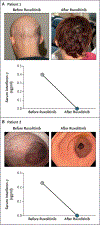
Figure 4.. Ruxolitinib-Induced Remission of Autoimmune Manifestations in Patients 1 and 2.
Panel A shows photographs of hair on the scalp of Patient 1 before and after ruxolitinib treatment. The graph shows serum levels of interferon-γ before and after treatment. Panel B shows the macroscopic appearance of gastric mucosa during endoscopy in Patient 2. The graph shows serum levels of interferon-γ before and after treatment.
Figure 5.
Figure 5.. Ruxolitinib-Induced Remission of Autoimmune Manifestations in Patients 3, 4, and 5.
Panel A shows photographs of hair on the scalp of Patient 3 before and after ruxolitinib treatment. The graph shows the patient’s change in weight after treatment. Panel B shows graphs of the number of bowel movements per day and body weight before and after treatment in Patient 4. Panel C shows graphs of body weight and serum levels of interferon-γ before and after treatment in Patient 5.

References

    1. Oftedal BE, Hellesen A, Erichsen MM, et al. Dominant mutations in the autoimmune regulator AIRE are associated with common organ-specific autoimmune diseases. Immunity 2015;42:1185–96. - PubMed
    1. Ricotta EE, Ferré EMN, Schmitt MM, DiMaggio T, Lionakis MS. Prevalence of APECED-like clinical disease in an electronic health record database, USA. J Clin Immunol 2022;42:904–6. - PMC - PubMed
    1. Constantine GM, Lionakis MS. Lessons from primary immunodeficiencies: autoimmune regulator and autoimmune polyendocrinopathy-candidiasis-ectodermal dystrophy. Immunol Rev 2019;287:103–20. - PMC - PubMed
    1. Ahonen P, Myllärniemi S, Sipilä I, Perheentupa J. Clinical variation of autoimmune polyendocrinopathy-candidiasis-ectodermal dystrophy (APECED) in a series of 68 patients. N Engl J Med 1990;322:1829–36. - PubMed
    1. Bruserud Ø, Oftedal BE, Landegren N, et al. A longitudinal follow-up of autoimmune polyendocrine syndrome type 1. J Clin Endocrinol Metab 2016;101:2975–83. - PMC - PubMed

Publication types

MeSH terms

Supplementary concepts