. 2024 Aug 1;327(2):L189-L202.
doi: 10.1152/ajplung.00407.2023. Epub 2024 May 29.

Sex-based differences in persistent lung inflammation following influenza infection of juvenile outbred mice

Affiliations

Sex-based differences in persistent lung inflammation following influenza infection of juvenile outbred mice

Brydie R Huckestein et al. Am J Physiol Lung Cell Mol Physiol. .

Abstract

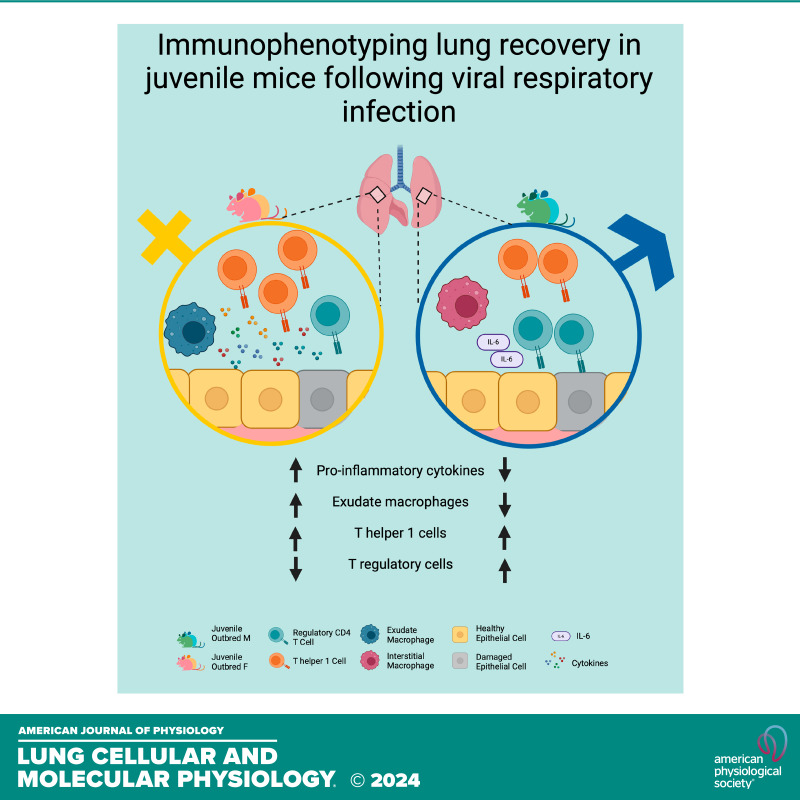
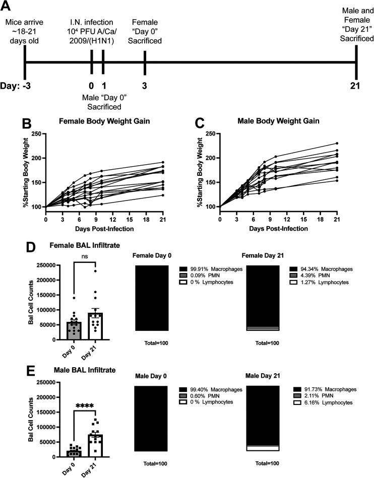
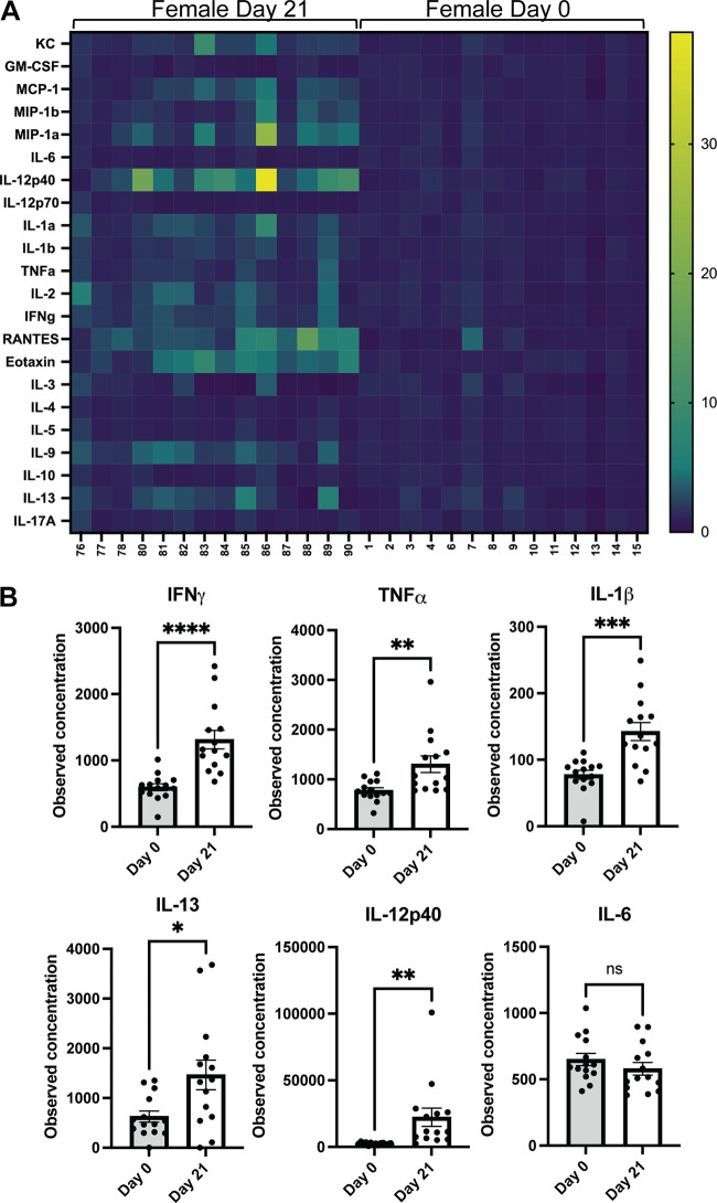
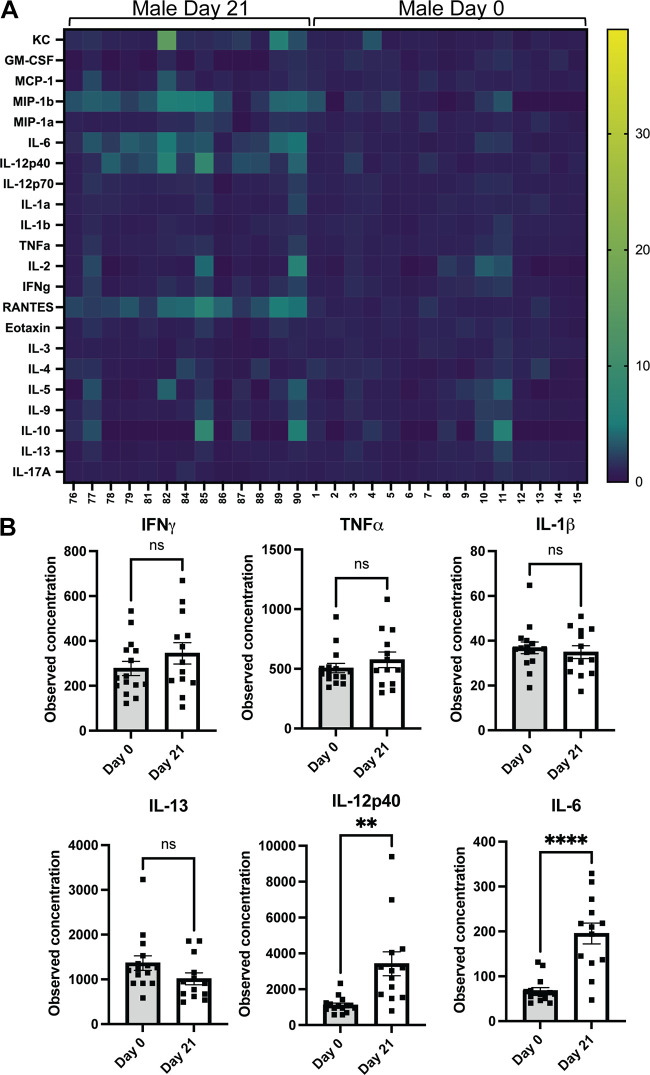
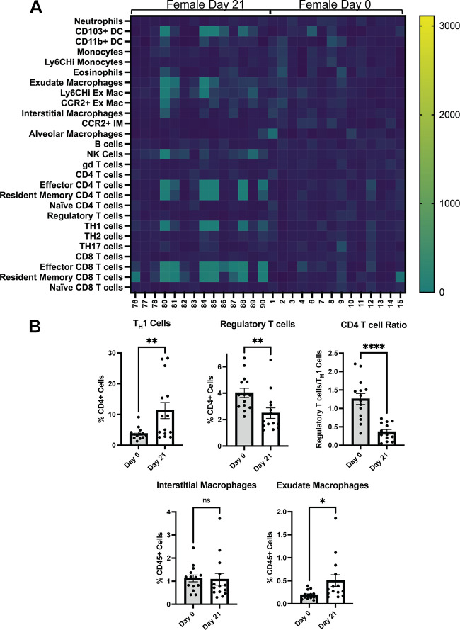
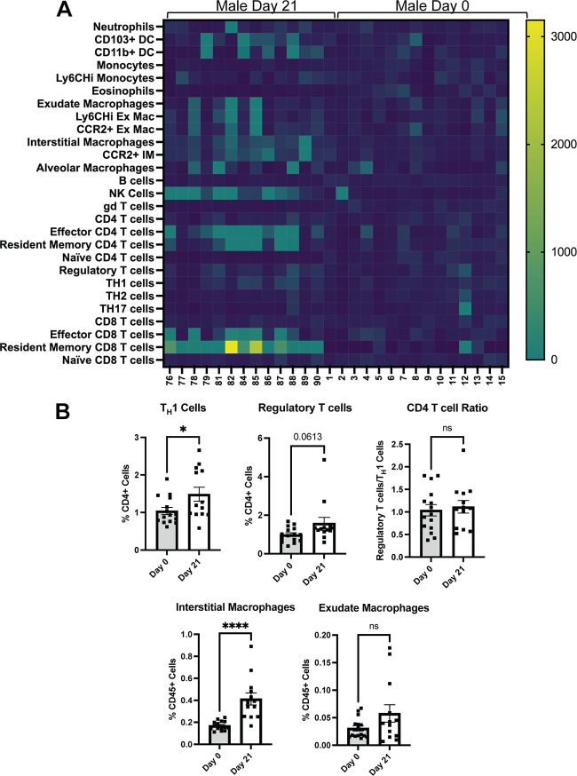
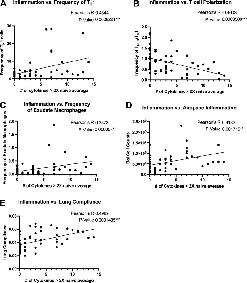
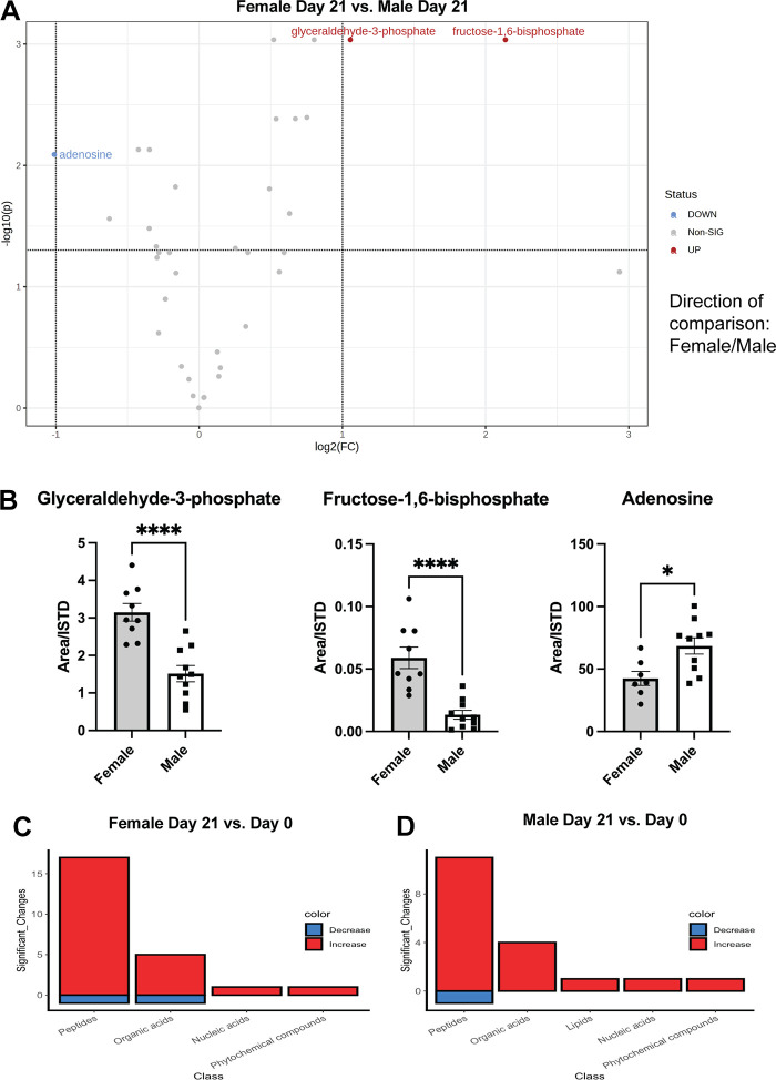
Children are susceptible to influenza infections and can experience severe disease presentation due to a lack of or limited pre-existing immunity. Despite the disproportionate impact influenza has on this population, there is a lack of focus on pediatric influenza research, particularly when it comes to identifying the pathogenesis of long-term outcomes that persist beyond the point of viral clearance. In this study, juvenile outbred male and female mice were infected with influenza and analyzed following viral clearance to determine how sex impacts the persistent inflammatory responses to influenza. It was found that females maintained a broader cytokine response in the lung following clearance of influenza, with innate, type I and type II cytokine signatures in almost all mice. Males, on the other hand, had higher levels of IL-6 and other macrophage-related cytokines, but no evidence of a type I or type II response. The immune landscape was similar in the lungs between males and females postinfection, but males had a higher regulatory T cell to TH1 ratio compared with female mice. Cytokine production positively correlated with the frequency of TH1 cells and exudate macrophages, as well as the number of cells in the bronchoalveolar lavage fluid. Furthermore, female lungs were enriched for metabolites involved in the glycolytic pathway, suggesting glycolysis is higher in female lungs compared with males after viral clearance. These data suggest juvenile female mice have persistent and excessive lung inflammation beyond the point of viral clearance, whereas juvenile males had a more immunosuppressive phenotype.NEW & NOTEWORTHY This study identifies sex-based differences in persistent lung inflammation following influenza infection in an outbred, juvenile animal model of pediatric infection. These findings indicate the importance of considering sex and age as variable in infectious disease research.

Keywords: T cell; cytokine; macrophage; pediatrics; pneumonia.


Conflict of interest statement

No conflicts of interest, financial or otherwise, are declared by the authors.

Figures

None
Graphical abstract
Figure 1.
Figure 1.
Characterization of weight gain and cellular infiltrate in influenza-infected male and female juvenile outbred mice. A: infection and euthanasia protocol for uninfected “day 0” mice and infected “day 21” mice. B and C: weight gain represented as percentage of starting body weight in female day 21 mice (B) and male day 21 mice (C). D and E: total number of cells in 1 mL of bronchoalveolar lavage fluid (BAL) represented as bar graph on left ± SE; frequency of macrophages, polymorphonuclear cells (PMN), and lymphocytes out of the total number of BAL cells represented as parts of whole for day 0 (middle) and day 21 (left) females (D) and males (E). Statistical significance determined by two-tailed Student’s t test, n = 15, 13 (B and C); n = 14, 13 (D, day 0, day 21); n = 15, 13 (E, day 0, day 21). ****P ≤ 0.0001, α = 0.05.
Figure 2.
Figure 2.
Cytokine profile in juvenile female lungs during influenza recovery. A: heatmap of relative cytokine expression, normalized to the average value of day 0 female mice for each cytokine. Each column represents one mouse, each row represents one cytokine. B: observed concentrations (pg/mL) of cytokines from day 0 and day 21 female mice. Data are represented as bar graph ± SE. Statistical significance was determined by two-tailed Student’s t test; n = 14. *P ≤ 0.05, **P ≤ 0.01, ***P ≤ 0.001, ****P ≤ 0.0001, α = 0.05. GM-CSF, granulocyte-macrophage colony-stimulating factor; KC, keratinocyte chemoattractant; MCP, monocyte chemoattractant protein; MIP, macrophage inflammatory protein; RANTES, regulated upon activation, normal T cell expressed and presumably secreted.
Figure 3.
Figure 3.
Cytokine profile in juvenile male lungs during influenza recovery. A: heatmap of relative cytokine expression, normalized to the average value of day 0 male mice for each cytokine. Each column represents one mouse, each row represents one cytokine. B: observed concentrations (pg/mL) of cytokines from day 0 and day 21 male mice. Data are represented as bar graph ± SE. Statistical significance was determined by two-tailed Student’s t test; n = 13, 15 (day 21, day 0). **P ≤ 0.01, ****P ≤ 0.0001, α = 0.05. KC, keratinocyte chemoattractant; MCP, monocyte chemoattractant protein; MIP, macrophage inflammatory protein; RANTES, regulated upon activation, normal T cell expressed and presumably secreted.
Figure 4.
Figure 4.
Immune cell population changes in the juvenile female lung during recovery from influenza infection. A: heatmap of the relative frequency of immune cells in the CD45+ population, normalized to the average value of day 0 female mice for each cell type. Each column represents one mouse, each row represents one cell type. B: frequency of T helper 1 cells (TH1, CD45+ CD90+ TCRb+ CD4+ Tbet+), regulatory T cells (CD45+ CD90+ TCRb+ CD4+ Foxp3+ CD25High), interstitial macrophages (CD25+ CD24− CD11b+ CD11c− CD64+), and exudate macrophages (CD25+ CD24− CD11b+ CD11c+ CD64+) in the CD45+ population from day 0 and day 21 female mice, and the ratio of regulatory T cells/TH1 cells (CD4 T cell ratio) in day 0 and day 21 female mice. Data are represented as bar graph ± SE. Statistical significance was determined by two-tailed Student’s t test; n = 14. *P ≤ 0.05, **P ≤ 0.01, ****P ≤ 0.0001, α = 0.05. DC, dendritic cells; NK, natural killer.
Figure 5.
Figure 5.
Immune cell population changes in the juvenile male lung during recovery from influenza infection. A: heatmap of the relative frequency of immune cells in the CD45+ population, normalized to the average value of day 0 male mice for each cell type. Each column represents one mouse and each row represents one cell type. B: frequency of T helper 1 cells (TH1, CD45+ CD90+ TCRb+ CD4+ Tbet+), regulatory T cells (CD45+ CD90+ TCRb+ CD4+ Foxp3+ CD25High), interstitial macrophages (CD25+ CD24− CD11b+ CD11c− CD64+), and exudate macrophages (CD25+ CD24− CD11b+ CD11c+ CD64+) in the CD45+ population from day 0 and day 21 male mice, and the ratio of regulatory T cells/TH1 cells (CD4 T cell Ratio) in day 0 and day 21 male mice. Data are represented as bar graph ± SE. Statistical significance was determined by two-tailed Student’s t test; n = 15, 13 (day 0, day 21). *P ≤ 0.05, ****P ≤ 0.0001, α = 0.05.
Figure 6.
Figure 6.
Correlates of lung inflammation. Pearson’s correlations are represented as scatter plots with the number of cytokines that were greater than twice the average of the naïve value for each sex (x-axis) vs. the frequency of T helper 1 cells (TH1, CD45+ CD90+ TCRb+ CD4+ Tbet+) (A) in the CD45+ population, the CD4 T cell ratio (frequency of regulatory T cells/TH1) (B), the frequency of exudate macrophages (CD25+ CD24− CD11b+ CD11c+ CD64+) in the CD45+ population (C), airspace inflammation [the total number of cells in 1 mL of bronchial alveolar lavage fluid (BAL)] (D), and lung compliance (E) (y-axis). Statistical significance determined by Pearson’s R, n = 55, **P ≤ 0.01, ***P ≤ 0.001, α = 0.05.
Figure 7.
Figure 7.
Metabolic profiling of juvenile male and female mice 21 days post-infection (dpi) with influenza. A: volcano plot display of metabolites that differ between males and females 21 dpi. Upregulated metabolites are higher in females (marked in red), downregulated metabolites are higher in males (marked in blue). False discovery rate is 0.05. B: concentrations of metabolites that were identified in (A) shown as peak area count relative to internal standard (area/ISTD). C and D: bar graph representation of the number of metabolites in each class that are either upregulated (red) or downregulated (blue) in day 21 females (C) or day 21 males (D) relative to the day 0 of each sex. Data are represented as bar graph ± SE. Statistical significance was determined by two-tailed Student’s t test; n = 14. *P ≤ 0.05, ****P ≤ 0.0001, α = 0.05.

References

    1. Jakab GJ, Astry CL, Warr GA. Alveolitis induced by influenza virus. Am Rev Respir Dis 128: 730–739, 1983. doi: 10.1164/arrd.1983.128.4.730. - DOI - PubMed
    1. Clohisey S, Baillie JK. Host susceptibility to severe influenza A virus infection. Crit Care 23: 303, 2019. doi: 10.1186/s13054-019-2566-7. - DOI - PMC - PubMed
    1. Albright FS, Orlando P, Pavia AT, Jackson GG, Cannon Albright LA. Evidence for a heritable predisposition to death due to influenza. J Infect Dis 197: 18–24, 2008. doi: 10.1086/524064. - DOI - PubMed
    1. Verhelst J, Parthoens E, Schepens B, Fiers W, Saelens X. Interferon-inducible protein Mx1 inhibits influenza virus by interfering with functional viral ribonucleoprotein complex assembly. J Virol 86: 13445–13455, 2012. doi: 10.1128/JVI.01682-12. - DOI - PMC - PubMed
    1. Ciancanelli MJ, Huang SX, Luthra P, Garner H, Itan Y, Volpi S, Lafaille FG, Trouillet C, Schmolke M, Albrecht RA, Israelsson E, Lim HK, Casadio M, Hermesh T, Lorenzo L, Leung LW, Pedergnana V, Boisson B, Okada S, Picard C, Ringuier B, Troussier F, Chaussabel D, Abel L, Pellier I, Notarangelo LD, García-Sastre A, Basler CF, Geissmann F, Zhang SY, Snoeck HW, Casanova JL. Infectious disease. Life-threatening influenza and impaired interferon amplification in human IRF7 deficiency. Science 348: 448–453, 2015. doi: 10.1126/science.aaa1578. - DOI - PMC - PubMed

MeSH terms

LinkOut - more resources