. 2024 Jul:73:103215.
doi: 10.1016/j.redox.2024.103215. Epub 2024 May 27.

FOXO1 regulates RUNX2 ubiquitination through SMURF2 in calcific aortic valve disease

Affiliations

FOXO1 regulates RUNX2 ubiquitination through SMURF2 in calcific aortic valve disease

Chen Jiang et al. Redox Biol. 2024 Jul.

Abstract

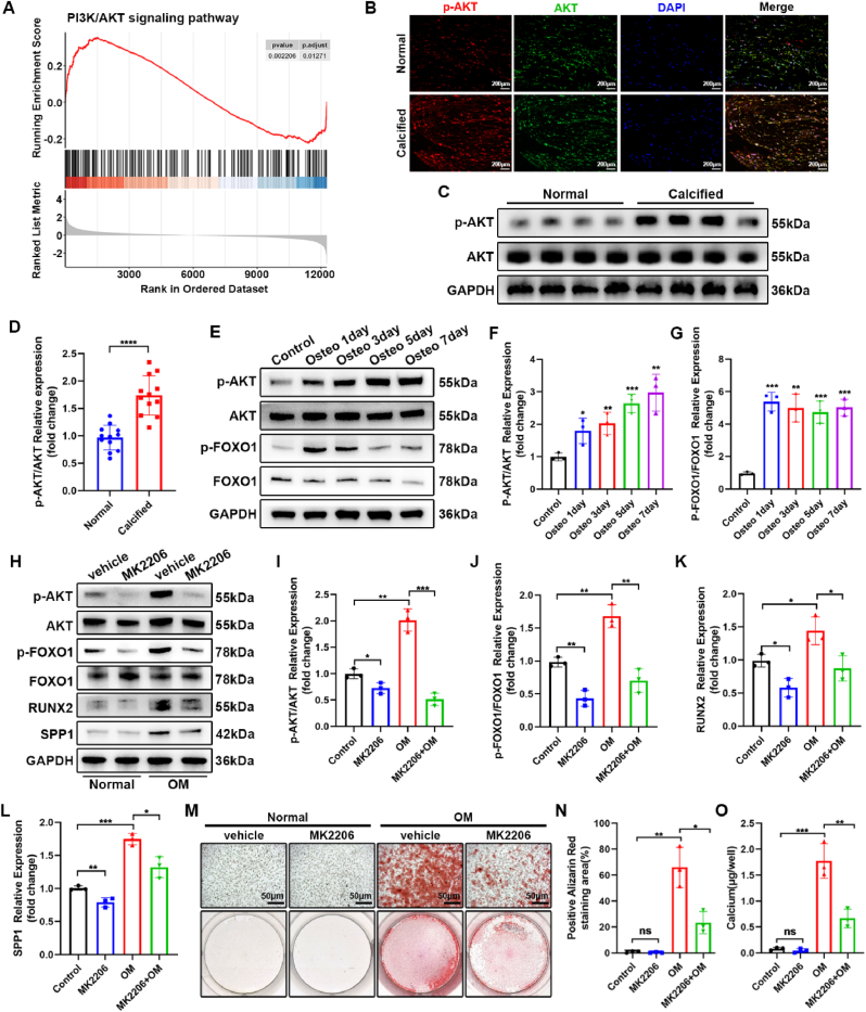
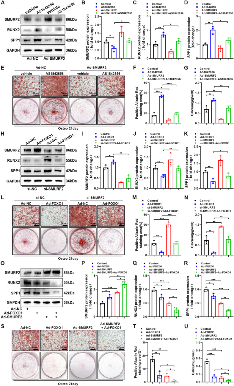
The prevalence of calcific aortic valve disease (CAVD) remains substantial while there is currently no medical therapy available. Forkhead box O1 (FOXO1) is known to be involved in the pathogenesis of cardiovascular diseases, including vascular calcification and atherosclerosis; however, its specific role in calcific aortic valve disease remains to be elucidated. In this study, we identified FOXO1 significantly down-regulated in the aortic valve interstitial cells (VICs) of calcified aortic valves by investigating clinical specimens and GEO database analysis. FOXO1 silencing or inhibition promoted VICs osteogenic differentiation in vitro and aortic valve calcification in Apoe-/- mice, respectively. We identified that FOXO1 facilitated the ubiquitination and degradation of RUNX2, which process was mainly mediated by SMAD-specific E3 ubiquitin ligase 2 (SMURF2). Our discoveries unveil a heretofore unacknowledged mechanism involving the FOXO1/SMURF2/RUNX2 axis in CAVD, thereby proposing the potential therapeutic utility of FOXO1 or SMURF2 as viable strategies to impede the progression of CAVD.

Keywords: Calcific aortic valve disease; Forkhead box O1; Phosphorylation; SMAD-Specific E3 ubiquitin ligase 2; Ubiquitination.


Conflict of interest statement

Declaration of competing interest The authors declare that they have no known competing financial interests or personal relationships that could have appeared to influence the work reported in this paper.

Figures

Image 1
Graphical abstract
Fig. 1
Fig. 1
FOXO1 is down-regulated in VICs of human calcific aortic valves. (A) H&E, Von Kossa, Alizarin Red staining and Immumohistochemical staining of FOXO1 in aortic valves, scale bar: 200 μm. (B–E) Western blot of FOXO1, RUNX2 and SPP1 expression in aortic valves (n = 12 independent human aortic valves per group). (F) Differential gene expression analysis showing up- and down-regulated genes in calcified valves compared to normal valves across three bulk RNA-seq datasets. (G) Venn diagram comparing the DEGs between three bulk RNA-seq datasets. (H) Volcano plot of the 648 common DEGs which were marked by blue or red dots. (I) Reduction dimplot of mice aortic valves scRNA-seq. (J) Featureplot revealed a remarkable down-regulation of FOXO1 in aortic valves from mice with hyperlipidemia. (K) Volcano plot showed that FOXO1 represented a significant down-regulation among transcriptional factors in VIC of hyperlipidemia mice. (L) Immunofluorescence staining of FOXO1 and vimentin in aortic valves, scale bar: 100 μm. Values are mean ± SD. Statistical differences were determined by Mann-Whitney test (D) and two-tailed unpaired Student's t-test (C and E). *p < 0.05, **p < 0.01, ***p < 0.001, ****p < 0.0001.
Fig. 2
Fig. 2
FOXO1 inhibits osteogenic differentiation of VICs. (A–D) Western blot analysis of protein levels in VICs with osteogenic medium at different time points (days 0, 1, 3, 5, 7, n = 3 independent experiments). (E–H) Western blot analysis of FOXO1, RUNX2 and SPP1 in VICs with FOXO1 overexpression and osteogenic medium (n = 3 independent experiments). Alizarin red staining of calcium deposition (I–J) and calcium content (K) in VICs overexpressing FOXO1 (n = 3 independent experiments). (L–O) Western blot analysis of FOXO1, RUNX2 and SPP1 in VICs following FOXO1 silencing (n = 3 independent experiments). Alizarin red staining of calcium deposition (P–Q) and calcium content (R) in VICs following FOXO1 silencing (n = 3 independent experiments). Scale bar: 50 μm. Values are mean ± SD. All statistical differences were determined using two-tailed unpaired Student's t-test. *p < 0.05, **p < 0.01, ***p < 0.001, ****p < 0.0001.
Fig. 3
Fig. 3
FOXO1 participates in VICs osteogenic differentiation mediated by AKT pathway. (A) GSEA of bulk RNA-seq showed an up-regulation of PI3K/AKT pathway. (B) Immunofluorescence staining of p-AKT and AKT in aortic valves, scale bar: 200 μm. (C–D) Western blot analysis of p-AKT and AKT expression in aortic valves. (n = 12 independent human aortic valves per group). (E–G) Western blot analysis of protein levels in VICs with osteogenic medium at different time points (days 0, 1, 3, 5, 7, n = 3 independent experiments). (H–L) Western blot analysis of protein levels in VICs with MK2206 and osteogenic medium (n = 3 independent experiments). Alizarin red staining of calcium deposition (M − N) and calcium content (O) in VICs with MK2206 and osteogenic medium (n = 3 independent experiments). Scale bar: 50 μm. Values are mean ± SD. Statistical differences were determined using Benjamini-Hochberg (BH)-procedure (Multiple comparison test) (A) and two-tailed unpaired Student's t-test (D, F, G, I-L, N and O). *p < 0.05, **p < 0.01, ***p < 0.001, ****p < 0.0001.
Fig. 4
Fig. 4
FOXO1 inhibitor AS1842856 promotes osteogenic differentiation of VICs by promoting FOXO1 phosphorylation. (A–D) Western blot analysis of protein levels in VICs with AS1842856 in 0, 0.5, 1, 3, 5 and 10 μM (n = 3 independent experiments). Alizarin red staining of calcium deposition (E–F) and calcium content (G) in VICs with AS1842856 and osteogenic medium (n = 3 independent experiments). (H–K) Western blot analysis of protein levels in VICs with FOXO1 silencing, AS1842856 and osteogenic medium (n = 3 independent experiments). Alizarin red staining of calcium deposition (L–M) and calcium content (N) in VICs with FOXO1 silencing, AS1842856 and osteogenic medium (n = 3 independent experiments). (O–Q) H&E, Von Kossa and Alizarin Red staining of aortic valves treated with AS1842856 and osteogenic medium in vitro (n = 3 independent experiments). scale bar: 200 μm. Values are mean ± SD. All statistical differences were determined using two-tailed unpaired Student's t-test. *p < 0.05, **p < 0.01, ***p < 0.001, ****p < 0.0001.
Fig. 5
Fig. 5
FOXO1 inhibition aggravates aortic valve calcification in vivo. Echocardiographic date of peak transvalvular jet velocity (A–B) and mean transvalvular pressure gradient (C) in Apoe−/− mice treated with or without AS1842856. H&E staining (D–E), Von Kossa staining (F–G), Alizarin Red staining (H–I) and Masson's trichrome staining (J–K) of aortic valve leaflets from Apoe−/− mice treated with or without AS1842856. scale bar: 200 μm or 100 μm. (L–M) Immunofluorescence staining of Runx2 (red) in aortic valves. DAPI was used for nuclear counterstaining (blue). scale bar: 200 μm. Values are the mean ± SD. All statistical differences were determined using two-tailed unpaired Student's t-test. *p < 0.05, **p < 0.01, ***p < 0.001, ****p < 0.0001. n = 10 independent mice per group.
Fig. 6
Fig. 6
FOXO1 interacts with RUNX2 and promotes its ubiquitination and degradation. (A) Co-immunoprecipitation (co-IP) to detect the interaction between Flag-tagged FOXO1 (Flag-FOXO1) and His-tagged RUNX2 (His-RUNX2). (B) Co-immunoprecipitation (co-IP) to detect the interaction between endogenous FOXO1 and RUNX2. (C–E) VICs were treated with Ad-NC or Ad-FOXO1 for 48 h. Before immunoblotting, cells were treated with the 26S proteasome inhibitor MG132 for 8 h (n = 3 independent experiments). (F–G) Western blot analysis of RUNX2 and GAPDH in VICs treated with Ad-Control or Ad-FOXO1 for 48 h, then treated with cycloheximide (CHX) for 0, 0.5, 1, 4 and 8 h (n = 3 independent experiments). (H) Co-IP to detect RUNX2 ubiquitination. VICs were treated with Ad-FOXO1 and AS1842856 for 48 h, then treated with MG132 for 8 h. The protein lysates were immunoprecipitated with an anti-RUNX2 antibody and immunoblotted with the indicated antibodies. (I) Western blot analysis of FOXO1 and RUNX2 in the nuclear, cytosolic and whole-cell of VICs treated with AS1842856. (J) Immunofluorescence staining of FOXO1 (Red) and RUNX2 (Green) in VICs treated with AS1842856. DAPI was used for nuclear counterstaining (blue). Scale bar: 20 μm. Values are mean ± SD. Statistical differences were determined using two-tailed unpaired Student's t-test (D and E) and Two-way ANOVA followed by Bonferroni post hoc test (G). *p < 0.05, **p < 0.01, ***p < 0.001, ****p < 0.0001.
Fig. 7
Fig. 7
FOXO1 promotes SMURF2-mediated RUNX2 ubiquitination in VICs. (A) E3 ubiquitin ligases of SMURF2 predicted by the UbiBrowser 2.0. (B) qRT-PCR analysis of the predicted E3 ubiquitin ligases of RUNX2 in VICs treated with AS1842856 (n = 3 independent experiments). (C–E) qRT-PCR and Western blot analysis of SMURF2 in VICs treated with FOXO1 overexpression, AS1842856 and osteogenic medium (n = 3 independent experiments). (F) Co-immunoprecipitation (co-IP) to detect the interaction between Myc-tagged SMURF2 (Myc-SMURF2) and His-tagged RUNX2 (His-RUNX2). (G) Co-immunoprecipitation (co-IP) to detect the interaction between endogenous SMERF2 and RUNX2. (H–J) Western blot analysis of VICs treated with FOXO1 overexpression and MG132 (n = 3 independent experiments). (K–L) Western blot analysis of RUNX2 and GAPDH in VICs treated with Ad-Control or Ad-FOXO1 for 48 h, then treated with CHX for 0, 0.5, 1, 4 and 8 h (n = 3 independent experiments). (M) Co-IP to detect RUNX2 ubiquitination. The protein lysates were immunoprecipitated with an anti-RUNX2 antibody and immunoblotted with the indicated antibodies. (N) Immunofluorescence staining of SMURF2 (Red) and RUNX2 (Green) in VICs. DAPI was used for nuclear counterstaining (blue). Scale bar: 20 μm. (O–P) Immunofluorescence staining of Smurf2 (red) in aortic valves (n = 10 independent mice per group). DAPI was used for nuclear counterstaining (blue). scale bar: 200 μm. Values are mean ± SD. Statistical differences were determined using two-tailed unpaired Student's t-test (B, D, E, I and J) and Two-way ANOVA followed by Bonferroni post hoc test (L). *p < 0.05, **p < 0.01, ***p < 0.001, ****p < 0.0001.
Fig. 8
Fig. 8
FOXO1 inhibits the osteogenic differentiation of VICs by up-regulating SMURF2. (A–D) Western blot analysis of SMURF2 and osteogenic markers (RUNX2 and SPP1) in VICs with AS1842856 and SMURF2 overexpression (n = 3 independent experiments). Alizarin red staining (E–F) and calcium content (G) in VICs with AS1842856 and SMURF2 overexpression (n = 3 independent experiments). (H–K) Western blot analysis of SMURF2 and osteogenic markers in VICs with FOXO1 overexpression and SMURF2 silencing (n = 3 independent experiments). Alizarin red staining (L–M) and calcium content (N) in VICs with FOXO1 overexpression and SMURF2 silencing (n = 3 independent experiments). (O–R) western blot analysis of SMURF2 and osteogenic markers in VICs with FOXO1 and SMURF2 overexpression (n = 3 independent experiments). Alizarin red staining (S–T) and calcium content (U) in VICs with FOXO1 and SMURF2 overexpression (n = 3 independent experiments). Values are mean ± SD. All statistical differences were determined using two-tailed unpaired Student's t-test. *p < 0.05, **p < 0.01, ***p < 0.001, ****p < 0.0001.

References

    1. Nkomo V.T., Gardin J.M., Skelton T.N., Gottdiener J.S., Scott C.G., Enriquez-Sarano M. Burden of valvular heart diseases: a population-based study. Lancet. 2006;368(9540):1005–1011. - PubMed
    1. Coffey S., Cox B., Williams M.J.A. The prevalence, incidence, progression, and risks of aortic valve sclerosis. J. Am. Coll. Cardiol. 2014;63(25):2852–2861. - PubMed
    1. Yadgir S., Johnson C.O., Aboyans V., Adebayo O.M., Adedoyin R.A., Afarideh M., et al. Global, regional, and national burden of calcific aortic valve and degenerative mitral valve diseases, 1990–2017. Circulation. 2020;141(21):1670–1680. - PubMed
    1. Liu Z., Wang K., Jiang C., Chen Y., Liu F., Xie M., et al. Morusin alleviates aortic valve calcification by inhibiting valve interstitial cell senescence through Ccnd1/Trim25/Nrf2 Axis. Adv. Sci (Weinh). 2024 - PMC - PubMed
    1. Goody P.R., Hosen M.R., Christmann D., Niepmann S.T., Zietzer A., Adam M., et al. Aortic valve stenosis. Arterioscler. Thromb. Vasc. Biol. 2020;40(4):885–900. - PubMed

MeSH terms

Supplementary concepts