. 2024 Jun 17;34(12):2728-2738.e6.
doi: 10.1016/j.cub.2024.04.079. Epub 2024 May 28.

CMTR-1 RNA methyltransferase mutations activate widespread expression of a dopaminergic neuron-specific mitochondrial complex I gene

Affiliations

CMTR-1 RNA methyltransferase mutations activate widespread expression of a dopaminergic neuron-specific mitochondrial complex I gene

Joshua D Meisel et al. Curr Biol. .

Abstract

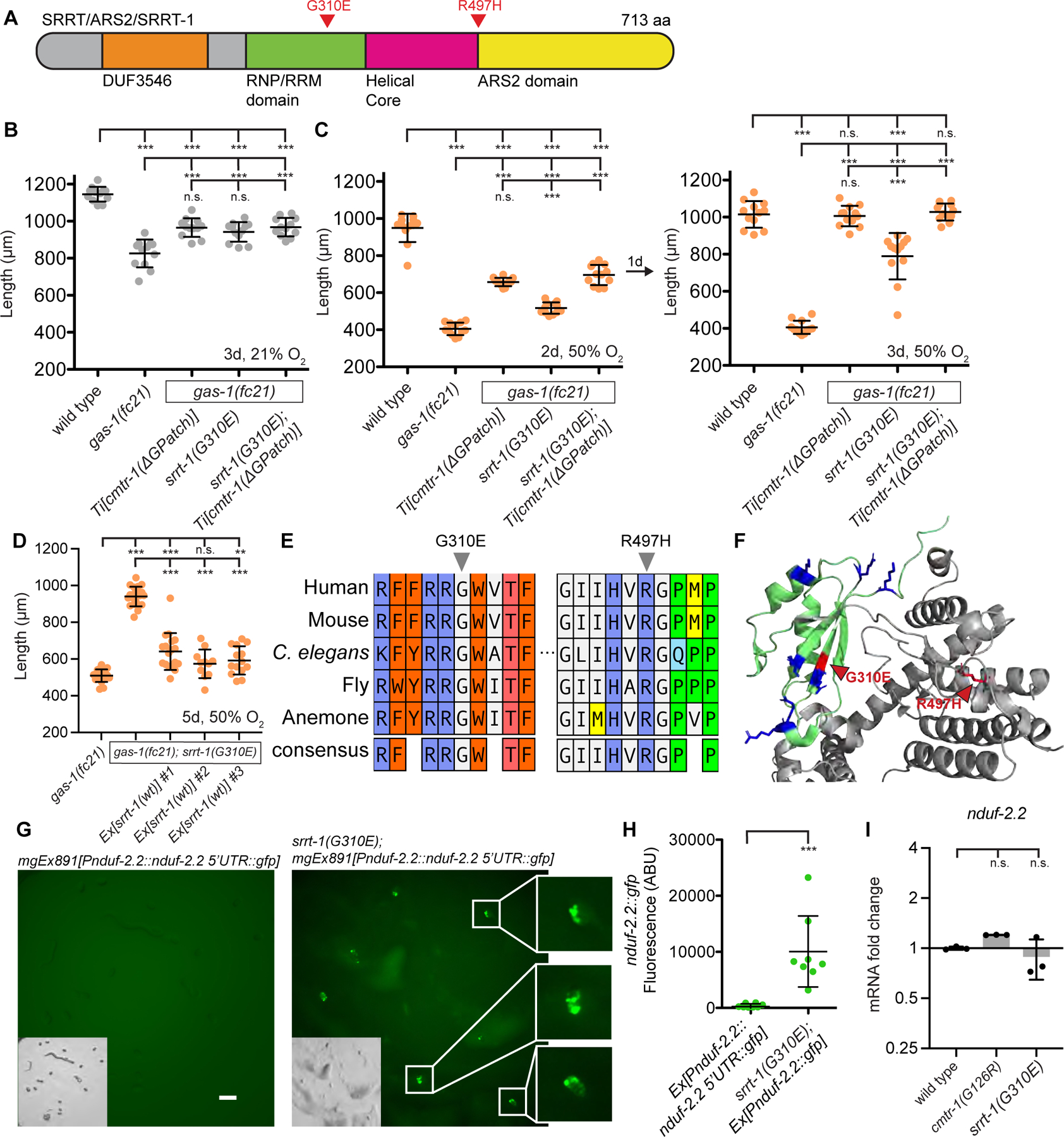
The mitochondrial proteome is comprised of approximately 1,100 proteins,1 all but 12 of which are encoded by the nuclear genome in C. elegans. The expression of nuclear-encoded mitochondrial proteins varies widely across cell lineages and metabolic states,2,3,4 but the factors that specify these programs are not known. Here, we identify mutations in two nuclear-localized mRNA processing proteins, CMTR1/CMTR-1 and SRRT/ARS2/SRRT-1, which we show act via the same mechanism to rescue the mitochondrial complex I mutant NDUFS2/gas-1(fc21). CMTR-1 is an FtsJ-family RNA methyltransferase that, in mammals, 2'-O-methylates the first nucleotide 3' to the mRNA CAP to promote RNA stability and translation5,6,7,8. The mutations isolated in cmtr-1 are dominant and lie exclusively in the regulatory G-patch domain. SRRT-1 is an RNA binding partner of the nuclear cap-binding complex and determines mRNA transcript fate.9 We show that cmtr-1 and srrt-1 mutations activate embryonic expression of NDUFS2/nduf-2.2, a paralog of NDUFS2/gas-1 normally expressed only in dopaminergic neurons, and that nduf-2.2 is necessary for the complex I rescue by the cmtr-1 G-patch mutant. Additionally, we find that loss of the cmtr-1 G-patch domain cause ectopic localization of CMTR-1 protein to processing bodies (P bodies), phase-separated organelles involved in mRNA storage and decay.10 P-body localization of the G-patch mutant CMTR-1 contributes to the rescue of the hyperoxia sensitivity of the NDUFS2/gas-1 mutant. This study suggests that mRNA methylation at P bodies may control nduf-2.2 gene expression, with broader implications for how the mitochondrial proteome is translationally remodeled in the face of tissue-specific metabolic requirements and stress.

Keywords: C. elegans; CMTR1; G-patch domain; Mitochondria; NADH:ubiquinone oxidoreductase; SRRT; complex I; hyperoxia; mRNA methylation; processing bodies.


Conflict of interest statement

Declaration of interests V.K.M. is listed as an inventor on patents filed by MGH on therapeutic uses of hypoxia. V.K.M. is a paid advisor to 5a.m. Ventures.

Figures

Figure 1.
Figure 1.. CMTR-1 G-patch mutations restore health of NDUFS2/gas-1(fc21) mutants
A. CMTR1/CMTR-1 is a SAM-dependent RNA methyltransferase that has been shown in mammalian cells to 2’-O-methylate the first transcribed nucleotide of nuclear-encoded mRNAs. B. Mutations in cmtr-1 that allow the NDUFS2/gas-1(fc21) mutant to survive hyperoxia are confined to conserved features of the G-patch domain (Pfam Signature PF01585). C. Growth of animals after 5 days exposure to 50% oxygen (left) or 5 days exposure to 100% oxygen followed by 3 days recovery at 21% oxygen (right). D. Mean intestinal fluorescence of the mitochondrial stress reporter hsp-6::gfp in L4 stage animals incubated at 21% or 100% oxygen for 1 day at 20°C. Exposure time = 100 ms, magnification = 69x, quantified from images in Figure S1B. E-F. Growth of animals following 4 days exposure to 50% oxygen. G. Images of animal growth and reproduction follow 4 days exposure to 50% oxygen. H. Total progeny produced from individual animals incubated at 21% oxygen. I. Growth of animals following 5 days exposure to 50% oxygen. J. Growth of animals following 2 days exposure to 21% oxygen. K. Growth of animals exposed to 3 days of 50% oxygen followed by recovery for 3 days at 21% oxygen. For all panels statistical significance was calculated using one-way ANOVA followed by Tukey’s Multiple Comparison Test. Error bars represent standard deviation. n.s. = not significant, * = p value <0.05, ** = p value <0.01, *** = p value <0.001. See also Figure S1 and Table S1.
Figure 2.
Figure 2.. G-patch mutant CMTR-1::GFP is ectopically localized to P-bodies, which is necessary for its rescue activity
A. Confocal microscopy of cmtr-1(wt)::gfp and cmtr-1(ΔG-patch)::gfp driven by the ribosomal promoter Prpl-28. Brightfield image is inset, white box corresponds to enlarged image on right. B. Confocal microscopy (left) and quantification (right) of cmtr-1(ΔG-patch)::gfp co-localization with the P-body marker dcap-1::DsRed. White box in brightfield image corresponds to fluorescent images on right. C. Bioinformatic analysis reveals three nuclear localization sequences in CMTR-1 and an N-terminal region predicted to form an intrinsically disordered domain. Deletion of amino acids 11–42 removes the first NLS and a portion of the IDD. D. Confocal microscopy of animals carrying extra-chromosomal arrays encoding G-patch mutant cmtr-1(G126R)::gfp or cmtr-1(Δ11–42 G126R)::gfp. E-F. Growth of animals following 5 days (E) or 2 days (F) exposure to 50% oxygen. Statistical significance was calculated using one-way ANOVA followed by Tukey’s Multiple Comparison Test. Error bars represent standard deviation. n.s. = not significant, *** = p value <0.001. See also Figure S2.
Figure 3.
Figure 3.. G-patch mutant CMTR-1 rescues NDUFS2/gas-1(fc21) by activating expression of the paralog NDUF-2.2
A. GFP reporter for nduf-2.2 expression was constructed using 3 kb of upstream promoter sequence and 67 bp of the endogenous 5’UTR which is the target for cap1 2’-O-methylation by CMTR-1. B. nduf-2.2::gfp reporter in wild-type animals is weakly expressed in a subset of head neurons indicated by white arrows. Exposure time = 2 seconds, magnification = 100x. C. nduf-2.2::gfp reporter (green arrow) in wild-type or cmtr-1(G126R) adult animals. myo-2::mCherry co-injection marker (red arrow) is visible. Exposure time = 2 seconds, magnification = 40x. D. nduf-2.2::gfp reporter in wild-type and cmtr-1(G126R) embryos. White arrows indicate embryos expressing no GFP. Exposure time = 2 seconds, magnification = 90x. E. Quantification of nduf-2.2::gfp fluorescence in embryos from wild type and cmtr-1(G126R) (panel D) and gas-1(fc21) (not pictured). Plotted are maximum fluorescence values with background subtracted. F. Total progeny produced from individual animals incubated at 21% oxygen. G. Images of nematode growth at 21% oxygen after 1 generation. Statistical significance was calculated using t-test (E) or one-way ANOVA followed by Tukey’s Multiple Comparison Test (F). Error bars represent standard deviation. n.s. = not significant, * = p value <0.05, ** = p value <0.01, *** = p value <0.001. See also Figure S3.
Figure 4.
Figure 4.. Mutation of the RNA binding protein Serrate activates nduf-2.2 and rescues gas-1(fc21)
A. Domain structure of RNA binding protein Serrate. B. Growth of animals following 3 days exposure to 21% oxygen. C. Growth of animals following 2 days (left) or 3 days (right) exposure to 50% oxygen. D. Growth of animals following 5 days exposure to 50% oxygen. E. Multiple sequence alignment of SRRT/srrt-1 homologs from animals made with ClustalW. Labelled residues correspond to suppressor mutations isolated in C. elegans srrt-1. F. Crystal structure of Human SRRT/ARS2 (PDB: 6F7J). Highlighted in green is the RNA-binding RRM domain. Blue residues correspond to positively charged amino acids; red residues correspond to mutations isolated in this study. G. nduf-2.2::gfp reporter in wild-type and srrt-1(G310E) embryos. White arrows indicate embryos expressing no GFP. Exposure time = 1 second, magnification = 90x. H. Quantification of GFP fluorescence in embryos from panel G. Plotted are maximum fluorescence values with background subtracted. I. Quantitative real-time PCR of nduf-2.2 mRNA normalized to the housekeeping gene rps-23. For all panels statistical significance was calculated using one-way ANOVA followed by Tukey’s Multiple Comparison Test. Error bars represent standard deviation. n.s. = not significant, * = p value <0.05, ** = p value <0.01, *** = p value <0.001. See also Figure S4.

References

    1. Rath S, Sharma R, Gupta R, Ast T, Chan C, Durham TJ, Goodman RP, Grabarek Z, Haas ME, Hung WHW, et al. (2020). MitoCarta3.0: an updated mitochondrial proteome now with sub-organelle localization and pathway annotations. Nucleic Acids Res 49, D1541–D1547. 10.1093/nar/gkaa1011. - DOI - PMC - PubMed
    1. Pagliarini DJ, Calvo SE, Chang B, Sheth SA, Vafai SB, Ong S-E, Walford GA, Sugiana C, Boneh A, Chen WK, et al. (2008). A Mitochondrial Protein Compendium Elucidates Complex I Disease Biology. Cell 134, 112–123. 10.1016/j.cell.2008.06.016. - DOI - PMC - PubMed
    1. Grimsrud PA, Carson JJ, Hebert AS, Hubler SL, Niemi NM, Bailey DJ, Jochem A, Stapleton DS, Keller MP, Westphall MS, et al. (2012). A Quantitative Map of the Liver Mitochondrial Phosphoproteome Reveals Posttranslational Control of Ketogenesis. Cell Metab 16, 672–683. 10.1016/j.cmet.2012.10.004. - DOI - PMC - PubMed
    1. Kuhn KM, DeRisi JL, Brown PO, and Sarnow P (2001). Global and Specific Translational Regulation in the Genomic Response of Saccharomyces cerevisiae to a Rapid Transfer from a Fermentable to a Nonfermentable Carbon Source. Mol Cell Biol 21, 916–927. 10.1128/mcb.21.3.916-927.2001. - DOI - PMC - PubMed
    1. Picard-Jean F, Brand C, Tremblay-Létourneau M, Allaire A, Beaudoin MC, Boudreault S, Duval C, Rainville-Sirois J, Robert F, Pelletier J, et al. (2018). 2’-O-methylation of the mRNA cap protects RNAs from decapping and degradation by DXO. PLoS ONE 13, e0193804. 10.1371/journal.pone.0193804. - DOI - PMC - PubMed

MeSH terms

LinkOut - more resources