. 2024 Jul 3;44(27):e1994232024.
doi: 10.1523/JNEUROSCI.1994-23.2024.

Ablation of Fatty Acid Transport Protein-4 Enhances Cone Survival, M-cone Vision, and Synthesis of Cone-Tropic 9- cis-Retinal in rd 12 Mouse Model of Leber Congenital Amaurosis

Affiliations

Ablation of Fatty Acid Transport Protein-4 Enhances Cone Survival, M-cone Vision, and Synthesis of Cone-Tropic 9- cis-Retinal in rd 12 Mouse Model of Leber Congenital Amaurosis

Songhua Li et al. J Neurosci. .

Abstract

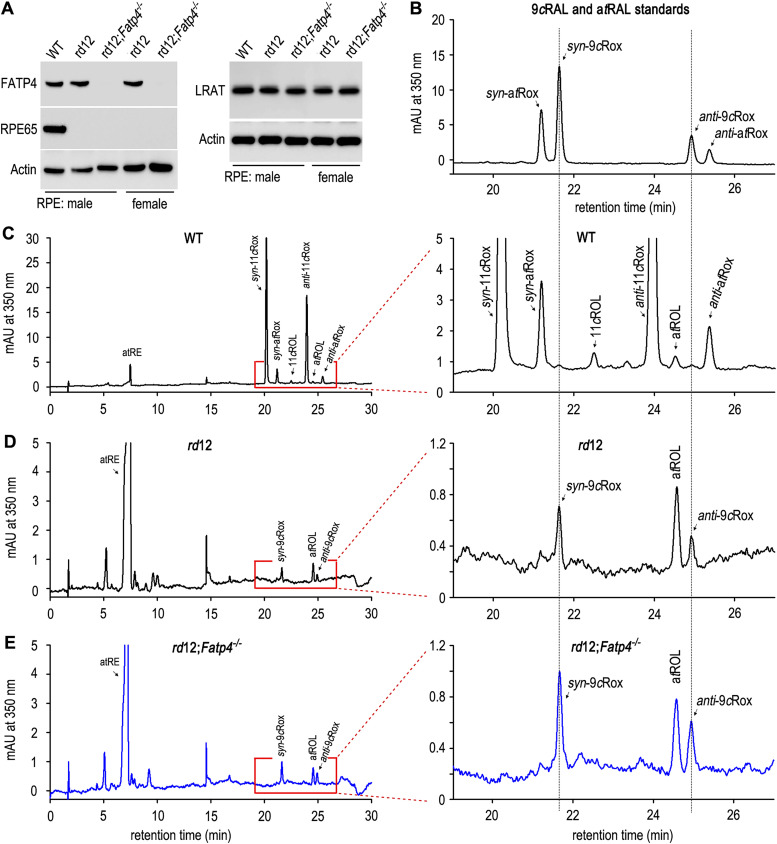
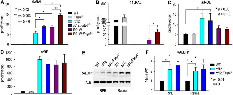
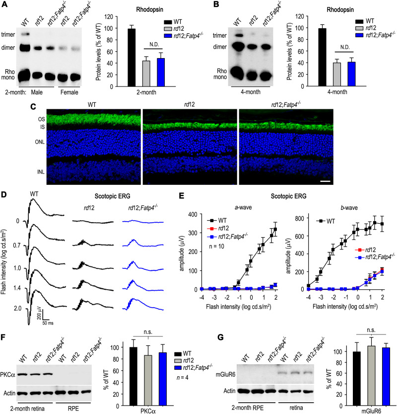
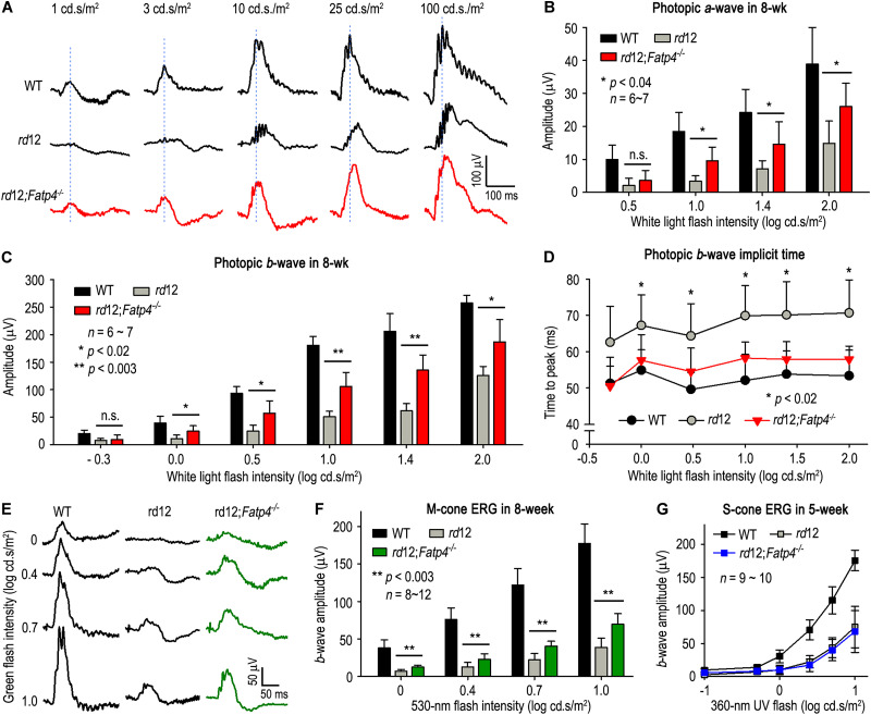
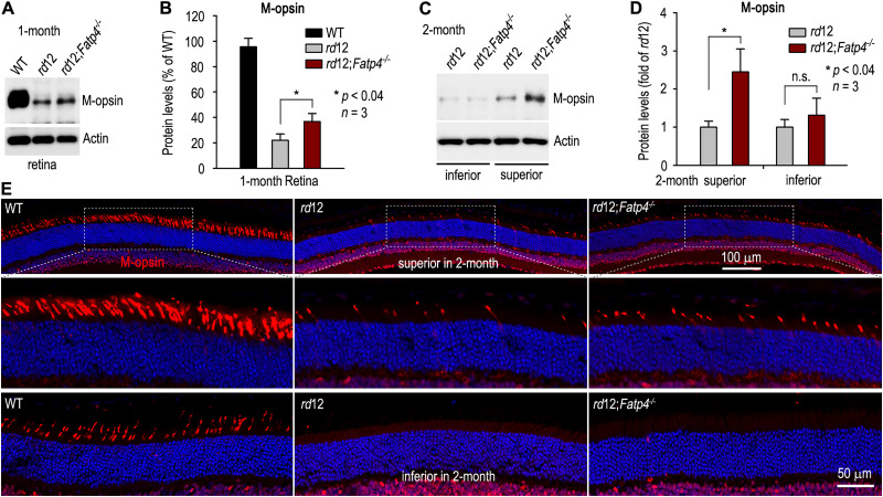
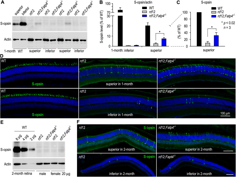
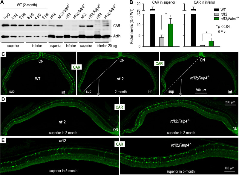
The canonical visual cycle employing RPE65 as the retinoid isomerase regenerates 11-cis-retinal to support both rod- and cone-mediated vision. Mutations of RPE65 are associated with Leber congenital amaurosis that results in rod and cone photoreceptor degeneration and vision loss of affected patients at an early age. Dark-reared Rpe65-/- mouse has been known to form isorhodopsin that employs 9-cis-retinal as the photosensitive chromophore. The mechanism regulating 9-cis-retinal synthesis and the role of the endogenous 9-cis-retinal in cone survival and function remain largely unknown. In this study, we found that ablation of fatty acid transport protein-4 (FATP4), a negative regulator of 11-cis-retinol synthesis catalyzed by RPE65, increased the formation of 9-cis-retinal, but not 11-cis-retinal, in a light-independent mechanism in both sexes of RPE65-null rd12 mice. Both rd12 and rd12;Fatp4-/- mice contained a massive amount of all-trans-retinyl esters in the eyes, exhibiting comparable scotopic vision and rod degeneration. However, expression levels of M- and S-opsins as well as numbers of M- and S-cones surviving in the superior retinas of rd12;Fatp4-/ - mice were at least twofold greater than those in age-matched rd12 mice. Moreover, FATP4 deficiency significantly shortened photopic b-wave implicit time, improved M-cone visual function, and substantially deaccelerated the progression of cone degeneration in rd12 mice, whereas FATP4 deficiency in mice with wild-type Rpe65 alleles neither induced 9-cis-retinal formation nor influenced cone survival and function. These results identify FATP4 as a new regulator of synthesis of 9-cis-retinal, which is a "cone-tropic" chromophore supporting cone survival and function in the retinas with defective RPE65.

Keywords: 9-cis-retinal; FATP4; RPE65 isomerase; cone photoreceptor; retinal dystrophy.


Conflict of interest statement

The authors declare no competing financial interests.

Figures

Figure 1.
Figure 1.
Identification of 9-cis-retinal increased in the eyes of rd12 mice lacking FATP4. A, Immunoblot analysis of FATP4, RPE65, and LRAT in the RPE with the indicated genotypes. Actin was detected as a loading control. B, HPLC chromatogram of 9cRAL and atRAL standards converted into the corresponding syn- and anti-isomers of 9-cis or all-trans retinaloximes (Rox). C–E, Representative HPLC chromatograms of retinoids extracted from the eyecups of dark-adapted WT (C), rd12 (D), and rd12;Fatp4−/− (E) mice. Peaks of all-trans-retinyl esters (atRE), syn-11-cis retinaloxime (syn-11cRox), syn-all-trans retinaloxime (syn-atRox), 11-cis-retinol (11cROL), anti-11-cis retinaloxime (anti-11cRox), all-trans-retinol (atROL), anti-all-trans retinaloxime (anti-atRox), syn-9-cis retinaloxime (syn-9cRox), and anti-9-cis retinaloxime (anti-9cRox) are marked.
Figure 2.
Figure 2.
Comparison of ocular retinoid and RALDH1 expression profiles in WT and rd12 mice with or without FATP4. Quantification of 9-cis-retinal (A), 11-cis-retinal (B), all-trans-retinol (C), and all-trans-retinyl esters (D) in the eyecups of overnight dark-adapted WT, Fatp4−/−, rd12, rd12;Fatp4−/−, R91W, and R91W;Fatp4−/− mice. E, Immunoblot analysis showing expression of RALDH1 in the retina and RPE of WT, rd12, and rd12;Fatp4−/− mice. F, Expression levels of RALDH1 in the retina or RPE of rd12 and rd12;Fatp4−/− mice are expressed as fold of RALDH1 expression level in the WT retina or RPE.
Figure 3.
Figure 3.
Comparable rod degeneration and scotopic ERGs in rd12 and rd12;Fatp4−/− mice. A, B, Immunoblot analysis of rhodopsin (Rho) in the retinas of 2-month-old (A) or 4-month-old (B) WT, rd12, and rd12;Fatp4−/− mice. Rho monomer, dimer, and trimer are indicated. Histograms show relative expression levels of rhodopsin (including monomer, dimer, and trimer) in WT, rd12, and rd12;Fatp4−/− mice at the indicated ages. C, Immunohistochemical analysis of Rho in 2-month-old WT, rd12, and rd12;Fatp4−/− retinal sections. The outer and inner nuclear layers (ONL and INL) were counterstained with DAPI (blue). OS, outer segments; IS, inner segments. D, Representative scotopic ERG responses of rods in overnight dark-adapted WT, rd12, and rd12;Fatp4−/− mice to the indicated light flashes. E, Amplitudes of scotopic ERG a- and b-waves evoked with the indicated flashes in 2-month-old WT, rd12, and rd12;Fatp4−/− mice. Error bars show SD (n = 10). F, G, Immunoblot analyses of PKCα (F) and mGluR6 (G) in the neural retina and RPE of 2-month-old WT, rd12, and rd12;Fatp4−/− mice. Actin was used as loading control. Histograms show relative expression levels of PKCα and mGluR6 in the neural retinas of the WT and mutant mice.
Figure 4.
Figure 4.
FATP4 deficiency improved M-cone but not S-cone vision in rd12 mice. A, Representative photopic ERG responses of 8-week-old WT, rd12, and rd12;Fatp4−/− mice to a series of increasing flashes (1–100 cd·s/m2) of white light under a rod-saturating background light. B, C, Amplitudes of photopic a- and b-waves evoked with the indicated white light flashes. D, Times from stimulus onset to peaks of photopic b-waves evoked with the indicated white light flashes in WT, rd12, and rd12;Fatp4−/− mice. Asterisks indicate statistically significant differences between rd12 and rd12;Fatp4−/− mice (*p < 0.02); error bars denote SD (n = 6–7). E, Representative ERG responses of M-cones in 8–10-week-old WT, rd12, and rd12;Fatp4−/− mice to the indicated flash intensities of 530 nm green light. The ERG responses were evoked with a series of increasing stimuli (log −1∼1 cd·s/m2) of 530 nm light. F, Amplitudes of M-cone b-waves evoked with the indicated flash intensities of the green light. G, Amplitudes of S-cone ERG b-waves evoked with the indicated flash intensities of 360 nm UV light.
Figure 5.
Figure 5.
Alleviation of M-cone degeneration in the superior retina of rd12;Fatp4−/− Mice. A, Immunoblot analysis of M-opsin in the retinas of 1-month-old WT, rd12, and rd12;Fatp4−/− mice. Actin was detected as loading control. B, Expression levels of M-opsin in rd12 and rd12;Fatp4−/− retinas are normalized with actin levels and shown as percent of M-opsin levels in the WT retina. C, Immunoblot analysis of M-opsin in the inferior or superior halves of 2-month-old WT, rd12, and rd12;Fatp4−/− mice. D, Normalized expression levels of M-opsin in the superior or inferior retinas of rd12;Fatp4−/− mice (in C) are shown as fold of M-opsin levels in the rd12 superior or inferior retinas. Asterisk indicates significant differences between rd12 and rd12;Fatp4−/− mice; n.s. denotes no significance. E, Immunohistochemistry of M-opsin in the superior and inferior retinas of 2-month-old WT, rd12, and rd12;Fatp4−/− mice.
Figure 6.
Figure 6.
FATP4 deficiency mitigated S-cone degeneration in the superior retina of rd12 mice. A, Immunoblot analysis of S-opsin in the inferior or superior halves of 1-month-old WT, rd12, and rd12;Fatp4−/− retinas. Note that the majority of S-opsin is distributed in the inferior half of WT retina, while in the rd12 and rd12;Fatp4−/− inferior retinas S-opsin is almost undetectable. B, Percentages of S-opsin included in the inferior or superior halves of WT retinas in A. Levels of S-opsin in the inferior and superior retinas of rd12 and rd12;Fatp4−/− mice are shown as percent of S-opsin levels in WT. C, S-opsin level in the WT superior retina is set as 100% and the levels of S-opsin in the superior retinas of rd12 and rd12;Fatp4−/− mice are shown as percent of S-opsin levels in WT. D, Immunohistochemical analysis of S-opsin in the superior and inferior retinas of 1-month-old WT, rd12, and rd12;Fatp4−/− mice. E, Immunoblot analysis of S-opsin in the indicated micrograms of total retinal homogenates from 2-month-old WT, rd12, and rd12;Fatp4−/− mice. F, Immunohistochemistry of S-opsin in the superior and inferior retinas of 2-month-old rd12 and rd12;Fatp4−/− mice. Scale bars denote 100 μm.
Figure 7.
Figure 7.
FATP4 deficiency slowed cone degeneration rates in rd12 mice. A, Immunoblot analysis of cone arrestin (CAR) in the indicated micrograms of the superior and inferior retinal homogenates from 2-month-old WT, rd12, and rd12;Fatp4−/− mice. Actin was detected as loading control. B, Normalized expression levels of CAR in the superior or inferior retinas of 2-month-old rd12 and rd12;Fatp4−/− mice are shown as percent of CAR levels in the superior or inferior halves of WT retina. C, Immunohistochemical analysis of CAR in retinal sections from 2-month-old WT, rd12, and rd12;Fatp4−/− mice. ON, optic nerve head; sup, superior; inf, inferior. D, Higher-magnification images of the areas of rectangles shown in C. E, Immunohistochemistry of CAR in the superior retinas of 5-month-old rd12 and rd12;Fatp4−/− mice.

Similar articles

References

    1. Amamoto R, Wallick GK, Cepko CL (2022) Retinoic acid signaling mediates peripheral cone photoreceptor survival in a mouse model of retina degeneration. Elife 11:e76389. 10.7554/eLife.76389 - DOI - PMC - PubMed
    1. Batten ML, Imanishi Y, Maeda T, Tu DC, Moise AR, Bronson D, Possin D, Van Gelder RN, Baehr W, Palczewski K (2004) Lecithin-retinol acyltransferase is essential for accumulation of all-trans-retinyl esters in the eye and in the liver. J Biol Chem 279:10422–10432. 10.1074/jbc.M312410200 - DOI - PMC - PubMed
    1. Bernstein PS, Li B, Vachali PP, Gorusupudi A, Shyam R, Henriksen BS, Nolan JM (2016) Lutein, zeaxanthin, and meso-zeaxanthin: the basic and clinical science underlying carotenoid-based nutritional interventions against ocular disease. Prog Retin Eye Res 50:34–66. 10.1016/j.preteyeres.2015.10.003 - DOI - PMC - PubMed
    1. Burkhardt DA (1994) Light adaptation and photopigment bleaching in cone photoreceptors in situ in the retina of the turtle. J Neurosci 14:1091–1105. 10.1523/JNEUROSCI.14-03-01091.1994 - DOI - PMC - PubMed
    1. Burns ME, Arshavsky VY (2005) Beyond counting photons: trials and trends in vertebrate visual transduction. Neuron 48:387–401. 10.1016/j.neuron.2005.10.014 - DOI - PubMed

MeSH terms

LinkOut - more resources