. 2024 May 29;15(5):374.
doi: 10.1038/s41419-024-06754-y.

GRP94 is an IGF-1R chaperone and regulates beta cell death in diabetes

Affiliations

GRP94 is an IGF-1R chaperone and regulates beta cell death in diabetes

Do-Sung Kim et al. Cell Death Dis. .

Abstract

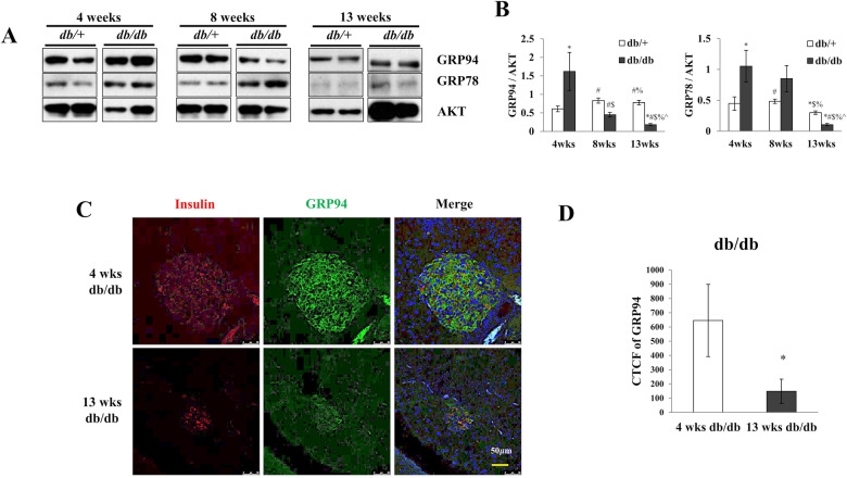
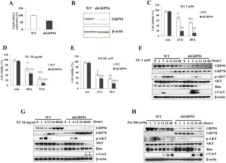
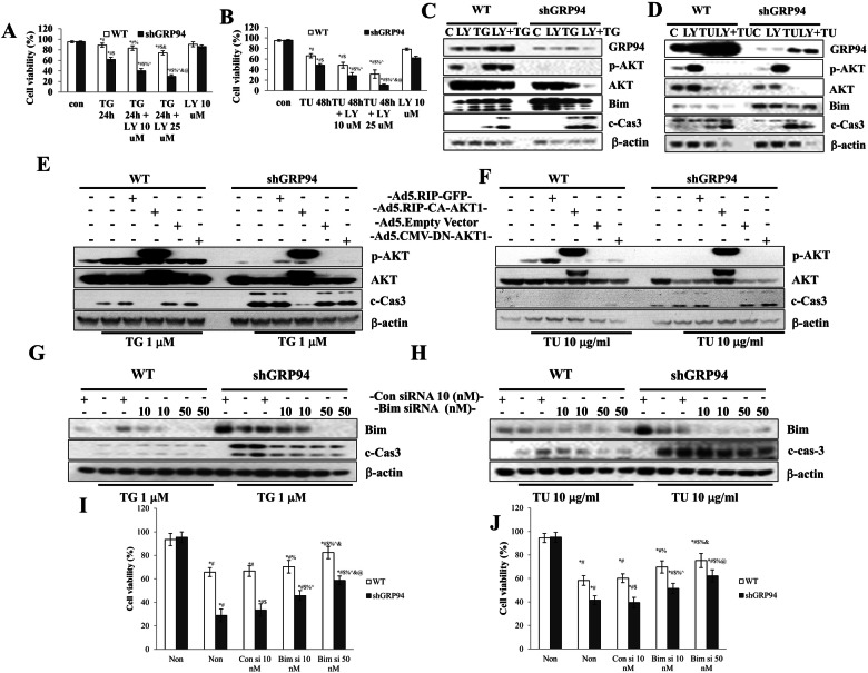
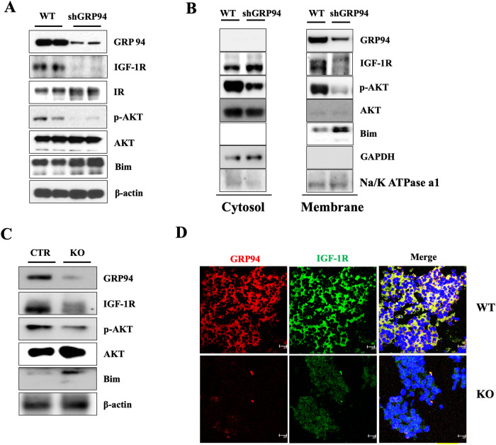
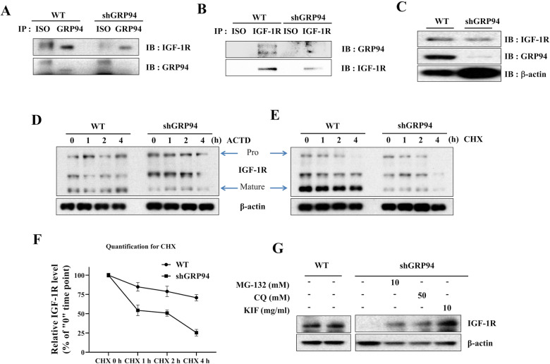
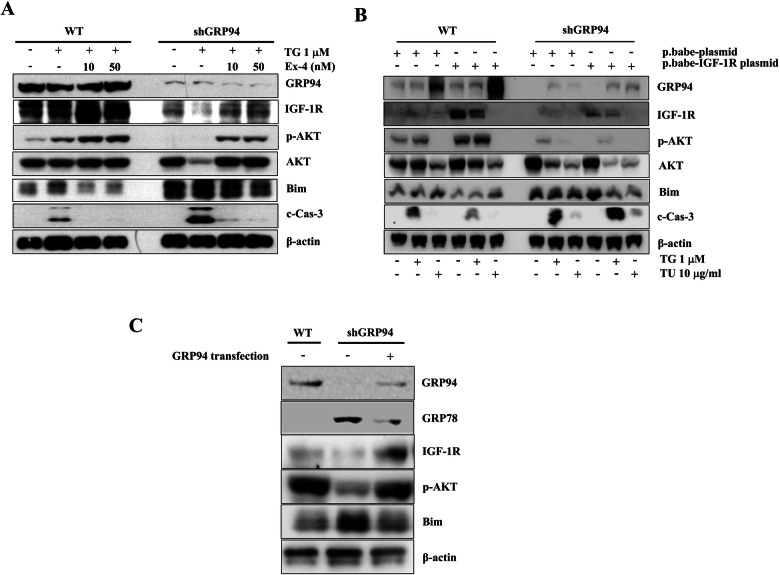
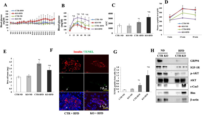
High workload-induced cellular stress can cause pancreatic islet β cell death and dysfunction, or β cell failure, a hallmark of type 2 diabetes mellitus. Thus, activation of molecular chaperones and other stress-response genes prevents β cell failure. To this end, we have shown that deletion of the glucose-regulated protein 94 (GRP94) in Pdx1+ pancreatic progenitor cells led to pancreas hypoplasia and reduced β cell mass during pancreas development in mice. Here, we show that GRP94 was involved in β cell adaption and compensation (or failure) in islets from leptin receptor-deficient (db/db) mice in an age-dependent manner. GRP94-deficient cells were more susceptible to cell death induced by various diabetogenic stress conditions. We also identified a new client of GRP94, insulin-like growth factor-1 receptor (IGF-1R), a critical factor for β cell survival and function that may mediate the effect of GRP94 in the pathogenesis of diabetes. This study has identified essential functions of GRP94 in β cell failure related to diabetes.


Conflict of interest statement

The authors declare no competing interests.

Figures

Fig. 1
Fig. 1. Dynamic change of GRP94 expression in db/db mice at different ages.
Expression of GRP94 and GRP78 in mouse islets isolated from 4, 8, and 13-week-old diabetic db/db mice and their heterozygous littermates controls by Western blot (A) and quantification (B). Immunoblots of GRP78, GRP94, and densitometry analyses are shown. *p < 0.05 versus 4wks db/+, #p < 0.05 versus 4wks db/db, $p < 0.05 versus 8wks db/+, %p < 0.05 versus 8wks db/db, ^p < 0.05 versus 4wks db/+, one-way ANOVA. C Immunostaining for GRP94 (green), insulin (red), and nucleus (blue) in sections from 4 and 13-week-old diabetic db/db mice. Scale bar, 50 µm. D Histograms show corrected total cell fluorescence (CTCF) of GRP94 fluorescence quantified by ImageJ software. The CTCF = Integrated Density – (Area of selected cell x Mean fluorescence of background readings). *p < 0.05, Student’s t-test.
Fig. 2
Fig. 2. GRP94 KD cells are more susceptible to stress-induced cell death.
A Relative GRP94 mRNA expression in WT control and Knockdown cells (shGRP94). B Protein expression of GRP94 in WT and KD cells. C Viability of WT control (white columns) and KD (black columns) cells after TG (1 μM)*p < 0.05 versus con WT, #p < 0.05 versus con shGRP94, $p < 0.05 versus 24 h WT, %p < 0.05 versus 24 h shGRP94, ^p < 0.05 versus 48 h WT, one-way ANOVA, D TU (10 μg/ml)*p < 0.05 versus con WT, #p < 0.05 versus con shGRP94, $p < 0.05 versus 48 h WT, %p < 0.05 versus 48 h shGRP94, ^p < 0.05 versus 72 h WT, one-way ANOVA, or E Pal (200 μM)*p < 0.05 versus con WT, #p < 0.05 versus con shGRP94, $p < 0.05 versus 24 h WT, %p < 0.05 versus 24 h shGRP94, ^p < 0.05 versus 48 h WT, one-way ANOVA, at different time after treatment. Cell death was measured by trypan blue (n = 3). FH Protein expression of GRP94, GRP78, p-AKT, AKT, Bim, cleaved Caspase-3 (c-Cas-3), and β-actin in WT control and GRP94 KD cells at indicated times after TG, TU, or Pal treatment as analyzed by immunoblot.
Fig. 3
Fig. 3. Regulation of AKT/Bim axis protects β cells from TG and TU-induced death.
A, B WT, and GRP94 KD Cells were exposed to LY for 30 min prior to and during the 24 h treatment with TG or 48 h treatment with TU. Cell viability was measured and compared. A *p < 0.05 versus con WT, #p < 0.05 versus con shGRP94, $p < 0.05 versus TG24h WT, %p < 0.05 versus TG24h shGRP94, ^p < 0.05 versus TG24h + LY10 μM WT, &p < 0.05 versus TG24h + LY10 μM shGRP94, @p < 0.05 versus TG24h + LY25 μM WT, one-way ANOVA. B *p < 0.05 versus con WT, #p < 0.05 versus con shGRP94, $p < 0.05 versus TU48h WT, %p < 0.05 versus TU48h shGRP94, ^p < 0.05 versus TU48h + LY10 μM WT, &p < 0.05 versus TU48h + LY10 μM shGRP94, @p < 0.05 versus TU48h + LY25 μM WT, one-way ANOVA, at indicated time after treatment with or without LY. C, D WT, and GRP94 KD cells were exposed to LY (10 µM) for 1 h, and treated with or without TG (1 μM) or TU (10 μg/ml). Immunoblots of GRP94, p-AKT, AKT, Bim, and caspase-3 cleavage. E, F WT and GRP94 KD cells were infected with the indicated adenovirus and then treated with TU or TG. Immunoblots of p-AKT, AKT, and caspase-3 cleavage. GJ WT and GRP94 KD cells were transfected with control siRNA or Bim siRNA, and then treated with TG for 6 h or TU for 48 h. G, H Total cell extracts analyzed by immunoblot of Bim and c-Cas-3. I, J Cell death was determined by trypan blue staining. *p < 0.05 versus con WT, #p < 0.05 versus con shGRP94, $p < 0.05 versus Consi10nM + TG24h or TU48h WT, %p < 0.05 versus Consi10nM + TG24h or TU48h shGRP94, ^p < 0.05 versus Bimsi10nM + TG24h or TU48h WT, &p < 0.05 versus Bimsi10nM + TG24h or TU48h WT, @p < 0.05 versus Bimsi50nM + TG24h or TU48h WT, one-way ANOVA.
Fig. 4
Fig. 4. GRP94 is required for membrane expression/maturation of IGF-1R.
A Immunoblot analysis of GRP94, IGF-1R, IR, p-AKT, AKT, Bim, and β-actin from total lysates of WT and GRP94 KD cells. B Immunoblot analysis of GRP94, IGF-1R, p-AKT, AKT, Bim, and GAPDH from cytosol or membrane of WT and GRP94 KD cells. C Immunoblot analysis of GRP94, IGF-1R, p-AKT, AKT, Bim, and β-actin in islets harvested from 8-weeks old control or GRP94 KO KO mice. D Immunofluorescence analysis of GRP94 (red), IGF-1R (green), and nucleus (blue) in WT or KO mouse islets. Scale bar, 25 µm.
Fig. 5
Fig. 5. GRP94 interacts with IGF-1R, and is a critical chaperone for the expression/maturation of IGF-1R.
A Immunoblot of IGF-1R and GRP94 following immunoprecipitation of GRP94 from lysates of WT or GRP94 KD cells. B Immunoblot of IGF-1R and GRP94 following immunoprecipitation of IGF-1R from lysates of WT or GRP94 KD cells. C Immunoblots of IGF-1R in WT and GRP94 knockdown cells. D WT and GRP94 knockdown cells were treated with actinomycin D (5 μg/ml, D) or cycloheximide (100 μg/ml, E) for indicated periods of time. After stimulation, the lysates were collected and IGF-1R levels were assayed by immunoblot. F The degradation/half-life after CHX treatment was calculated as (value at 1 h - value at 4 h)/Value at 1 h. Degradation rate was calculated based on three independent experiments using ImageJ quantification. Pro-IGF-1R degraded faster than the mature form of IGF-1R. G Immunoblot analysis for IGF-1R expression in WT and GRP94 KD cells after treatment with proteasome inhibitor (MG-132), autophagy inhibitor (CQ), or ERAD inhibitor (kifunensine, KIF).
Fig. 6
Fig. 6. Treatment with Exendin-4 or overexpression of IGF-1R or GRP94 protects β cells from TG-induced apoptosis.
A WT and GRP94 KD cells were treated with 1 μM TG in the absence or presence of 10 nM or 50 nM Exendin-4 for 6 h. Total cell extracts were analyzed by immunoblot for GRP94, IGF-1R, p-AKT, AKT, Bim, c-Cas-3, and β-actin. B WT and GRP94 KD cells were transfected with control plasmid (p.babe plasmid) or IGF-1R overexpression (p.babe-IGF-1R) plasmid and then treated with TG for 6 h or TU for 48 h. Total cell extracts were then analyzed by immunoblot for GRP94, IGF-1R, p-AKT, AKT, Bim, c-Cas-3, and β-actin. C GRP94 KD cells were transfected with GRP94 WT plasmid. Total cell extracts were then analyzed by immunoblot for GRP94, IGF-1R, p-AKT, Bim, and β-actin.
Fig. 7
Fig. 7. GRP94 deletion increases β cell susceptibility to HFD-induced β cell death and diabetes progression.
GRP94 KO (n = 12) and Cre control (n = 12) mice fed normal diet (ND) or HFD for 20 weeks. Random-fed blood glucose levels (A), blood glucose levels during an IPGTT (B), and area under the curve (AUC) during an IPGTT after HFD (C). *p < 0.05 versus control-ND, #p < 0.05 versus KO-ND, $p < 0.05 versus control-HFD, one-way ANOVA. D C-peptide secretion during an IPGTT measured before (0 min), 15 min, and 30 min after glucose injection. *p < 0.05 versus control-ND, #p < 0.05 versus KO-ND, $p < 0.05 versus control-HFD, one-way ANOVA. E β cell mass was analyzed in ND-fed and HFD-fed mice. Ten pancreatic sections from each individual mouse (N = 4 per group) were analyzed. *p < 0.05 versus control-ND, #p < 0.05 versus KO-ND, $p < 0.05 versus control-HFD, one-way ANOVA. F Fluorescence analysis from triple staining for TUNEL, insulin, and DAPI. White arrows point to TUNEL+ cells. G Histogram shows percentages of TUNEL-positive β cells in each group. Scale bar, 50 µm. *p < 0.05 versus control-ND, #p < 0.05 versus KO-ND, $p < 0.05 versus control-HFD; one-way ANOVA. H Protein expression in mouse islets isolated from all 4 treatment groups at week 21. Immunoblot shows relative protein expression of GRP94, IGF-1R, p-AKT, AKT, c-Cas-3, and β-actin. *P < 0.05.

Similar articles

Cited by

References

    1. Ardestani A, Paroni F, Azizi Z, Kaur S, Khobragade V, Yuan T, et al. MST1 is a key regulator of beta cell apoptosis and dysfunction in diabetes. Nat Med. 2014;20:385–97. doi: 10.1038/nm.3482. - DOI - PMC - PubMed
    1. Rhodes CJ. Type 2 diabetes-a matter of beta-cell life and death? Science. 2005;307:380–4. doi: 10.1126/science.1104345. - DOI - PubMed
    1. Butler AE, Janson J, Bonner-Weir S, Ritzel R, Rizza RA, Butler PC. Beta-cell deficit and increased beta-cell apoptosis in humans with type 2 diabetes. Diabetes. 2003;52:102–10. doi: 10.2337/diabetes.52.1.102. - DOI - PubMed
    1. Eletto D, Dersh D, Argon Y. GRP94 in ER quality control and stress responses. Semin Cell Dev Biol. 2010;21:479–85. doi: 10.1016/j.semcdb.2010.03.004. - DOI - PMC - PubMed
    1. Zhang Y, Wu BX, Metelli A, Thaxton JE, Hong F, Rachidi S, et al. GP96 is a GARP chaperone and controls regulatory T cell functions. J Clin Invest. 2015;125:859–69. doi: 10.1172/JCI79014. - DOI - PMC - PubMed

Publication types

MeSH terms