. 2024 May 29;15(1):4527.
doi: 10.1038/s41467-024-48320-2.

Pancreatic beta-cell IL-22 receptor deficiency induces age-dependent dysregulation of insulin biosynthesis and systemic glucose homeostasis

Affiliations

Pancreatic beta-cell IL-22 receptor deficiency induces age-dependent dysregulation of insulin biosynthesis and systemic glucose homeostasis

Haressh Sajiir et al. Nat Commun. .

Abstract

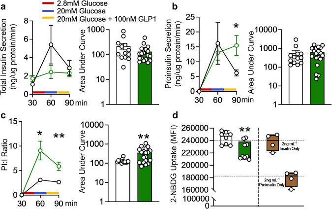
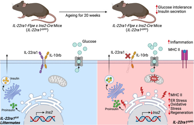
The IL-22RA1 receptor is highly expressed in the pancreas, and exogenous IL-22 has been shown to reduce endoplasmic reticulum and oxidative stress in human pancreatic islets and promote secretion of high-quality insulin from beta-cells. However, the endogenous role of IL-22RA1 signaling on these cells remains unclear. Here, we show that antibody neutralisation of IL-22RA1 in cultured human islets leads to impaired insulin quality and increased cellular stress. Through the generation of mice lacking IL-22ra1 specifically on pancreatic alpha- or beta-cells, we demonstrate that ablation of murine beta-cell IL-22ra1 leads to similar decreases in insulin secretion, quality and islet regeneration, whilst increasing islet cellular stress, inflammation and MHC II expression. These changes in insulin secretion led to impaired glucose tolerance, a finding more pronounced in female animals compared to males. Our findings attribute a regulatory role for endogenous pancreatic beta-cell IL-22ra1 in insulin secretion, islet regeneration, inflammation/cellular stress and appropriate systemic metabolic regulation.


Conflict of interest statement

S.Z.H, M.A.M and J.B.P are inventors on a patent relating to IL-22 use in metabolic disease. The remaining authors declare no other competing interests.

Figures

Fig. 1
Fig. 1. Endogenous IL-22RA1 signaling regulates oxidative homeostasis and insulin quality control in human islets.
a mRNA fold change of IL-22RA1 gene expression in healthy lean versus T2D human donor islets, relative to control GAPDH. b Total insulin, c proinsulin secretion (ng/10 islets/30 min), and d proinsulin: insulin ratio from human donor islets during glucose stimulated insulin secretion following treatment 10 µg mL−1 anti-IL22RA1 (p = 0.0341). e mRNA fold change of spliced XBP-1 (sXBP-1; p = 0.0225) and f NOS2 in human donor islets, relative to control GAPDH following treatment with 10 µg mL−1 anti-IL22RA1. g Intracellular nitrite production (µM) in human donor islets following 24 h treatment with 10 µg mL−1 anti-IL22RA1. All graphs presented as Mean ± SEM. a n = 3 biologically independent donors, Two-tailed Mann-Whitney Test; b–g n = 3 biologically independent human islet donors, Kruskal-Wallis test with Dunn’s multiple comparisons test. *p < 0.05; n.s., non-significant. *versus vehicle (anti-IgG) control. Source data are provided as a Source Data file.
Fig. 2
Fig. 2. Ablation of endogenous IL-22ra1 signaling in pancreatic β-cells leads to impaired glycemic tolerance with age.
a Experimental schematic, created with BioRender.com, released under a Creative Commons Attribution-NonCommercial-NoDerivs 4.0 International license. b Pancreatic sections of β-cell IL-22ra1 knockout animals and their wildtype littermate counterparts stained for cre-recombinase, IL-22ra1 and Insulin. c Changes in glucose tolerance following oral glucose administration in animals with age. d Area under the curve during oral glucose tolerance tests in animals with age (16 weeks p = 0.0380; 20 weeks p = 0.0146). All graphs are presented as Mean ± SEM. Female animals; n = 7 biologically independent animals (8–16 weeks), 9 biologically independent animals (20 weeks), Two-tailed unpaired Student’s t-test. *p < 0.05; n.s., non-significant. *versus wildtype (IL-22rafl/fl) littermates. Scale Bar: 20 um. Source data are provided as a Source Data file.
Fig. 3
Fig. 3. Endogenous pancreatic β-cell IL-22ra1 signaling is a key regulator of insulin biosynthesis.
a Pancreatic sections stained for insulin and proinsulin. b Mean intensity per islet area of insulin and proinsulin. c Total serum insulin (p = 0.0175) and proinsulin (p = 0.0452) in animals at 20 weeks of age. d Serum proinsulin: insulin ratio (e) Heatmap showing mRNA fold change of insulin secretion and (f) insulin signaling markers, in whole pancreatic tissue, relative to control housekeeping gene Ywhaz. Box plots in (b–d) display the median (central line), 25th to 75th percentile (box) and minimum to maximum values (whiskers). Female animals; (ab) n = 12 independent islets from 3 biologically independent animals (insulin), n = 15 independent islets from 3 biologically independent animals (WT, proinsulin), n = 12 independent islets from 3 biologically independent animals (IL-22ra1ΔβKO, proinsulin). c, d n = 8 biologically independent wildtype (IL-22rafl/fl) and 9 biologically independent IL-22ra1ΔβKO animals; e, f n = 7 biologically independent animals. Two-tailed unpaired Student’s t-test; *p < 0.05, **p < 0.01; n.s., non-significant. *versus wildtype (IL-22rafl/fl) littermates. Scale bar: 20 um. Source data are provided as a Source Data file.
Fig. 4
Fig. 4. Endogenous pancreatic β-cell IL-22ra1 signaling modulates islet growth and regeneration.
a H&E sections from pancreatic tissue. b Pancreatic islet area (p < 0.0001), and c frequency distribution of islet areas by size from serial pancreatic sections in animals at 20 weeks of age. d Absolute islet counts in animals following pancreatic islet isolation at 20 weeks of age (p < 0.0001). e Heatmap showing mRNA fold change of islet growth/regeneration markers in whole pancreatic tissue, relative to control housekeeping gene Ywhaz. f Immunohistochemical staining of pancreatic sections for Ki-67 at 8 weeks of age. g Number of Ki-67+ cells per islet area (p = 0.0090). All graphs are presented as Mean ± SEM. Female animals; ac n = 165 independent islets (all islets in serial sections) from 3 biologically independent wildtype (IL-22rafl/fl), and 115 independent islets (all islets in serial sections) from 3 biologically independent IL-22ra1ΔβKO animals. d n = 3 biologically independent wildtype (IL-22rafl/fl) and 4 biologically independent IL-22ra1ΔβKO animals. f, g n = 16 independent islets (all islets in one section) from 3 biologically independent wildtype (IL-22rafl/fl), and 10 independent islets (all islets in one section) from 3 biologically independent IL-22ra1ΔβKO animals. e n = 7 biologically independent animals; Two-tailed unpaired Student’s t-test. **p < 0.01, ****p < 0.0001; n.s., non-significant. * versus wildtype (IL-22rafl/fl) littermates. Scale Bar: 50 um. Source data are provided as a Source Data file.
Fig. 5
Fig. 5. Lack of endogenous pancreatic β-cell IL-22ra1 signaling causes islet dysfunction due to increased inflammation and cellular stress.
a Heatmap showing mRNA fold change in cellular stress markers and, b inflammatory markers in whole pancreatic tissue, relative to control housekeeping gene Ywhaz. c Mean intensity of Grp-78 per islet area (p = 0.0386) and representative image. d Mean intensity of 4-Hne per islet area (p = 0.0252) and representative image. e Percentage of islet area stained by Iba-1 (p = 0.0024) and representative image. f Percentage of islet area stained by MHC II (p = 0.0034) and representative image. All bar graphs are presented as Mean ± SEM. Female animals; a, b n = 7 biologically independent animals. c n = 15 independent islets (all islets in one section) from 3 biologically independent wildtype (IL-22rafl/fl), and 12 independent islets (all islets in one section) from 3 biologically independent IL-22ra1ΔβKO animals. d n = 7 independent islets (all islets in one section) from 3 biologically independent wildtype (IL-22rafl/fl), and 6 independent islets (all islets in one section) from 3 biologically independent IL-22ra1ΔβKO animals. e n = 13 independent islets (all islets in one section) from 3 biologically independent wildtype (IL-22rafl/fl), and 10 independent islets (all islets in one section) from 3 biologically independent IL-22ra1ΔβKO animals. f n = 14 independent islets (all islets in one section) from 3 biologically independent wildtype (IL-22rafl/fl), and 10 independent islets (all islets in one section) from 3 biologically independent IL-22ra1ΔβKO animals. Two-tailed unpaired Student’s t-test. *p < 0.05, **p < 0.01; n.s., non-significant. *versus wildtype (IL-22ra1fl/fl) littermates. Scale bar: 20 um. Source data are provided as a Source Data file.
Fig. 6
Fig. 6. Ablation of endogenous pancreatic β-cell IL-22ra1 signaling leads to poor quality insulin secretion and reduced glucose uptake.
a Total insulin secretion (ng/ug protein/min) (b) proinsulin secretion (ng/ug protein/min) (90 min; p = 0.0459), and c proinsulin: insulin ratio of mouse islets during in-vitro glucose stimulated insulin secretion, following stimulation with 2.8 mM glucose, 20 mM glucose and 20 mM glucose + 100 nM GLP-1. (60 min; p = 0.0182, 90 min; p = 0.0056, AUC; p = 0.0076). d 2-NBDG uptake in 3T3-L1 adipocytes exposed to 2 ng/mL islet insulin secretion following stimulation with 20 mM glucose + 100 nM GLP-1, p = 0.0054. All graphs in (a-c) are presented as Mean ± SEM, box plots in (d) display the median (central line), 25th to 75th percentile (box) and minimum to maximum values (whiskers). Female animals; ac n = 13 independent samples (10 islets/sample) from 3 biologically independent wildtype (IL-22rafl/fl), and 19 independent samples (10 islets/sample) from 4 biologically independent IL-22ra1ΔβKO animals. RM two-way ANOVA with the Geisser-Greenhouse correction and Sidak’s multiple comparisons test (line graphs); two-tailed Mann-Whitney Test (bar/box plots). d n = 8 (IL-22rafl/fl) and 9 (IL-22ra1ΔβKO) independent samples. *p < 0.05; n.s., non-significant; **p < 0.01; n.s., non-significant. *versus wildtype (IL-22rafl/fl) control. Source data are provided as a Source Data file.
Fig. 7
Fig. 7. Summary of findings.
Graphical abstract of findings showing that ablation of pancreatic beta-cell IL-22ra1 signaling leads to increased islet cellular stress and MHC II expression, reduced islet regeneration and insulin biosynthesis, and hypersecretion of proinsulin during glucose stimulation. These factors contribute to the age-related hyperglycaemia observed in IL-22ra1ΔβKO animals. Figure created with BioRender.com, released under a Creative Commons Attribution-NonCommercial-NoDerivs 4.0 International license.

References

    1. Rutz S, Wang X, Ouyang W. The IL-20 subfamily of cytokines — from host defence to tissue homeostasis. Nat. Rev. Immunol. 2014;14:783–795. doi: 10.1038/nri3766. - DOI - PubMed
    1. Hasnain SZ, et al. Glycemic control in diabetes is restored by therapeutic manipulation of cytokines that regulate beta cell stress. Nat. Med. 2014;20:1417–1426. doi: 10.1038/nm.3705. - DOI - PubMed
    1. Sabat R, Ouyang W, Wolk K. Therapeutic opportunities of the IL-22–IL-22R1 system. Nat. Rev. Drug Discov. 2014;13:21–38. doi: 10.1038/nrd4176. - DOI - PubMed
    1. Jones BC, Logsdon NJ, Walter MR. Structure of IL-22 Bound to Its High-Affinity IL-22R1 Chain. Structure. 2008;16:1333–1344. doi: 10.1016/j.str.2008.06.005. - DOI - PMC - PubMed
    1. Lonsdale J, et al. The Genotype-Tissue Expression (GTEx) project. Nat. Genet. 2013;45:580–585. doi: 10.1038/ng.2653. - DOI - PMC - PubMed