

A multistage framework for respiratory disease detection and assessing severity in chest X-ray images
- PMID: 38811599
- PMCID: PMC11137152
- DOI: 10.1038/s41598-024-60861-6
A multistage framework for respiratory disease detection and assessing severity in chest X-ray images
Abstract
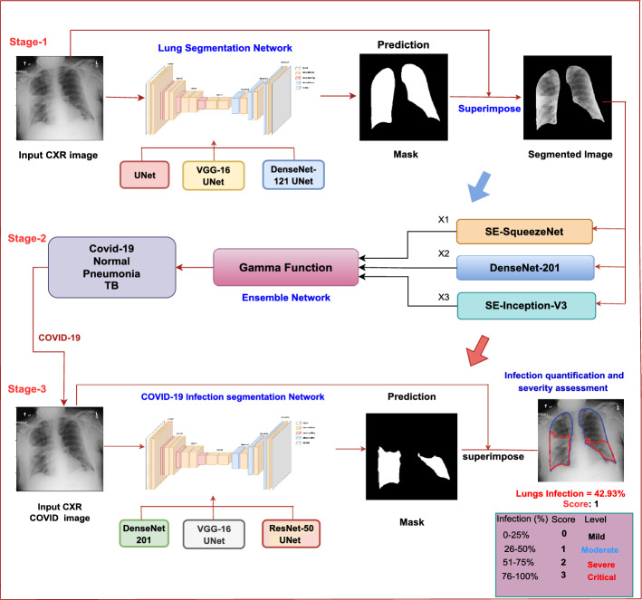
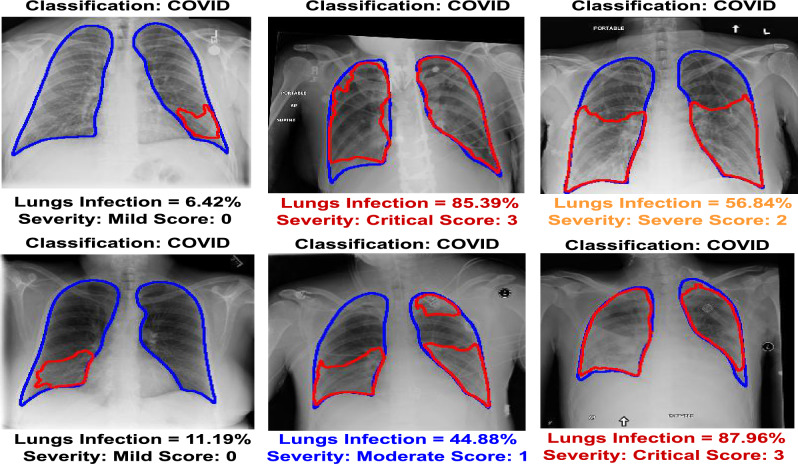
Chest Radiography is a non-invasive imaging modality for diagnosing and managing chronic lung disorders, encompassing conditions such as pneumonia, tuberculosis, and COVID-19. While it is crucial for disease localization and severity assessment, existing computer-aided diagnosis (CAD) systems primarily focus on classification tasks, often overlooking these aspects. Additionally, prevalent approaches rely on class activation or saliency maps, providing only a rough localization. This research endeavors to address these limitations by proposing a comprehensive multi-stage framework. Initially, the framework identifies relevant lung areas by filtering out extraneous regions. Subsequently, an advanced fuzzy-based ensemble approach is employed to categorize images into specific classes. In the final stage, the framework identifies infected areas and quantifies the extent of infection in COVID-19 cases, assigning severity scores ranging from 0 to 3 based on the infection's severity. Specifically, COVID-19 images are classified into distinct severity levels, such as mild, moderate, severe, and critical, determined by the modified RALE scoring system. The study utilizes publicly available datasets, surpassing previous state-of-the-art works. Incorporating lung segmentation into the proposed ensemble-based classification approach enhances the overall classification process. This solution can be a valuable alternative for clinicians and radiologists, serving as a secondary reader for chest X-rays, reducing reporting turnaround times, aiding clinical decision-making, and alleviating the workload on hospital staff.
© 2024. The Author(s).
Conflict of interest statement
The authors declare no competing interests.
Figures
Similar articles
-
Disease Localization and Severity Assessment in Chest X-Ray Images using Multi-Stage Superpixels Classification.Comput Methods Programs Biomed. 2022 Jul;222:106947. doi: 10.1016/j.cmpb.2022.106947. Epub 2022 Jun 9. Comput Methods Programs Biomed. 2022. PMID: 35749885 Free PMC article.
-
Improving the performance of CNN to predict the likelihood of COVID-19 using chest X-ray images with preprocessing algorithms.Int J Med Inform. 2020 Dec;144:104284. doi: 10.1016/j.ijmedinf.2020.104284. Epub 2020 Sep 23. Int J Med Inform. 2020. PMID: 32992136 Free PMC article.
-
COVID-19 severity detection using chest X-ray segmentation and deep learning.Sci Rep. 2024 Aug 27;14(1):19846. doi: 10.1038/s41598-024-70801-z. Sci Rep. 2024. PMID: 39191941 Free PMC article.
-
A review on lung boundary detection in chest X-rays.Int J Comput Assist Radiol Surg. 2019 Apr;14(4):563-576. doi: 10.1007/s11548-019-01917-1. Epub 2019 Feb 7. Int J Comput Assist Radiol Surg. 2019. PMID: 30730032 Free PMC article. Review.
-
Thoracic imaging tests for the diagnosis of COVID-19.Cochrane Database Syst Rev. 2020 Nov 26;11:CD013639. doi: 10.1002/14651858.CD013639.pub3. Cochrane Database Syst Rev. 2020. Update in: Cochrane Database Syst Rev. 2021 Mar 16;3:CD013639. doi: 10.1002/14651858.CD013639.pub4. PMID: 33242342 Updated.
References
-
- Rahman T, et al. Reliable tuberculosis detection using chest x-ray with deep learning, segmentation and visualization. IEEE Access. 2020;8:191586–191601. doi: 10.1109/ACCESS.2020.3031384. - DOI
-
- Varshni D, Thakral K, Agarwal L, Nijhawan R, Mittal A. Pneumonia detection using cnn based feature extraction. In: Varshni D, editor. 2019 IEEE International Conference on Electrical, Computer and Communication Technologies (ICECCT) IEEE; 2019. pp. 1–7.
-
- Deng J, et al. Imagenet: A large-scale hierarchical image database. In: Deng J, et al., editors. 2009 IEEE Conference on Computer Vision and Pattern Recognition. IEEE; 2009. pp. 248–255.
-
- Rajpurkar, P. et al. Chexnet: Radiologist-level pneumonia detection on chest x-rays with deep learning. Preprint at arXiv:1711.05225 (2017).
MeSH terms
LinkOut - more resources
Full Text Sources
Medical
Miscellaneous
