. 2024 May 29;7(1):658.
doi: 10.1038/s42003-024-06366-4.

Vimentin is a key regulator of cell mechanosensing through opposite actions on actomyosin and microtubule networks

Affiliations

Vimentin is a key regulator of cell mechanosensing through opposite actions on actomyosin and microtubule networks

Farid Alisafaei et al. Commun Biol. .

Abstract

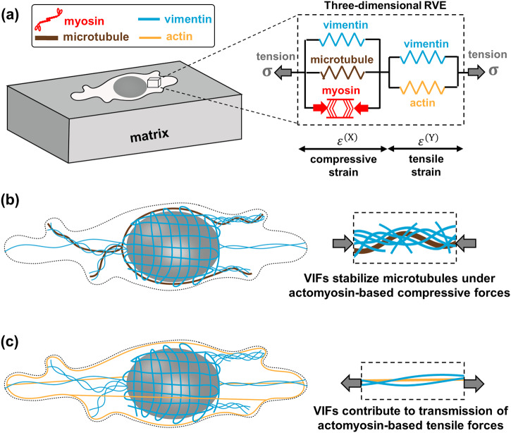
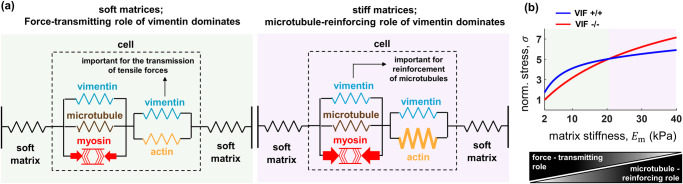
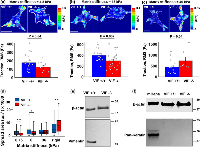
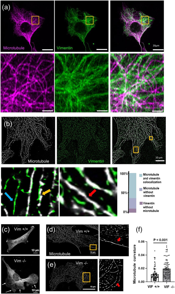
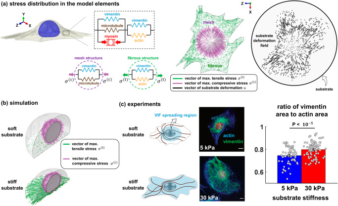
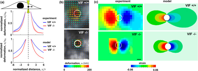
The cytoskeleton is a complex network of interconnected biopolymers consisting of actin filaments, microtubules, and intermediate filaments. These biopolymers work in concert to transmit cell-generated forces to the extracellular matrix required for cell motility, wound healing, and tissue maintenance. While we know cell-generated forces are driven by actomyosin contractility and balanced by microtubule network resistance, the effect of intermediate filaments on cellular forces is unclear. Using a combination of theoretical modeling and experiments, we show that vimentin intermediate filaments tune cell stress by assisting in both actomyosin-based force transmission and reinforcement of microtubule networks under compression. We show that the competition between these two opposing effects of vimentin is regulated by the microenvironment stiffness. These results reconcile seemingly contradictory results in the literature and provide a unified description of vimentin's effects on the transmission of cell contractile forces to the extracellular matrix.


Conflict of interest statement

The authors declare no competing interests.

Figures

Fig. 1
Fig. 1. Mechanical crosstalk between cytoskeletal components.
a The cell model includes (i) an active force-generating contractile element representing myosin motors, (ii) the actin filament network, (iii) the microtubule network, and (iv) two elements of the vimentin intermediate filament network. b The first vimentin element laterally reinforces and stabilizes microtubules under contractility-based compressive forces. c The second vimentin element interacts with actin filaments and is involved in the transmission of contractility-based tensile forces to the matrix.
Fig. 2
Fig. 2. Disruption of the vimentin network can decrease or increase cell contractility depending on microenvironment stiffness (model predictions).
a On the one hand, vimentin intermediate filaments (VIF) are involved in the transmission of actomyosin-based tensile forces to the matrix and therefore disruption of vimentin can decrease cell contractility and cell traction stress. On the other hand, vimentin filaments reinforce microtubules under compression and resist cell contraction. As a result, disruption of vimentin can lead to destabilization of microtubules and reduce the resistance against cell contraction, which can in turn increase cell traction stress. Cells on soft substrates are characterized by a weak actomyosin network and vimentin plays an important role in the transmission of forces to the substrate (force-transmitting role of VIF), while on stiff substrates, microtubules experience high compression, and they require reinforcement from the vimentin network to withstand the compression and resist cell contraction (microtubule-reinforcing role of VIF). b The model shows how the two opposing effects of vimentin compete in a matrix stiffness-dependent manner. For low matrix stiffness (low cellular contractility), the force-transmitting role of vimentin overpowers its microtubule-reinforcing role and therefore disruption of vimentin decreases cell contractile force. In contrast, for high matrix stiffness (high cellular contractility), disruption of vimentin increases cell contractile force as the microtubule-reinforcing role of vimentin becomes more important with increasing matrix stiffness.
Fig. 3
Fig. 3. Matrix-stiffness dependent effect of vimentin on traction forces and spreading area (experimental validations).
ac VIF −/− cells generate lower traction forces on soft substrates (4.5 kPa elastic modulus with n = 21 and 11, and 15 kPa elastic modulus with n = 21 and 28), while they generate higher forces on stiff substrates (40 kPa elastic modulus with n = 15 and 7). The unpaired Student’s t-test was used. The height of the bars and the error bars indicate the mean and the standard error, respectively. Traction stresses are the root mean square (RMS) values of cellular contractile forces per unit area. Scale bar: 20 µm. d Similarly, compared with wild-type cells, VIF −/− cells spread less on soft substrates, whereas they spread more on rigid substrates (all n > 100). Panel (d) was reproduced from our previously published data in ref. . These results are consistent with the model predictions and confirm that vimentin is a key modulator of cell responses to ECM mechanics. e Western blot for vimentin from wild-type (VIF +/+) and vimentin-null (VIF −/−) mouse embryonic fibroblast cells. No vimentin was detected in VIF −/− cells. β-actin was used as a loading control. f Western blot for keratin from hepatocyte cells, wild-type (VIF +/+), and vimentin-null (VIF −/−) mouse embryonic fibroblast cells. No keratin was detected in either mouse embryonic fibroblast cell. Keratin from hepatocytes was used as a positive control, and β-actin was used as a loading control.
Fig. 4
Fig. 4. Disruption of vimentin filaments negatively affects microtubule stability causing abnormal buckling of microtubules.
a Airy-scan images of VIF +/+ cells show that microtubules interpenetrate a dense network of vimentin filaments. b STORM images of VIF +/+ cells show that a large fraction of microtubules colocalizes with vimentin filaments. Yellow, red, and blue arrows denote vimentin-microtubule colocalization, microtubule track without vimentin, and vimentin track without microtubule, respectively. c Representative confocal images of microtubules in VIF +/+ and VIF −/− cells. Arrows in VIF −/− image highlight long curved microtubule filaments or bundles not seen in VIF +/+ cells. d STORM images of microtubules in VIF +/+ and (e) VIF −/− cells. Zoomed-in images show differences in microtubule curvature. f Microtubule filament curvature analysis from STORM images. Approximately 100 microtubule filaments were analyzed from 10 cells (10 filaments per cell) with 2 independent trials per condition. Two-sided unpaired Student’s t-test was used.
Fig. 5
Fig. 5. The effect of substrate stiffness on vimentin organization.
a Using finite element simulations, a fibroblast with a random shape is simulated on a stiff substrate as a 3D continuum of representative volume elements (RVEs), each of which is composed of five elements. As the active element (representing myosin motors) generates internal contractile forces, the elements in parallel with the active element experience compression, while the elements in series undergo tensile stresses. We determine the maximum compressive stress σ(c) and the maximum tensile stress σ(t) that the compressive and tensile elements experience, respectively, at each RVE. b To study how substrate stiffness impacts σ(c) and σ(t), we simulate cells on soft and stiff circular micropatterned substrates. Our simulations show that, while the tensile stress σ(t) is remarkedly disrupted on soft substrates, cells on both substrates experience high compressive stress σ(c) around the nucleus in the direction perpendicular to the nuclear envelope, expecting the existence of wavy and compressed VIFs in the juxtanuclear region of cells on both substrates. c In agreement with the simulations, our experiments show formation of mesh-like vimentin networks around the nucleus on both soft and stiff substrates. Furthermore, concomitant with the increased tension in the tensile vimentin element predicted by the model, our experiments show that cells on the stiff substrate form vimentin fibers that can reach the cell periphery, indicating that vimentin fibers extend toward the cell periphery with increasing cytoskeletal tension. (n = 70 and 99. The unpaired Student’s t-test was used. The height of the bars and the error bars indicate the mean and the standard error, respectively. Scale bars: 10 μm).
Fig. 6
Fig. 6. The effect of vimentin on the propagation of local forces in the cytoplasm.
The involvement of vimentin in the propagation of both tensile and compressive forces is further illustrated in optical tweezer experiments where a bead with a radius of r = 1 μm is dragged in the x-direction in the cytoplasm over u0 = 200 nm. Visualizing the movement of surrounding fluorescently-labeled mitochondria shows the effect of vimentin in the transmission of local forces in the cytoplasm. a The displacement field along the x-direction in the cytoplasm is plotted as a function of the distance from the bead for VIF −/− and VIF +/+ cells. The solid lines and the semitransparent areas around the solid lines represent the mean and standard error, respectively, with at least n = 10 for each curve. b A representative image of the displacement field in the cytoplasm around the bead (white circle). c A representative image of the strain field around the bead (white circle) determined as the derivative of the displacement field. As the bead moves in the cytoplasm in the x-direction, compressive (negative strain) and tensile (positive strain) fields are generated in the front and back of the bead, respectively, which extend farther in wild-type fibroblasts than in VIF −/− cells. Scale bars: 1 μm.

References

    1. Eyckmans J, Boudou T, Yu X, Chen CS. A Hitchhiker’s Guide to Mechanobiology. Dev. Cell. 2011;21:35–47. doi: 10.1016/j.devcel.2011.06.015. - DOI - PMC - PubMed
    1. Tzima E, et al. A mechanosensory complex that mediates the endothelial cell response to fluid shear stress. Nature. 2005;437:426–431. doi: 10.1038/nature03952. - DOI - PubMed
    1. Harris AK, Wild P, Stopak D. Silicone rubber substrata: a new wrinkle in the study of cell locomotion. Science. 1980;208:177–179. doi: 10.1126/science.6987736. - DOI - PubMed
    1. Legant WR, et al. Measurement of mechanical tractions exerted by cells in three-dimensional matrices. Nat. Methods. 2010;7:969–971. doi: 10.1038/nmeth.1531. - DOI - PMC - PubMed
    1. Oakes PW, Banerjee S, Marchetti MC, Gardel ML. Geometry Regulates Traction Stresses in Adherent Cells. Biophys. J. 2014;107:825–833. doi: 10.1016/j.bpj.2014.06.045. - DOI - PMC - PubMed

Publication types