. 2024 May 29;14(1):12383.
doi: 10.1038/s41598-024-63271-w.

PRSS3/mesotrypsin as a putative regulator of the biophysical characteristics of epidermal keratinocytes in superficial layers

Affiliations

PRSS3/mesotrypsin as a putative regulator of the biophysical characteristics of epidermal keratinocytes in superficial layers

Moeko Kida et al. Sci Rep. .

Abstract

Mesotrypsin, encoded by the PRSS3 gene, is a distinctive trypsin isoform renowned for its exceptional resistance to traditional trypsin inhibitors and unique substrate specificity. Within the skin epidermis, this protein primarily expresses in the upper layers of the stratified epidermis and plays a crucial role in processing pro-filaggrin (Pro-FLG). Although prior studies have partially elucidated its functions using primary cultured keratinocytes, challenges persist due to these cells' differentiation-activated cell death program. In the present study, HaCaT keratinocytes, characterized by minimal endogenous mesotrypsin expression and sustained proliferation in differentiated states, were utilized to further scrutinize the function of mesotrypsin. Despite the ready degradation of the intact form of active mesotrypsin in these cells, fusion with Venus, flanked by a peptide linker, enables evasion from the protein elimination machinery, thus facilitating activation of the Pro-FLG processing system. Inducing Venus-mesotrypsin expression in the cells resulted in a flattened phenotype and reduced proliferative capacity. Moreover, these cells displayed altered F-actin assembly, enhanced E-cadherin adhesive activity, and facilitated tight junction formation without overtly influencing epidermal differentiation. These findings underscore mesotrypsin's potentially pivotal role in shaping the characteristic cellular morphology of upper epidermal layers.

Keywords: Cell shape; Epidermis; Growth arrest; Keratinocyte; PRSS3/mesotrypsin; Tight junction.


Conflict of interest statement

The authors declare no competing interests.

Figures
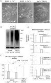
Figure 1
Figure 1
Expression of Pro-FLG and its processing protease mesotrypsin in NHEK and HaCaT cells. (A) Morphological characterization of model cells. NHEK cells were treated with (+ Ca2+) or without (− Ca2+) 2 mM CaCl2 for 2 days. Right, HaCaT cells cultured in DH10 medium. NHEK cells subjected to Ca2+ treatment undergo differentiation and exhibit a flattened morphology. Scale bars, 200 µm. (B) Expression of Pro-FLG and its processing enzyme mesotrypsin. The Pro-FLG protein is abundantly expressed in both differentiated NHEK and HaCaT cells. (C) Analysis of the mRNA expression of the mesotrypsinogen gene (PRSS3) revealed that HaCaT cells demonstrate the considerably lower transcriptional activity of the mesotrypsin gene compared to NHEK cells. n = 4, *p < 0.05. There are significant variations in error bars among NHEK cell samples, attributed to substantial differences in the basal expression of PRSS3 in NHEK (lot 1) and NHEK (lot 2). (D) The expression of PRSS3 was evaluated in each experiment, elucidating that its upregulation associated with differentiation was exclusively observed in NHEK (lot 2).
Figure 2
Figure 2
The regulation of mesotrypsin expression in HaCaT cells is governed by a protein elimination system. (A) Schematic representation delineating doxycycline-inducible expression constructs for active mesotrypsin (left) and subsequent analyses of the transgene product in Cos7 or HaCaT cells (right). TRE, tetracycline/doxycycline-responsive element. T7-tagged active mesotrypsin was successfully generated in Cos7 cells, while its production was notably absent in HaCaT cells, despite its mRNA being expressed. n = 3, **p < 0.01. (B) Upper, Presentation of the expression construct featuring a Venus-mesotrypsin fusion protein linked by a peptide linker and TEV recognition site. Lower, Assessment of the exogenous Venus-mesotrypsin fusion protein in both Cos7 and HaCaT cells. Cos7 cells exclusively express the fusion protein (~ 60 kDa), whereas HaCaT cells produce an additional 38 kDa protein, recognized by anti-Venus/GFP antibodies, following doxycycline treatment. Antibodies targeting the T7 tag associated with mesotrypsin were used to identify the 60 kDa fusion protein, but the protein was not reactive toward the ~ 25 kDa Venus-free mesotrypsin in HaCaT cells. (C) Treatment with the proteasome inhibitor MG132 effectively prevented the time-dependent reduction in the Venus-mesotrypsin fusion protein (60 kDa). n = 3, *p < 0.05.
Figure 3
Figure 3
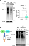
The Venus/GFP-mesotrypsin fusion protein exhibited pro-FLG-processing activity. (A) HaCaT cells expressing the Venus-mesotrypsin fusion protein exhibit a reduction in Pro-FLG levels. n = 3, *p < 0.05. (B) Recombinant mesotrypsin fused with GFP, an intrinsic derivative of Venus, degrades pro-FLG while leaving β-actin unaffected in the cellular extracts derived from HaCaT cells.
Figure 4
Figure 4
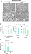
Mesotrypsin induces a flattened morphology and growth arrest in HaCaT cells. (A) Phenotypic depiction of HaCaT cells expressing Venus-mesotrypsin (right) and those without expression (left). Scale bars, 200 µm. (B) Left, Quantitative assessment of cell size. The expression of Venus-mesotrypsin, as opposed to Venus alone, results in a significant increase in cell size. Middle and right, Determination of cell thickness using z-stack images. n = 3, **p < 0.01. ***p < 0.001. Randomly selected 20 cells were analyzed in each experiment. In contrast to Cos7 cells, HaCaT cells exhibit a flattened morphology upon Venus-mesotrypsin expression. (C) The Alamar Blue assay revealed growth arrest in HaCaT cells expressing Venus-mesotrypsin. n = 5, ***p < 0.001.
Figure 5
Figure 5
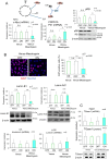
Mesotrypsin enhances adherens junctions (AJs). (A) Assessment of E-cadherin mRNA (left, n = 3) and protein (right, n = 3, **p < 0.01) expression. In addition, HaCaT cells expressing Venus-mesotrypsin exhibited upregulated E-cadherin. (B) Upper left, A schematic representation of the cell aggregation assay designed to evaluate cadherin activity. Cells subjected to trypsin treatment in the presence of calcium (TC treatment) eliminate most cell adhesion molecules, leaving cadherins intact. Disrupted cells in rotational culture reassembled into aggregates, the size of which reflects the activity of E-cadherin. Middle and lower, Representative images of reaggregated cells. Scale bars, 200 µm. Right, Quantification of aggregate sizes. n = 3, *p < 0.05. The expression of Venus-mesotrypsin, as opposed to Venus alone, results in the formation of large aggregates, indicating heightened cadherin activity.
Figure 6
Figure 6
Mesotrypsin plays a supportive role in forming adherens junctions (AJs) and tight junctions (TJs). (A) The experimental timeline for the analysis of TJ formation is depicted at the top. Immunolocalization of E-cadherin (green) and ZO-1 (red) in HaCaT cells expressing Venus only (left) or Venus-mesotrypsin (right). In the middle, HaCaT cells without transgene expression were generated. The cells were counterstained with Hoechst 33258. Scale bars, 50 µm. (B) Quantification of the accumulation of E-cadherin (left) and ZO-1 (right) in HaCaT cells expressing Venus-mesotrypsin. The signal intensities of E-cadherin (left) and ZO-1 (right) per unit length of the cell-edge lines were measured. n = 3, **p < 0.01. Randomly selected 30 (left) or 29 cells (right) were analyzed in each experiment. The presence of Venus-mesotrypsin significantly accelerates the accumulation of E-cadherin and ZO-1 at cell–cell contact sites. Treatment with only the JNK inhibitor was insufficient for promoting TJ formation in HaCaT cells expressing Venus alone or lacking Venus-mesotrypsin.
Figure 7
Figure 7
Mesotrypsin induces G1 growth arrest and cell cycle exit without impacting epidermal differentiation. (A) Expression profile of key elements involved in cell cycle progression. Venus-mesotrypsin, as opposed to Venus alone, leads to the downregulation of MYC (c-Myc) and upregulation of CDKN1A (p21) (left and middle). Concurrently, there was a reduction in RB phosphorylation. n = 3, *p < 0.05. (B) Upper, Immunostaining and quantification of the Ki67-positive cell population (depicted in red in the upper left panels, n = 3, ***p < 0.001.). The cells were counterstained with Hoechst 33258. Scale bars, 50 µm. Lower, when Lamin B1 was downregulated while lamin A/C was unaffected, the cell cycle was arrested in HaCaT cells expressing Venus-mesotrypsin. n = 3, **p < 0.01. (C) Expression analysis of various markers associated with epidermal differentiation, such as IVL (involucrin) (n = 5), LOR (loricrin) (n = 5), and TGase-1 (mRNA; n = 4, protein; n = 3), revealed no significant changes in HaCaT cells upon induction with Venus-mesotrypsin.

Similar articles

References

    1. Hoober JK, Eggink LL. The discovery and function of filaggrin. Int. J. Mol. Sci. 2022;23:1455. doi: 10.3390/ijms23031455. - DOI - PMC - PubMed
    1. Ipponjima S, Umino Y, Nagayama M, Denda M. Live imaging of alterations in cellular morphology and organelles during cornification using an epidermal equivalent model. Sci. Rep. 2020;10:62240. doi: 10.1038/s41598-020-62240-3. - DOI - PMC - PubMed
    1. Matsui T, et al. A unique mode of keratinocyte death requires intracellular acidification. Proc. Natl. Acad. Sci. USA. 2021;118:2020722118. doi: 10.1073/pnas.2020722118. - DOI - PMC - PubMed
    1. Rübsam M, et al. E-cadherin integrates mechanotransduction and EGFR signaling to control junctional tissue polarization and tight junction positioning. Nat. Commun. 2017;8:01170. doi: 10.1038/s41467-017-01170-7. - DOI - PMC - PubMed
    1. Dreesen O, et al. Lamin B1 fluctuations have differential effects on cellular proliferation and senescence. J. Cell. Biol. 2013;200:605–617. doi: 10.1083/jcb.201206121. - DOI - PMC - PubMed