. 2024 Oct;45(10):2119-2133.
doi: 10.1038/s41401-024-01305-9. Epub 2024 May 29.

A novel small-molecule PCSK9 inhibitor E28362 ameliorates hyperlipidemia and atherosclerosis

Affiliations

A novel small-molecule PCSK9 inhibitor E28362 ameliorates hyperlipidemia and atherosclerosis

Wei-Zhi Wang et al. Acta Pharmacol Sin. 2024 Oct.

Abstract

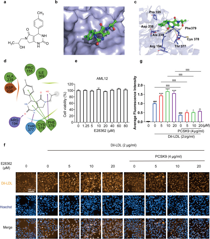
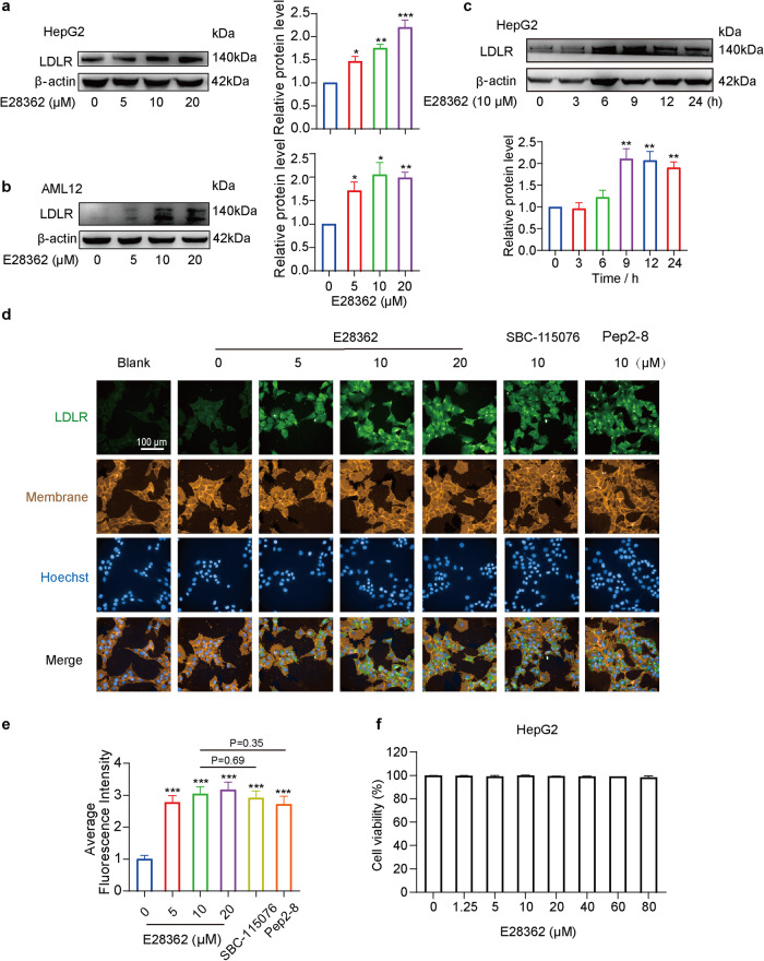
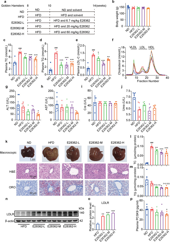
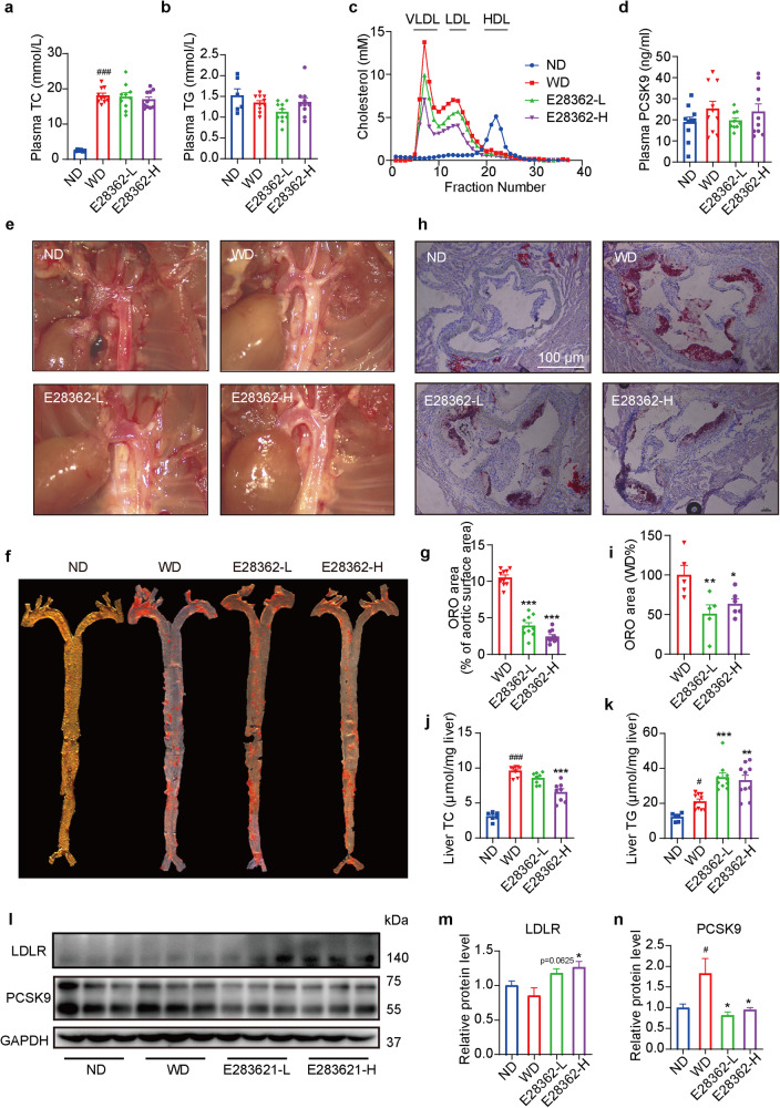
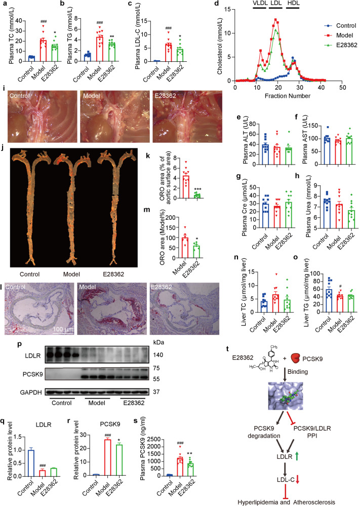
Proprotein convertase subtilisin/kexin type 9 (PCSK9) binds to the epidermal growth factor precursor homologous domain A (EGF-A) of low-density lipoprotein receptor (LDLR) in the liver and triggers the degradation of LDLR via the lysosomal pathway, consequently leading to an elevation in plasma LDL-C levels. Inhibiting PCSK9 prolongs the lifespan of LDLR and maintains cholesterol homeostasis in the body. Thus, PCSK9 is an innovative pharmacological target for treating hypercholesterolemia and atherosclerosis. In this study, we discovered that E28362 was a novel small-molecule PCSK9 inhibitor by conducting a virtual screening of a library containing 40,000 compounds. E28362 (5, 10, 20 μM) dose-dependently increased the protein levels of LDLR in both total protein and the membrane fraction in both HepG2 and AML12 cells, and enhanced the uptake of DiI-LDL in AML12 cells. MTT assay showed that E28362 up to 80 μM had no obvious toxicity in HepG2, AML12, and HEK293a cells. The effects of E28362 on hyperlipidemia and atherosclerosis were evaluated in three different animal models. In high-fat diet-fed golden hamsters, administration of E28362 (6.7, 20, 60 mg·kg-1·d-1, i.g.) for 4 weeks significantly reduced plasma total cholesterol (TC), triglyceride (TG), low-density lipoprotein-cholesterol (LDL-C) and PCSK9 levels, and reduced liver TC and TG contents. In Western diet-fed ApoE-/- mice (20, 60 mg·kg-1·d-1, i.g.) and human PCSK9 D374Y overexpression mice (60 mg·kg-1·d-1, i.g.), administration of E28362 for 12 weeks significantly decreased plasma LDL-C levels and the area of atherosclerotic lesions in en face aortas and aortic roots. Moreover, E28362 significantly increased the protein expression level of LDLR in the liver. We revealed that E28362 selectively bound to PCSK9 in HepG2 and AML12 cells, blocked the interaction between LDLR and PCSK9, and induced the degradation of PCSK9 through the ubiquitin-proteasome pathway, which finally resulted in increased LDLR protein levels. In conclusion, E28362 can block the interaction between PCSK9 and LDLR, induce the degradation of PCSK9, increase LDLR protein levels, and alleviate hyperlipidemia and atherosclerosis in three distinct animal models, suggesting that E28362 is a promising lead compound for the treatment of hyperlipidemia and atherosclerosis.

Keywords: E28362; LDLR; PCSK9 inhibitor; atherosclerosis; hyperlipidemia.


Conflict of interest statement

The authors declare no competing interests.

Figures

Fig. 1
Fig. 1. Identification of novel PCSK9 inhibitors using virtual screening assays.
a The structure of E28362 (C16H19O3N3). b The E28362 binding pocket of PCSK9. c The key amino acids of PCSK9 interacting with E28362. d The binding mode of E28362 was predicted with induced-fit docking. e The cytotoxic effects of E28362 on AML12 cells after incubation with E28362 (0–80 μM) for 24 h. f, g E28362 increased the uptake of DiI-LDL in AML12 cells. AML12 cells were treated with DMSO or E28362 (5, 10 or 20 μM) in DMEM with or without human PCSK9 protein (4 μg/mL) for 18 h, and then the medium was replaced with DMEM containing 2 μg/mL DiI-LDL for another 4 h. Nuclei were stained with Hoechst. Representative images are shown (f). Scale bars: 100 μm. The average fluorescence intensity of DiI-LDL (g) in AML12 cells was calculated. Values are presented as mean ± SEM. Statistical significance was calculated using the unpaired two-tailed Student’s t-test. ∗∗∗P < 0.001 vs E28362 (0 μM) + DiI-LDL (2 μg/mL) group; ^P < 0.05, ^^P < 0.01 vs E28362 (0 µM) + PCSK9 (4 μg/mL) + DiI-LDL (2 μg/mL) group; ###P < 0.001 vs E28362 (0 μM) group; $$$P < 0.001.
Fig. 2
Fig. 2. E28362 increases LDLR protein expression in hepatocytes.
The total LDLR protein level was measured by WB (ac) and the LDLR protein level in the membrane (d, e) was determined by immunofluorescence staining. a, b HepG2 (a) and AML12 (b) cells were treated with DMSO or E28362 at 0, 5, 10, and 20 μM for 18 h. c HepG2 cells were treated with E28362 (10 μM) for 0, 3, 6, 9, 12, 24 h. d, e HepG2 cells were treated with E28362 (0, 5, 10, and 20 μM), SBC-115076 (10 μM), or Pep2-8 (10 μM) for 18 h and then incubated with Alexa Fluor 488-conjugated goat anti-rabbit IgG (H + L). The blank group cells were only treated with DMSO. d Representative immunoblot and images are shown. Green, membrane LDLR proteins stained with Alexa Fluor 488-conjugated goat anti-rabbit IgG; red, membranes stained with DiI; blue, nuclei stained with DAPI. Scale bars: 100 μm. e The average fluorescence intensity of LDLR was calculated by ImageJ. f The cytotoxic effects of E28362 (0–80 μM) on HepG2 cells after incubation for 24 h. Values are presented as means ± SEM. Statistical significance was calculated using an unpaired 2-tailed Student’s t-test. *P < 0.05, **P < 0.01, ***P < 0.001 vs E28362 (0 μM).
Fig. 3
Fig. 3. E28362 improves the plasma profile and alleviates hepatic lipid accumulation in golden hamsters.
Male golden hamsters were intragastrically administered with vehicle or E28362 (6.7, 20, or 60 mg/kg per day) for 4 weeks (n = 6 per group). a Schematic diagram of the experimental design and treatment. b The body weight of golden hamsters at the end of the experiment. ce Plasma TC (c), TG (d), and LDL-C (e) levels. f Plasma was pooled per group and the distribution of cholesterol over the individual lipoproteins was analyzed after separation by fast protein liquid chromatography (FPLC). gj Plasma ALT (g), AST (h), Cre (i), and urea (j) levels. k Representative pictures of macroscopic appearance (Scale bars: 1 cm), hematoxylin and eosin (H&E) staining (Scale bars: 50 μm), and Oil red O (ORO) staining (Scale bars: 50 μm). l, m Hepatic TC and TG contents. n, o The hepatic protein expression of LDLR was determined by WB. Representative images are shown. Protein levels were normalized to β-actin. p Plasma PCSK9 content was measured by a commercial ELISA kit. Values are presented as means ± SEM. n = 6 per group. Blue, red, green, purple and orange-yellow colors represented ND, HFD, E28362-L, E28362-M, and E28362-H, respectively. Statistical significance was calculated with one-way ANOVA. #P < 0.05, ###P < 0.001 vs ND group; *P < 0.05, **P < 0.01, ***P < 0.001 vs HFD group.
Fig. 4
Fig. 4. E28362 inhibits the development of atherosclerosis in ApoE−/− mice.
Male ApoE−/− mice were intragastrically administered with vehicle or E28362 (30 or 60 mg/kg per day) for 12 weeks. a, b Plasma TC (a) and TG (b) levels were measured (n = 6 for ND group; n = 10 for WD, E28362-L, and E28362-H group). c Plasma was pooled per group and the distribution of cholesterol over the individual lipoproteins was analyzed after separation by FPLC (n = 6 for ND group; n = 10 for WD, E28362-L, and E28362-H group). d The plasma PCSK9 content was measured by a commercial ELISA kit (n = 6 for ND group; n = 10 for WD, E28362-L, and E28362-H group). e Representative gross lesions of the aortic arch. f, g Representative ORO-stained images of the full length of the aorta (f) and quantification of the percentage of the plaque area vs. the area of the full-length aorta (g) (n = 10 per group). h, i Representative image of the ORO-stained sections from the aortic root (h) and quantification of aortic root lesion areas (n = 5-6 for per group) (i). j, k Hepatic TC and TG contents (n = 6 for ND group; n = 10 for WD, E28362-L, and E28362-H group). ln The hepatic protein expression of PCSK9 and LDLR was determined by WB (n = 6 for ND group; n = 10 for WD, E28362-L, and E28362-H group). Representative images are shown. Protein levels were normalized to GAPDH. Values are presented as means ± SEM. For all data, blue, red, green and purple lines showed the fractions of ND, WD, E28362-L, and E28362-H, respectively. Statistical significance was calculated with one-way ANOVA. #P < 0.05, ###P < 0.001 vs ND group; *P < 0.05, **P < 0.01, ***P < 0.001 vs WD group.
Fig. 5
Fig. 5. E28362 binds to PCSK9 and blocks the interaction between PCSK9 and LDLR.
a Surface plasmon resonance sensorgram for the interaction between E28362 and human PCSK9 protein. The dissociation constant (KD) value is shown. b The amount of PCSK9 rather than LDLR retained increased with the increase of E28362 concentration in the cellular thermal shift assay. Protein band intensities were quantified by ImageJ. c E28362 improved the thermal stability of PCSK9 rather than LDLR in the cellular thermal shift assay. Protein band intensities were quantified by ImageJ. Blot intensities were normalized to the intensity obtained for the 4 °C sample. Values are presented as means ± SEM. Statistical significance was calculated using an unpaired 2-tailed Student’s t-test. b *P < 0.05 vs E28362 (0 μM). c P < 0.05, E28362 (20 μM) at 59 °C vs DMSO at 59 °C. d Schematic diagram of the PCSK9-LDLR BiFC model. e HEK293a cells were co-transfected with or without pBiFC-VC155-his LDLR and pBiFC-VN173-flag PCSK9 plasmids, and then the transfected cells were treated with DMSO or E28362 (2.5, 5, 10, and 20 μM) for 18 h. The average fluorescence intensity of Venus was calculated. f The cytotoxic effects of E28362 (0–80 μM) on HEK293a cells after incubation for 24 h. e, f Values are presented as means ± SEM. Statistical significance was calculated using an unpaired 2-tailed Student’s t-test. ∗∗∗P < 0.0001 vs E28362 (0 μM).
Fig. 6
Fig. 6. E28362 induces PCSK9 degradation through the ubiquitin-proteasome pathway.
a E28362 decreased the PCSK9 protein level in HepG2 and AML12 cells. HepG2 or AML12 cells were treated with E28362 (0, 5, 10, and 20 μM) for 18 h. *P < 0.05, **P < 0.01 vs E28362 (0 μM). b E28362 (10 μM) decreased the PCSK9 protein level when treated for the indicated times in HepG2 cells. *P < 0.05, **P < 0.01, ***P < 0.001 vs E28362 treated duration (0 h). c E28362 decreased the secretion of PCSK9 protein into the media in HepG2 cells. **P < 0.01, ***P < 0.001 vs E28362 (0 μM) (d, e) HepG2 cells were treated with or without DSMO, cycloheximide (CHX, 10 μg/mL), MG132 (10 μM), and E28362 (10 μM) for the indicated periods. P < 0.05, ∗∗P < 0.01 vs HepG2 treated with DMSO for the indicated time when CHX was present. f HepG2 cells were treated with (CHX, 10 μg/mL), MG132 (10 μM), and E28362 (10 μM) or DMSO for 18 h and lysed, and then the cell lysates were incubated with anti-PCSK9 for 24 h. After that, the mixtures were incubated with A + G agarose immunoprecipitation (IP) beads for 2 h. Samples were then separated and proteins were detected by WB. For all data, values are presented as means ± SEM; statistical significance was calculated using an unpaired 2-tailed Student’s t-test.
Fig. 7
Fig. 7. E28362 decreased PCSK9 levels and alleviated the development of atherosclerosis in PCSK9 overexpression mice.
Male C57BL/6 J mice were injected with PCSK9 D374Y overexpression virus or the corresponding control virus through the tail vein, and then the mice were fed a WD and intragastrically administered with vehicle or E28362 (60 mg/kg per day) for 12 weeks. ac Plasma TC (a), TG (b), and LDL-C (c) levels were measured (n = 10 per group). d Plasma was pooled per group and the distribution of cholesterol over the individual lipoproteins was analyzed after separation by FPLC (n = 10 per group). eh Plasma ALT (e), AST (f), Cre (g), and urea (h) levels were measured (n = 10 per group). i Representative gross lesions of the aortic arch. j Representative ORO-stained full length of the aorta. k Percentage of the plaque area vs. the area of the full-length aorta (n = 10 per group). l Representative image of ORO-stained sections from the aortic root sections. Scale bars: 100 μm. m Aortic root lesion areas (n = 10 per group). n, o Hepatic TC and TG contents (n = 10 per group). pr WB analysis was conducted to determine the hepatic protein expression levels of PCSK9 and LDLR (n = 10 per group). Representative images are shown and the relative protein level was calculated by ImageJ. s The plasma PCSK9 content was measured by a commercial ELISA kit (n = 10 per group). For all data, blue, red and green lines showed the fractions of control, model, and E28362, respectively. Values are presented as means ± SEM. #P < 0.05; ###P < 0.001 vs Control group; *P < 0.05, **P < 0.01, ***P < 0.001 vs Model group. ac, e, h, ns Statistical significance was calculated with one-way ANOVA; k, m Statistical significance was calculated using an unpaired 2-tailed Student’s t-test. t Schematic diagram shows the hypolipidemic and anti-atherosclerotic mechanism of E28362. Small-molecule compound E28362 binds to PCSK9, blocks the protein-protein interaction (PPI) between PCSK9 and LDLR, and induces the degradation of PCSK9, which leads to increased LDLR protein level, decreased LDL-C level, and finally exerts beneficial effects on hyperlipidemia and atherosclerosis.

References

    1. Virani SS, Alonso A, Benjamin EJ, Bittencourt MS, Callaway CW, Carson AP, et al. Heart disease and stroke statistics-2020 update: a report from the American Heart Association. Circulation. 2020;141:e139–e596. - PubMed
    1. Nayor M, Brown KJ, Vasan RS. The molecular basis of predicting atherosclerotic cardiovascular disease risk. Circ Res. 2021;128:287–303. - PMC - PubMed
    1. Arvanitis M, Lowenstein CJ. Dyslipidemia. Ann Intern Med. 2023;176:ITC81–ITC96. - PubMed
    1. Herrington W, Lacey B, Sherliker P, Armitage J, Lewington S. Epidemiology of atherosclerosis and the potential to reduce the global burden of atherothrombotic disease. Circ Res. 2016;118:535–46. - PubMed
    1. Borén J, Chapman MJ, Krauss RM, Packard CJ, Bentzon JF, Binder CJ, et al. Low-density lipoproteins cause atherosclerotic cardiovascular disease: Pathophysiological, genetic, and therapeutic insights: a consensus statement from the european atherosclerosis society consensus panel. Eur Heart J. 2020;41:2313–30. - PMC - PubMed