. 2024 Oct;31(10):1567-1579.
doi: 10.1038/s41594-024-01322-6. Epub 2024 May 29.

MYC phase separation selectively modulates the transcriptome

Affiliations

MYC phase separation selectively modulates the transcriptome

Junjiao Yang et al. Nat Struct Mol Biol. 2024 Oct.

Erratum in

Abstract

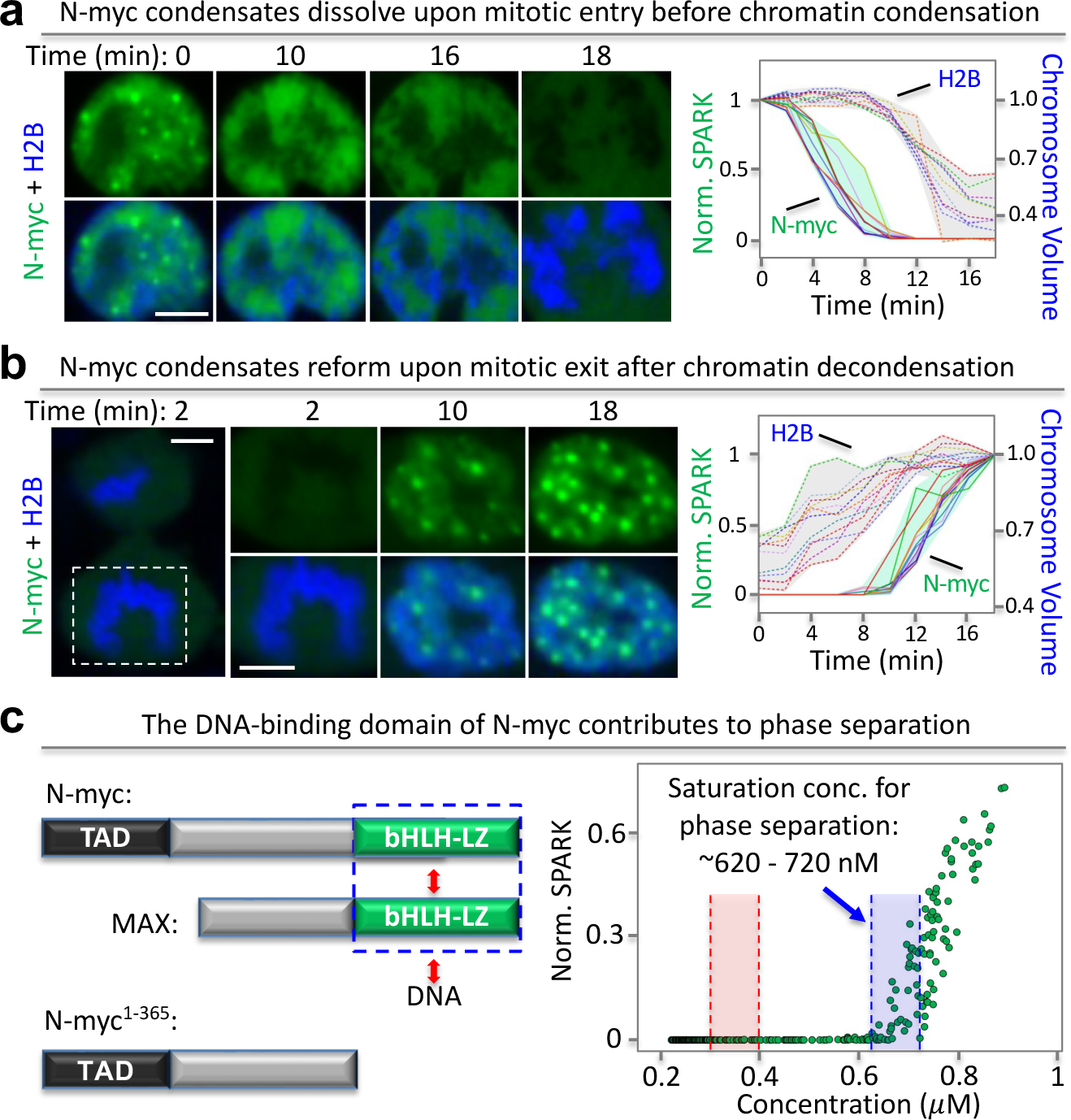
Dysregulation and enhanced expression of MYC transcription factors (TFs) including MYC and MYCN contribute to the majority of human cancers. For example, MYCN is amplified up to several hundredfold in high-risk neuroblastoma. The resulting overexpression of N-myc aberrantly activates genes that are not activated at low N-myc levels and drives cell proliferation. Whether increasing N-myc levels simply mediates binding to lower-affinity binding sites in the genome or fundamentally changes the activation process remains unclear. One such activation mechanism that could become important above threshold levels of N-myc is the formation of aberrant transcriptional condensates through phase separation. Phase separation has recently been linked to transcriptional regulation, but the extent to which it contributes to gene activation remains an open question. Here we characterized the phase behavior of N-myc and showed that it can form dynamic condensates that have transcriptional hallmarks. We tested the role of phase separation in N-myc-regulated transcription by using a chemogenetic tool that allowed us to compare non-phase-separated and phase-separated conditions at equivalent N-myc levels, both of which showed a strong impact on gene expression compared to no N-myc expression. Interestingly, we discovered that only a small percentage (<3%) of N-myc-regulated genes is further modulated by phase separation but that these events include the activation of key oncogenes and the repression of tumor suppressors. Indeed, phase separation increases cell proliferation, corroborating the biological effects of the transcriptional changes. However, our results also show that >97% of N-myc-regulated genes are not affected by N-myc phase separation, demonstrating that soluble complexes of TFs with the transcriptional machinery are sufficient to activate transcription.


Conflict of interest statement

Competing interests: X.S. and W.A.W. are co-founders of Granule Therapeutics. The remaining authors declare no competing interests.

Figures

Extended Data Fig. 1 |
Immunofluorescence images of Kelly cells treated with DMSO or MYC/MAX dimerization inhibitor MYCi975. (Left) Kelly cells were incubated with DMSO or MYCi975 (30 μM) for 20 hours, followed by immunostaining with N-myc antibody. Stars (*) indicate representative cells. Triangle (Δ) indicates few (that is un-representative) cells with high MYCN expression. Scale bar, 5 μm. (Right) Normalized average N-myc immunofluorescence intensity in DMSO or MYCi975 treated cells. Data are shown as mean ± SD. N = 3 biological replicates.
Extended Data Fig. 2 |
Neuroblastoma cell lines with various degrees of MYC (N-myc, c-MYC) protein expression. Western blot analysis of MYC proteins in various neuroblastoma cells.
Extended Data Fig. 3 |
Estimation of N-myc concentration in the Kelly cells. The overall procedure is (a) calculation of an mEGFP concentration-brightness standard curve using purified mEGFP protein. (b) Western blot analysis to determine relative amount of N-myc in Kelly vs SH-EP cells. (c) Fluorescence imaging (Hoechst)-based measurement of the nuclear volume of SH-EP and Kelly cells. The N-myc-mEGFP concentration in SH-EP cells is determined by comparing the fluorescence intensity of N-myc-mEGFP versus that of the purified mEGFP. (d) Estimation of the N-myc concentration in the nucleus of Kelly cells. Data are mean + SD (n = 20 cells). Scale bar, 10 μm.
Extended Data Fig. 4 |
Relationship of N-myc condensate number and size to the N-myc protein levels. (a) Number of N-myc condensates was plotted against the N-myc protein level per nucleus. SH-EP cells expressing N-mycmEGFP were imaged under the spinning disc confocal microscope. (b) Size of N-myc condensates was plotted against the N-myc protein level in single nucleus. (c) SH-EP cells with different expressing levels of N-myc-mEGFP. Images are max-projected to show all condensates. Dim cells are shown with brightness adjustment as listed. Scale bar: 5 μm.
Extended Data Fig. 5 |
Endogenous N-myc condensates contain DNA-binding and dimerization partner, transcriptional machinery and nascent RNA but do not co-localize with c-Jun. Immunostained N-myc condensates colocalized with MED1 (a), Pol II S5p (b), MAX (c), nascent RNA (d) and c-Jun (e). Kelly cells were stained with antibody against N-myc, and antibodies against MED1, Pol II S5p, or c-Jun, or co-expressed with MAX-mKO3, or labeled by 5-ethynyluridine for labeling nascent RNA. The arrows point to representative condensates. The fluorescence intensity profile (right panel) is extracted from the position shown by the dashed line. Co-localization assessment by Pearson’s correlation coefficient is shown in right panels. Center lines show the median values. Green boxes contain the 25th to 75th percentiles of dataset. Black whiskers mark the 10th and 90th percentiles. Outliers are marked with grey dots. NMED1 = 76, NPol II-S5P = 67, NMAX = 61, Nnascent RNA = 82 cells, Nc-Jun = 67 cells. Scale bars, 5 μm.
Extended Data Fig. 6 |
Condensate number and size related to protein levels of the truncated N-myc lacking the DNA-binding domain. (a) Number of N-myc1–365 condensates was plotted against the N-myc protein level per nucleus. (b) Size of N-myc1–365 condensates was plotted against the N-myc protein level in single nucleus. (c) SH-EP cells with different expressing levels of N-myc1–365- mEGFP. Images are max-projected to show all condensates. Dim cells are shown with brightness adjustment as listed. Scale bar: 5 μm.
Extended Data Fig. 7 |
MAX contributes to N-myc phase separation. (a) MAX was knocked out by sgRNA in SH-EP cells expressing MYCN-mEGFP. Top left: SPARK value against N-myc concentration in wild type SH-EP cells expressing MYCN-mEGFP. Top middle: SPARK value against N-myc concentration in MAX-KO SH-EP cells expressing MYCN-mEGFP. Lower left: western blot showing MAX protein level in wild type (WT) and MAX-KO (sgMAX) SH-EP cells. Lower middle: SPARK value in MAX-KO SH-EP cells expressing MYCN-mEGFP and MAX-mKO3. Right panel: N-myc saturation concentration in WT, MAX-KO and rescued SH-EP cells. The MAX-KO SH-EP cells were rescued by expressing MAX-mKO3. Center lines show the median values. Boxes contain the 25th to 75th percentiles of dataset. Whiskers mark the minimum and maximum values. NWT = 40, NsgMAX = 49, NsgMAX+MAX-mKO3 = 54. P value, two-sided non-paired t-test. (b) Left: SPARK value against N-myc concentration in SH-EP cells expressing MYCN-mEGFP. Right: SPARK value against N-myc concentration in SH-EP cells expressing MYCNmEGFP and MAX-mKO3.
Extended Data Fig. 8 |
Phase separation of full-length N-myc protein is enhanced by DNA oligonucleotides containing non-canonical Myc E-box sequences. (a) Fluorescence anisotropy assay showing binding of the N-Myc / MAX heterodimer to non-canonical E-box DNA in 20 mM HEPES, 150 mM KCl, 10 mM MgCl2, 1 mM DTT, and 0.01% NP-40 containing buffer. The concentration of fluorophore-labeled oligonucleotide DNA containing 1 NCE-box sequence (CATCTG or CATATG) was fixed at 50 nM and a ratio of Myc / Max of 3:1 was used. The dissociation constant (Kd) of the interaction between proteins and DNA was determined by fitting the experimental data in Origin Pro 8.0 {Roehrl, 2004 #1}. MAX/CEbox and N-myc/MAX/Cebox data are the same as in Fig. 4b. (b) Brightfield microscopy images of solutions containing the N-myc / Max complex (10 .M Myc / 3 .M Max) in the presence of 1NCEbox (26-mer), and 7NCEbox (182 mer) DNA in 20 mM HEPES, 150 mM KCl, 10 mM MgCl2, 1 mM DTT. Purple boxes highlights the images with N-myc phase separation observed. The images show similar phase separation as with canonical E-box sequences. The scale bar is 5 μm.
Extended Data Fig. 9 |
Saturation concentration of SparkDrop-tagged N-myc and the stable N-myc/SparkDrop SH-EP cells used for the RNAseq experiments. (a) Saturation concentration of SparkDrop-tagged N-myc with and without lenalidomide. Each green dot represents data from a single cell (~200 cells for each group). The concentration was calculated based on the green fluorescence of mEGFP in SparkDrop. (b) Representative images of stable SparkDrop-MYCN cells before and 30 minutes after 1 .M lenalidomide (lena) incubation. Scale bar: 20 .m. (c) N-myc concentration is 0.20 Å} 0.01 .M (Mean Å} SD). N = 200 cells.
Extended Data Fig. 10 |
RT-qPCR of SERINC2 mRNA in SH-EP cells expressing N-myc/SparkDrop treated with 1 μM lenalidomide for different duration. Data are shown as mean Å} SD. N = 3 biological independent replicates. P value, two-sided non-paired t-test.
Fig. 1.
Fig. 1.. N-myc undergoes phase separation, which requires the intrinsically disordered transactivation domain.
(a) Immunofluorescence images of N-myc in MYCN-amplified neuroblastoma Kelly cells (top left panel) and the MYCN-nonamplified SH-EP cells (bottom left panel). N-myc forms puncta in the Kelly cells but not in the SH-EP cells that express no or little N-myc (Fig. S3). Right panel, average N-myc antibody fluorescence intensity in Kelly and SH-EP cells (N ~ 100 cells from 3 biological replicates). Data are shown as mean±SD. Each dot represents one cell. Scale bar, 10 μm. (b) Expression of mEGFP fused N-myc in the neuroblastoma SH-EP cells that have no or little endogenous N-myc protein expression. Left: quantitative analysis of N-myc puncta formation against its protein level in single cells. Each green circle corresponds to an individual cell, i.e. ~300 cells were analyzed. The concentration of the fusion protein was estimated based on purified mEGFP (see details in Methods). The red dashed lines depict a range of saturation concentrations for phase separation. Right: representative fluorescence images; the upper panel is brightened, the lower is not. Scale bar, 5 μm. (c) Coalescence events between N-myc condensates. Top: time-lapse fluorescence images. Scale bar, 1 μm. Bottom-left: Aspect ratio over time. Quantitative analysis of the coalescence events was plotted. Each line represents the best fit for each event. Gray shade indicates the range. Bottom-right: inverse capillary velocity derived from coalescence events. Data are shown as mean+SD (n = 12). (d) Quantitative analysis of the truncated N-myc lacking N-terminal TAD, which is an IDR. Each green circle corresponds to an individual cell (~200 cells). Disorder prediction by PONDR is shown at the bottom. Scale bar, 5 μm.
Fig. 2.
Fig. 2.. N-myc condensates contain transcriptional markers including DNA-binding and dimerization partner, genomic DNA, transcriptional machinery and nascent RNA.
(a) Fluorescence images of N-myc-mEGFP and MAX-mKO3 in SH-EP cells. Representative condensates are indicated by arrows. (b) Fluorescence images of N-myc condensates with single molecule DNA FISH against TP53. (c) Immunofluorescence (IF) images of MED1 with N-myc-mEGFP condensates. (d) IF-imaged Pol II S5p with fluorescence images of N-myc-mEGFP. (e) 5-ethynyluridine-labeled nascent RNA with fluorescence images of N-myc-mEGFP condensates. (f) Co-localization assessment by Pearson’s correlation coefficient. C enter lines show the median values. Green boxes contain the 25th to 75th percentiles of dataset. Black whiskers mark the 10th and 90th percentiles. Outliers are marked with grey dots. Data are mean ± SD. The profile of fluorescence intensity corresponds to the position indicated by the dashed line. Arrows in the profile plot point to the condensates with colocalization. NMAX = 63, NMED1 = 70, NPol II-S5P = 63, Nnascent RNA = 67 cells. Scale bars, 5 μm (a – e).
Fig. 3.
Fig. 3.. Disassembly and assembly of N-myc condensates during mitotic entry and exit, respectively.
(a) Time-lapse fluorescence images of SH-EP cells expressing N-myc-mEGFP when cells enter mitosis. mIFP-tagged histone 2B (H2B, in blue) was co-expressed and used to calculate the chromosome volume. Right panel: plots of quantitatively analyzed condensate dissolution and chromosome condensation over time. Each line indicates a single-cell trace (n = 11 cells). Shades show the maximum and minimum values of data. (b) Time-lapse fluorescence images of SH-EP cells expressing N-myc-mEGFP when cells exit mitosis. Right panel: plots of quantitatively analyzed reformation of N-myc condensates and chromosome de-condensation. Each line indicates a single-cell trace (n = 12 cells). Shades show the maximum and minimum values of data. (c) Expression of the truncated N-myc lacking the DNA-binding domain in SH-EP cells. The blue box depicts a range of saturation concentrations for N-myc1-365 phase separation. The red box depicts a range of saturation concentrations for full length N-myc phase separation (see Fig. 1b). Scale bars, 5 μm (a, b).
Fig. 4.
Fig. 4.. Phase separation of full-length N-myc protein is enhanced by Max and DNA oligonucleotides containing Myc E-box sequences.
(a) Far UV circular dichroism (CD) spectra for N-myc, MAX, oligonucleotide DNA containing 1 E-box sequence (CACGTG, referred to as 1EBox DNA) and for a complex of all three. The sum of the individual spectra is shown for comparison. The minima at 208 and 222 nm indicate helical structure. (b) Fluorescence anisotropy assay showing binding of the N-Myc / MAX heterodimer to 50 nM fluorophore-labeled 1Ebox DNA. The ratio of Myc / Max was fixed at 3:1. The data are the mean of 3 replicates, the error bars ± SD. (c) Differential Interference Contrast (DIC) microscopy images of N-myc / Max complex (10 μM Myc / 3 μM Max) in presence of 1Ebox (26-mer), 3Ebox (78-mer) and 7Ebox (182 mer) DNA. Purple boxes highlights the images with N-myc phase separation observed. Non-Ebox DNA (with sequence TTAGCA instead of the E-box; 182-mer) did not induce any phase separation under the concentrations evaluated. Solution conditions were 20 mM HEPES, 150 mM KCl, 10 mM MgCl2, 1 mM DTT. (d) Quantification of dilute phase concentration of 25 μM N-myc by analytical HPLC. No phase separation was seen without 150 mM KCl (blue bar). N-myc phase separation is progressively enhanced by Max and the further addition of 1Ebox DNA (at 5 and 10 μM; green and magenta bars, respectively). Non-Ebox DNA (at 5 and 10 μM; cyan bars) did not change the N-myc dilute phase concentration and therefore did not cause phase separation. Data are mean ± SD (n = 2 samples). (e) Phase-separated condensates contain N-myc, Max and DNA oligonucleotides as evidenced by fluorescence imaging of LD-655-labeled N-myc, Rhodamine Red X-labeled Max and Alexa 488-labeled DNA oligonucleotides. (f) DIC images of 1Ebox, 3Ebox and 7Ebox DNA only (without N-myc and Max proteins), which did not phase separate. Solution conditions were 20 mM HEPES, 150 mM KCl, 10 mM MgCl2, 1 mM DTT.
Fig. 5.
Fig. 5.. th TAD and DNA-binding domain are required for transcriptional activity of N-myc condensates.
Bo (a) Lenalidomide-activatable SparkDrop drives phase separation of a truncated N-myc lacking TAD. (b-c) Fluorescence images of N-myc138-464/SparkDrop with MAX-mKO3 (b) and MED1 (c). The profile of fluorescence intensity corresponds to the position indicated by the dashed line. Arrows in the profile plot point to the condensates with colocalization. (d) Co-localization assessment by Pearson’s correlation coefficient. Center lines show the median values. Green boxes contain the 25th to 75th percentiles of dataset. Black whiskers mark the 10th and 90th percentiles. Outliers are marked with grey dots. NMAX = 71, NMED1 = 68, NPol II-S5P = 67. Data are mean ± SD. (e) Lenalidomide-activatable SparkDrop drives phase separation of a truncated N-myc lacking DNA-binding domain. (f) Fluorescence images showing SparkDrop-driven N-myc1-365 condensates do not colocalize with MED1. The profile of fluorescence intensity corresponds to the position indicated by the dashed line. Arrows in the profile plot point to the condensates with colocalization. (g) Co-localization assessment by Pearson’s correlation coefficient. Data are mean ± SD. NMAX = 68, NMED1 = 64, NPol II-S5P = 65 cells. Center lines show the median values. Green boxes contain the 25th to 75th percentiles of dataset. Black whiskers mark the 10th and 90th percentiles. Outliers are marked with grey dots. Scale bars: 5 μm (a – c, e, f).
Fig. 6.
Fig. 6.. Transcriptional role of N-myc phase separation revealed using the chemogenetic tool SparkDrop.
(a) SparkDrop drives N-myc phase separation without change of protein abundance. From left to right: schematic and representative images of N-myc/SparkDrop, SPARK value and total fluorescence change upon lenalidomide incubation. (b) Fluorescence images of N-myc/SparkDrop and a no-HOTag6 control with MAX-mKO3. The profile of fluorescence intensity corresponds to the position indicated by the dashed line. (c – e) Fluorescence images showing SparkDrop-driven N-myc condensates contain transcriptional machinery components like MED1 (c), RNA Pol II S5p (d), and nascent RNA (e). The profile of fluorescence intensity corresponds to the position indicated by the dashed line. Arrows in the profile plot point to the condensates with colocalization. (f) Co-localization assessment by Pearson’s correlation coefficient. Center lines show the median values. Green boxes contain the 25th to 75th percentiles of dataset. Black whiskers mark the 10th and 90th percentiles. Outliers are marked with grey dots. NMAX = 64, NMED1 = 67, NPol II-S5P = 68, Nnascent RNA = 64 cells. (g) Quantitative analysis of SH-EP cell proliferation using CellTiter-Glo with N-myc in non-phase-separated vs phase-separated state. Luminescence was measured after the cells were treated with DMSO or lenalidomide (1 μM) for 72 hrs. Data are mean ± SD (n = 5). P value, two-sided non-paired t-test. (h) Confocal fluorescence images of cells stably expressing SparkDrop-tagged N-myc or the control. The cells were incubated with lenalidomide or DMSO for 16 hrs. RT-qPCR was then performed. PS: phase separation. (i) Western blot of N-myc from cells treated with and without lenalidomide. (j) RT-qPCR of N-myc-regulated genes SERINC2 and ANXA8 in cells without and with N-myc PS. Data are mean ± SD (n = 3). P value, two-sided non-paired t-test. Scale bars: 5 μm (a – e, h).
Fig. 7.
Fig. 7.. SparkDrop-induced phase separation of N-myc regulates transcription of a small percentage of genes.
(a) Schematic of SparkDrop-based N-myc phase separation without change of protein abundance levels. (b) Heatmap of genes regulated in response to N-myc/SparkDrop-expression (without phase separation) showing expression levels of core target genes of N-myc. (c) Volcano plot showing DEGs that are regulated by phase-separated N-myc compared to no N-myc expression. The significantly up- and down-regulated mRNAs (|FC| ≥ 1.5, p-value < 0.01, FDR < 0.1) are marked in red and blue, respectively. Black dots indicate no significant changes. P values and fold change came from DESeq2 with default setting. (d) GO enrichment analysis of biological processes that are regulated by phase-separated N-myc. P value, double sided hypergeometric test. (e) Heat map showing DEGs regulated by diffuse N-myc (non-phase separated) and phase-separated N-myc. The color represents z-scores. SH-EP cells expressing N-myc/SparkDrop were treated with DMSO or lenalidomide for 16 hours, followed by processing for RNA-seq. (f) Volcano plot showing DEGs that are regulated by SparkDrop-induced phase separation of N-myc (i.e., phase-separated N-myc compared to non-phase separated N-myc). The mRNAs with significant up- and down-regulation (|FC| ≥ 1.5, p-value < 0.01, FDR < 0.1) are marked in red and blue, respectively. Black dots represent mRNAs with no significant changes. P values and fold change came from DESeq2 with default setting. (g) Schematic showing transcriptional regulation by diffuse N-myc (no phase separation), phase separated-N-myc, as well as N-myc phase separation (induced by SparkDrop), i.e., the difference between them.

References

    1. Mastop M et al. Characterization of a spectrally diverse set of fluorescent proteins as FRET acceptors for mTurquoise2. Sci. Rep. 1–18 (2017). doi:10.1038/s41598-017-12212-x - DOI - PMC - PubMed
    1. Tsutsui H, Karasawa S, Okamura Y & Miyawaki A Improving membrane voltage measurements using FRET with new fluorescent proteins. Nat Methods 5, 683–685 (2008). - PubMed
    1. Baluapuri A, Wolf E & Eilers M Target gene-independent functions of MYC oncoproteins. Nat Rev Mol Cell Biol 1–13 (2020). doi:10.1038/s41580-020-0215-2 - DOI - PMC - PubMed
    1. Dang CV MYC on the Path to Cancer. Cell 149, 22–35 (2012). - PMC - PubMed
    1. Meyer N & Penn LZ Reflecting on 25 years with MYC. Nat Rev Cancer 8, 976–990 (2008). - PubMed

Methods-only Reference

    1. Jao CY & Salic A Exploring RNA transcription and turnover in vivo by using click chemistry. Proceedings of the National Academy of Sciences 105, 15779–15784 (2008). - PMC - PubMed
    1. Pillai-Kastoori L, Schutz-Geschwender AR & Harford JA A systematic approach to quantitative Western blot analysis. Analytical Biochemistry 593, 113608 (2020). - PubMed
    1. Farina A, Faiola F & Martinez E Reconstitution of an E box-binding Myc:Max complex with recombinant full-length proteins expressed in Escherichia coli. Protein Expr Purif 34, 215–222 (2004). - PMC - PubMed
    1. Zeid R et al. Enhancer invasion shapes MYCN-dependent transcriptional amplification in neuroblastoma. Nat Genet 1–16 (2018). doi:10.1038/s41588-018-0044-9 - DOI - PMC - PubMed
    1. Bremer A et al. Quantifying Coexistence Concentrations in Multi-Component Phase-Separating Systems Using Analytical HPLC. Biomolecules 12, 1480 (2022). - PMC - PubMed

MeSH terms