

Effect of umbilical cord blood-mononuclear cells on knee osteoarthritis in rabbits
- PMID: 38811966
- PMCID: PMC11138004
- DOI: 10.1186/s13018-024-04815-8
Effect of umbilical cord blood-mononuclear cells on knee osteoarthritis in rabbits
Abstract
Background: To investigate the effect and underlying mechanism of umbilical cord blood-mononuclear cells (UCB-MNCs) in treating knee osteoarthritis (KOA) in rabbits.
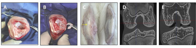
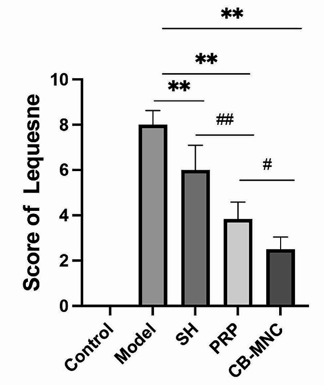
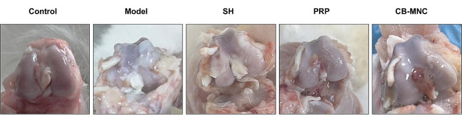
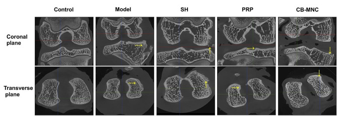
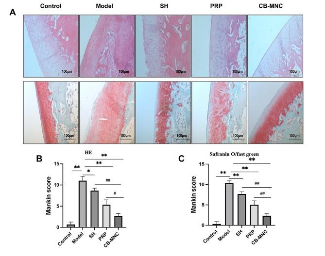
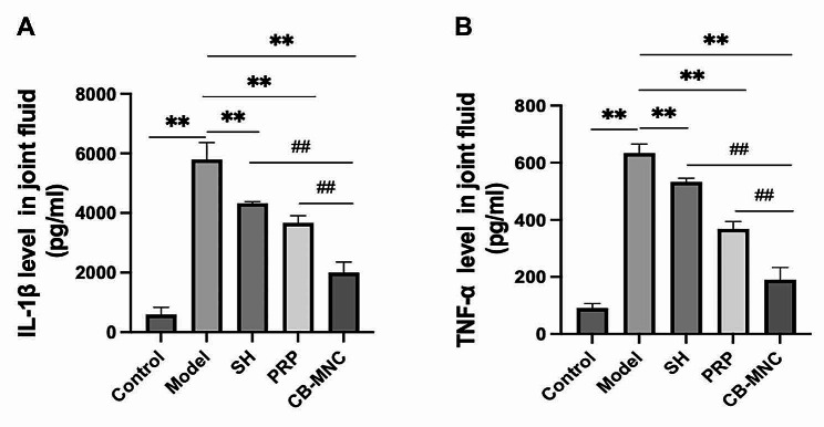
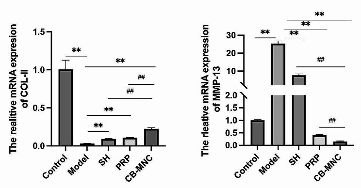
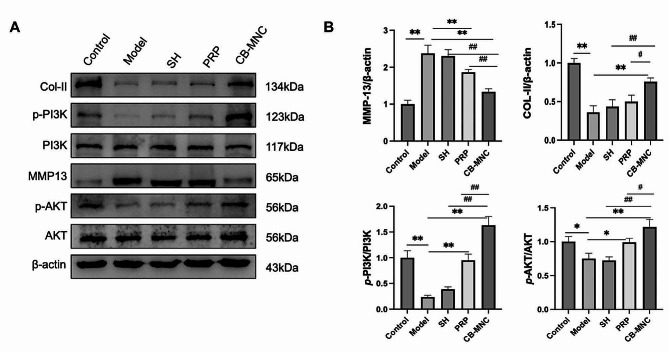
Methods: A rabbit KOA model was prepared by anterior cruciate ligament transection (ACLT). Fifty New Zealand white rabbits were randomly divided into the control group, model group, sodium hyaluronate (SH) group, platelet-rich plasma (PRP) group and UCB-MNC group. Knee injections were performed once a week for five consecutive weeks. The gross view of the knee joint, morphology of knee cartilage and structural changes in the knee joint were observed on CT scans, and graded by the Lequesne MG behavioral score and the Mankin score. TNF-α and IL-1β levels in the synovial fluid of the knee were measured by the enzyme-linked immunosorbent assay (ELISA). Expression levels of MMP-13 and COL-II in the knee cartilage were detected by Western blotting and qRT-PCR.
Results: The Lequesne MG behavioral score and the Mankin score were significantly higher in the model group than those in the control group (P < 0.05). Rabbits in the SH, PRP and UCB-MNC groups had sequentially lower scores than those in the model group. Imaging features of KOA were more pronounced in the model group than in the remaining groups. CB-MNC significantly relieved KOA, compared to SH and PRP. Significantly higher levels of TNF-α and IL-1β in the synovial fluid of the knee, and up-regulated MMP-13 and down-regulated COL-II in the knee cartilage were detected in the model group than in the control group. These changes were significantly reversed by the treatment with SH, PRP and UCB-MNCs, especially UCB-MNCs.
Conclusion: Injections of UCB-MNCs into knees protect the articular cartilage and hinder the progression of KOA in rabbits by improving the local microenvironment at knee joints.
Keywords: Osteoarthritis; Platelet-rich plasma; Umbilical cord blood-mononuclear cells.
© 2024. The Author(s).
Conflict of interest statement
The authors declare no competing interests.
Figures
Similar articles
-
[Warm acupuncture stimulation improves cartilage damage and motor function by regulating JAK2/STAT3 signaling pathway in rabbits with knee osteoarthritis].Zhen Ci Yan Jiu. 2022 Dec 25;47(12):1088-94. doi: 10.13702/j.1000-0607.20211331. Zhen Ci Yan Jiu. 2022. PMID: 36571224 Chinese.
-
Effects of Platelet-Rich Plasma Combined with Physical Therapy on IL-1β, TGF-β1, and MMP-3 in Patients with Knee Osteoarthritis.Mol Biotechnol. 2025 May;67(5):1991-2001. doi: 10.1007/s12033-024-01177-8. Epub 2024 May 21. Mol Biotechnol. 2025. PMID: 38771422
-
Intra-Articular Platelet-Rich Plasma Combined With Hyaluronic Acid Injection for Knee Osteoarthritis Is Superior to Platelet-Rich Plasma or Hyaluronic Acid Alone in Inhibiting Inflammation and Improving Pain and Function.Arthroscopy. 2021 Mar;37(3):903-915. doi: 10.1016/j.arthro.2020.10.013. Epub 2020 Oct 20. Arthroscopy. 2021. PMID: 33091549 Clinical Trial.
-
Advantages of Pure Platelet-Rich Plasma Compared with Leukocyte- and Platelet-Rich Plasma in Treating Rabbit Knee Osteoarthritis.Med Sci Monit. 2016 Apr 17;22:1280-90. doi: 10.12659/msm.898218. Med Sci Monit. 2016. PMID: 27086145 Free PMC article.
-
The Effect of Platelet-Rich Plasma on Synovial Fibrosis and Cartilage Degeneration in Knee Osteoarthritis.Am J Sports Med. 2025 May;53(6):1428-1439. doi: 10.1177/03635465251324942. Epub 2025 Mar 20. Am J Sports Med. 2025. PMID: 40114320
Cited by
-
Reinforcement of osteochondoral defects repair with leukocyte platelet-rich fibrin and bone marrow-derived mononuclear cells in a rabbit model.BMC Musculoskelet Disord. 2025 Jul 25;26(1):707. doi: 10.1186/s12891-025-08952-x. BMC Musculoskelet Disord. 2025. PMID: 40713617 Free PMC article.
References
-
- Abbasi J. Can exercise prevent knee osteoarthritis? [J]. JAMA. 2017;318(22):2169–71. - PubMed
MeSH terms
Substances
LinkOut - more resources
Full Text Sources
Research Materials
