. 2024 May 29;25(1):227.
doi: 10.1186/s12931-024-02769-3.

Expression of glucocorticoid receptor and HDACs in airway smooth muscle cells is associated with response to steroids in COPD

Affiliations

Expression of glucocorticoid receptor and HDACs in airway smooth muscle cells is associated with response to steroids in COPD

Liang Zhou et al. Respir Res. .

Abstract

Background: Steroid insensitivity in Chronic Obstructive Pulmonary Disease (COPD) presents a problem for controlling the chronic inflammation of the airways. The glucocorticoid receptor (GR) mediates the intracellular signaling of inhaled corticosteroids (ICS) by interacting with transcription factors and histone deacetylases (HDACs). The aim of this study was to assess if COPD patients' response to ICS in vivo, may be associated with the expression of GR, the complex of GR with transcription factors, and the expression of various HDACs in vitro.

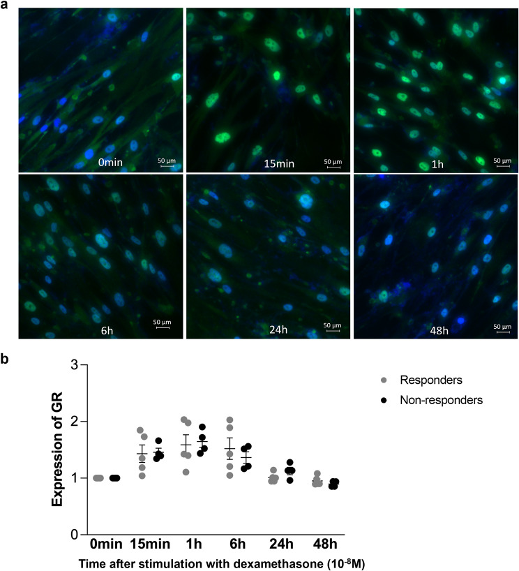
Methods: Primary airway smooth muscle cells (ASMC) were established from endobronchial biopsies obtained from patients with asthma (n = 10), patients with COPD (n = 10) and subjects that underwent diagnostic bronchoscopy without pathological findings and served as controls (n = 6). ASMC were also established from 18 COPD patients, 10 responders and 8 non-responders to ICS, who participated in the HISTORIC study, an investigator-initiated and driven clinical trial that proved the hypothesis that COPD patients with high ASMC in their endobronchial biopsies respond better to ICS than patients with low ASMC. Expression of GR and its isoforms GRα and GRβ and HDACs was investigated in primary ASMC in the absence or in the presence of dexamethasone (10- 8M) by western blotting. The complex formation of GR with transcription factors was assessed by co-immunoprecipitation.

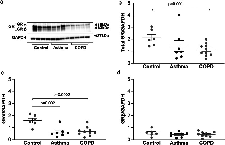
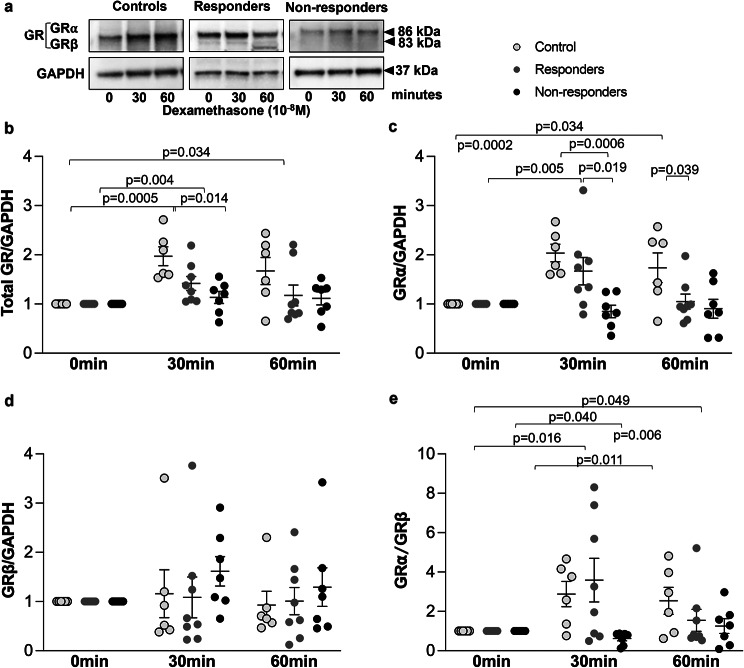
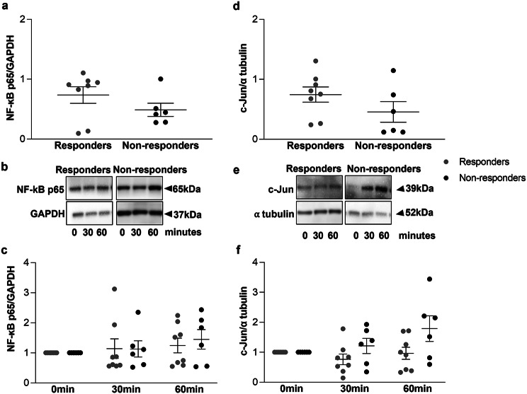
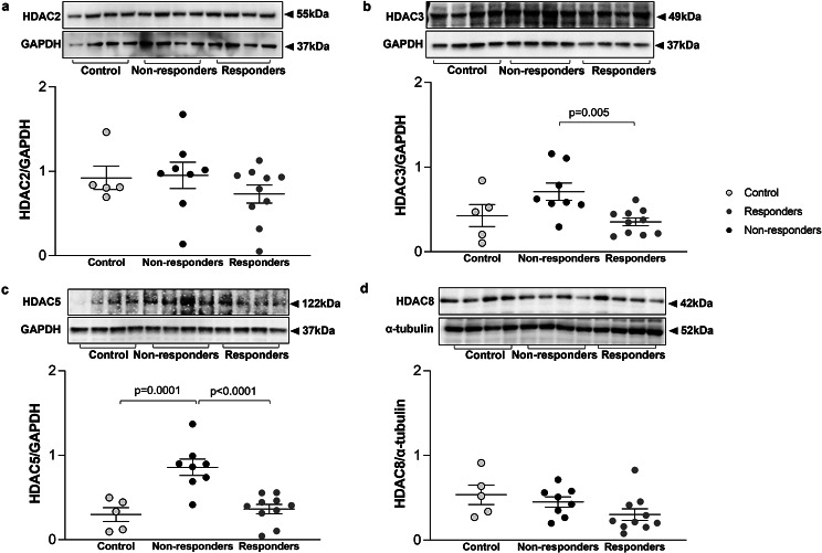
Results: Expression of GR and its isoform GRα but not GRβ was significantly reduced in ASMC from COPD patients as compared to controls. There were no significant differences in the expression of GR, GRα and GRβ between responders and non-responders to ICS. However, treatment with dexamethasone upregulated the expression of total GR (p = 0.004) and GRα (p = 0.005) after 30 min in responders but not in non-responders. Τhe formation of the complex GR-c-Jun was increased 60 min after treatment with dexamethasone only in responders who exhibited significantly lower expression of HDAC3 (p = 0.005) and HDAC5 (p < 0.0001) as compared to non-responders.

Conclusions: These data suggest that ASMC from COPD patients who do not respond to treatment with ICS, are characterized by reduced GR-c-Jun complex formation and increased expression of HDAC3 and HDAC5.

Trial registration: ISRCTN11017699 (Registration date: 15/11/2016).

Keywords: Airway smooth muscle cells; Chronic obstructive pulmonary disease; Glucocorticoid receptor; Glucocorticoid sensitivity; Histone deacetylases.


Conflict of interest statement

The authors declare no competing interests.

Figures

Fig. 1
Fig. 1
Expression of total GR, GRα and GRβ in primary ASMC. (a) Representative Western-blots of the expression of total GR, GRα and GRβ in primary ASMC established from controls (n = 6), patients with asthma (n = 7 ) and patients with COPD (n = 10). b-d Quantitation of the western blots performed by Image J. Expression of total GR represents the sum of the expression of GRα and GRβ isoforms. Error bars represent mean ± SEM
Fig. 2
Fig. 2
Expression of total GR, GRα and GRβ in primary ASMC from COPD patients. (a) Representative Western-blots of the expression of total GR, GRα and GRβ in primary ASMC from 8 patients responded to treatment with ICS (responders) and 8 patients did not respond to treatment with ICS (non-responders). (b-g) Quantitation of the western blots performed by Image J. Among the 16 COPD patients, 6 patients had high ASMC (> 20% of the bronchial tissue area) and 10 patients had low ASMC (⩽20% of the bronchial tissue area), in their endobronchial biopsy (e, f, g). Expression of total GR represents the sum of the expression of GRα and GRβ isoforms. Error bars represent mean ± SEM. Statistical analysis revealed no significant differences between the groups
Fig. 3
Fig. 3
Expression of total GR, GRα and GRβ in response to dexamethasone within 60 min. (a) Representative Western-blots of the effect of dexamethasone (10-8M) on the expression of total GR, GRα and GRβ in primary ASMC from controls (n = 6), COPD patients who responded to treatment with ICS (n = 8) and COPD patients who did not response to treatment with ICS (n = 7). (b-e) Quantitation of the western blots performed by Image J. Expression of total GR represents the sum of the expression of GRα and GRβ isoforms. Results are expressed as fold change to time 0. Error bars represent mean ± SEM
Fig. 4
Fig. 4
Activation of GR in primary ASMC in response to dexamethasone within 48 h. The activation of the GR (green/turqoise) was determined by its translocation into the nucleus (blue). (a) Representative immunofluorescence photographs showing the activation of GR and its translocation into the nucleus in response to dexamethasone (1 × 10-8M). Photographs with larger cell area are provided in supplemantary Fig. 1. (b) Quantitation of the staining in ASMC from COPD patients, 5 responders to ICS and 4 non-responders to ICS. Images were acquired by ECLIPSE Ti2 (Nikon, Tokyo, Japan) and documented by imaging software NIS-Elements (Nikon, Japan). Results represent the expression of GR measured by the intensity of green colour as fold change to time 0. Error bars represent mean ± SEM. Green: GR; Blue: nuclei
Fig. 5
Fig. 5
Expression of transcription factors in primary ASMC in response to dexamethasone. Expression of NF-kB (a) and c-Jun (d) in primary ASMC from responders and non-responders to ICS. Results represent the quantitation of respective western blots from 8 responders and 6 non-responders. Error bars represent mean ± SEM. Representative Western-blots of the effect of dexamethasone (10-8M) on the expression of total NF-kB (b) and c-Jun (e) in primary ASMC from 8 responders and 6 non-responders to ICS. (c and f) Quantitation of the western blots performed by Image J. Results represent fold change to time 0. Error bars represent mean ± SEM
Fig. 6
Fig. 6
Complex of GR and transcription factors in primary ASMCs in response to dexamethasone within 60 min. Complex of GR and NF-kB (a) and GR and c-Jun (d) transcription factors in primary ASMC from COPD patients who responded or did not respond to ICS assessed by co-immunoprecipitation. Results represent the quantitation of respective western blots from 6 responders and 6 non-responders. Error bars represent mean ± SEM. Representative Western-blots of the effect of dexamethasone (1 × 10-8M) on the expression of GR-NF-kB complex (b) and GR-c-Jun complex (e) in primary ASMC from 6 responders and 6 non-responders to ICS. c and f Quantitation of the western blots performed by Image J. Dots represent fold change to time 0. Error bars represent mean ± SEM
Fig. 7
Fig. 7
Expression of HDACs in primary ASMC from controls and from COPD patients. Representative western blots for the expression of HDAC2 (a), HDAC3 (b), HDAC5 (c) and HDAC8 (d) in primary ASMC from controls (n = 5) and patients with COPD who responded (n = 10) or did not respond to ICS (n = 8). GAPDH was used as loading control for large molecular weight HDACs and α-tubulin for small molecular weight HADC8. Quantitation of the western blots performed by Image J. Error bars represent mean ± SEM

Similar articles

Cited by

References

    1. Wedzicha JA, Banerji D, Chapman KR, Vestbo J, Roche N, Ayers RT, et al. Indacaterol-Glycopyrronium versus Salmeterol-Fluticasone for COPD. N Engl J Med. 2016;374:2222–34. doi: 10.1056/NEJMoa1516385. - DOI - PubMed
    1. Barnes PJ. Inhaled corticosteroids are not beneficial in chronic obstructive pulmonary disease. Am J Respir Crit Care Med. 2000;161:342–4. doi: 10.1164/ajrccm.161.2.16125_2. - DOI - PubMed
    1. Barnes PJ. New anti-inflammatory targets for chronic obstructive pulmonary disease. Nat Rev Drug Discov. 2013;12:543–59. doi: 10.1038/nrd4025. - DOI - PubMed
    1. Yang IA, Ferry OR, Clarke MS, Sim EH, Fong KM. Inhaled corticosteroids versus placebo for stable chronic obstructive pulmonary disease. Cochrane Database Syst Rev. 2023;3:CD002991. - PMC - PubMed
    1. Timmermans S, Souffriau J, Libert C. A General introduction to Glucocorticoid Biology. Front Immunol. 2019;10:1545. doi: 10.3389/fimmu.2019.01545. - DOI - PMC - PubMed

MeSH terms