

Clinical evaluation of deep learning-enhanced lymphoma pet imaging with accelerated acquisition
- PMID: 38812107
- PMCID: PMC11492391
- DOI: 10.1002/acm2.14390
Clinical evaluation of deep learning-enhanced lymphoma pet imaging with accelerated acquisition
Abstract
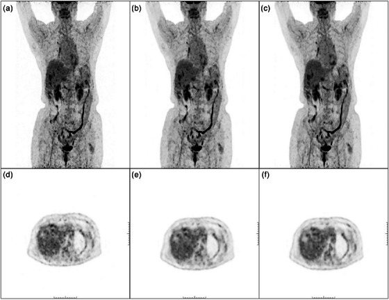
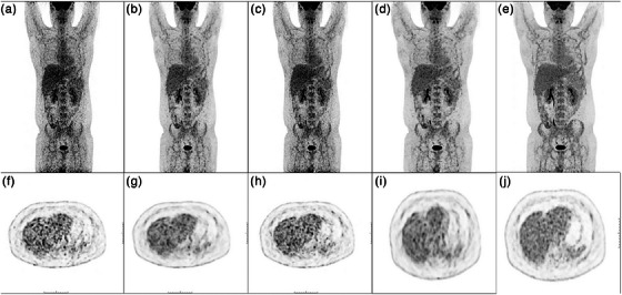
Purpose: This study aims to evaluate the clinical performance of a deep learning (DL)-enhanced two-fold accelerated PET imaging method in patients with lymphoma.
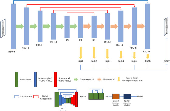
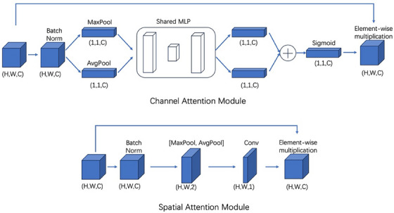
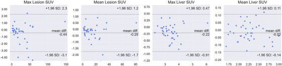
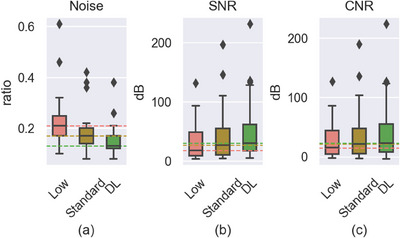
Methods: A total of 123 cases devoid of lymphoma underwent whole-body 18F-FDG-PET/CT scans to facilitate the development of an advanced SAU2Net model, which combines the advantages of U2Net and attention mechanism. This model integrated inputs from simulated 1/2-dose (0.07 mCi/kg) PET acquisition across multiple slices to generate an estimated standard dose (0.14 mCi/kg) PET scan. Additional 39 cases with confirmed lymphoma pathology were utilized to evaluate the model's clinical performance. Assessment criteria encompassed peak-signal-to-noise ratio (PSNR), structural similarity index (SSIM), a 5-point Likert scale rated by two experienced physicians, SUV features, image noise in the liver, and contrast-to-noise ratio (CNR). Diagnostic outcomes, including lesion numbers and Deauville score, were also compared.
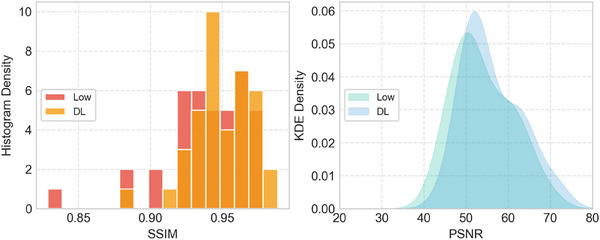
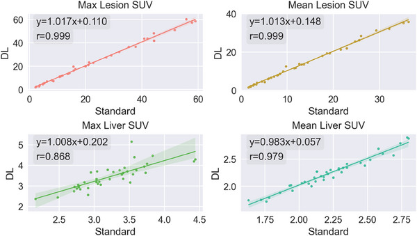
Results: Images enhanced by the proposed DL method exhibited superior image quality (P < 0.001) in comparison to low-dose acquisition. Moreover, they illustrated equivalent image quality in terms of subjective image analysis and lesion maximum standardized uptake value (SUVmax) as compared to the standard acquisition method. A linear regression model with y = 1.017x + 0.110 ( ) can be established between the enhanced scans and the standard acquisition for lesion SUVmax. With enhancement, increased signal-to-noise ratio (SNR), CNR, and reduced image noise were observed, surpassing those of the standard acquisition. DL-enhanced PET images got diagnostic results essentially equavalent to standard PET images according to two experienced readers.
Conclusion: The proposed DL method could facilitate a 50% reduction in PET imaging duration for lymphoma patients, while concurrently preserving image quality and diagnostic accuracy.
Keywords: PET; deep learning; low‐dose imaging; lymphoma.
© 2024 The Author(s). Journal of Applied Clinical Medical Physics published by Wiley Periodicals LLC on behalf of American Association of Physicists in Medicine.
Conflict of interest statement
Author BP, ZP work for RadioDynamic Healthcare. Author NG is a stock share holder of RadioDynamic Healthcare.
Figures
References
MeSH terms
Substances
LinkOut - more resources
Full Text Sources
Medical
