

SRS 16-86 promotes diabetic nephropathy recovery by regulating ferroptosis
- PMID: 38812118
- PMCID: PMC11215488
- DOI: 10.1113/EP091520
SRS 16-86 promotes diabetic nephropathy recovery by regulating ferroptosis
Abstract
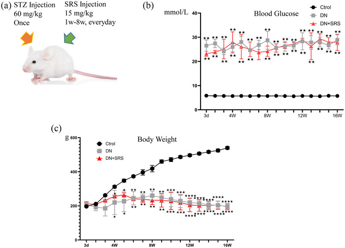
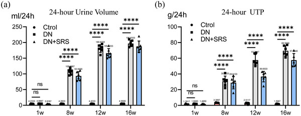
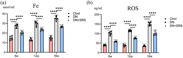
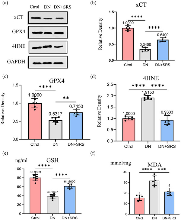
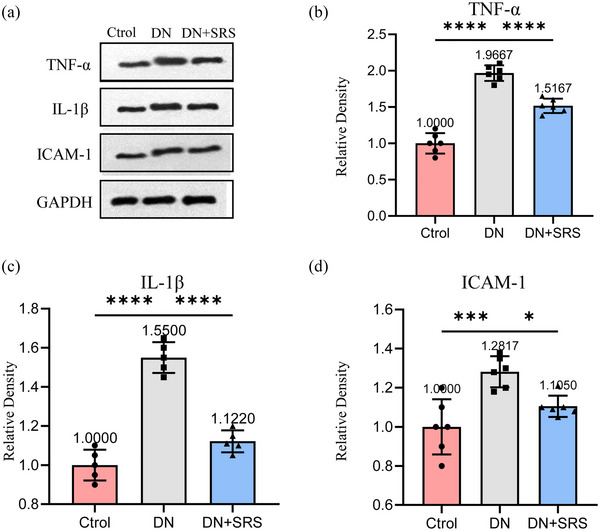
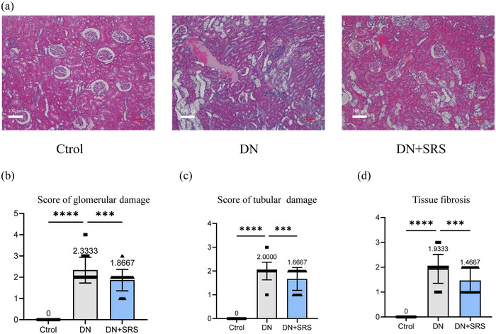
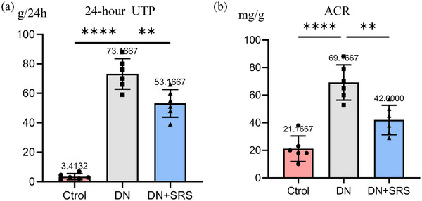
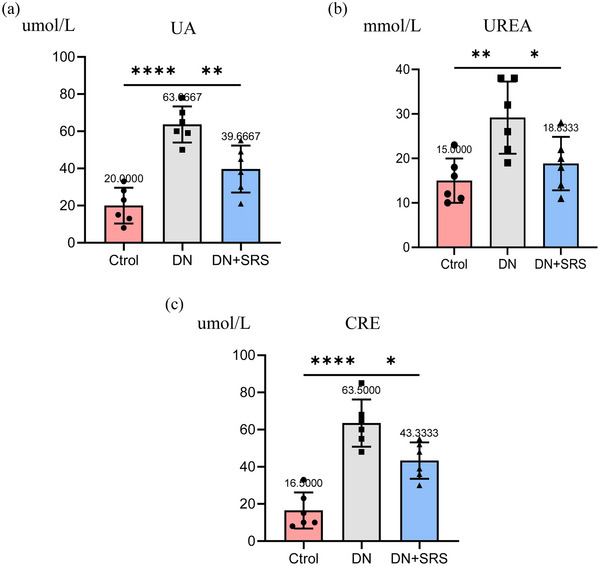
Diabetic nephropathy (DN) is a common complication of diabetes mellitus (DM), and cell death plays an important role. Ferroptosis is a recently discovered type of iron-dependent cell death and one that is different from other kinds of cell death including apoptosis and necrosis. However, ferroptosis has not been described in the context of DN. This study explored the role of ferroptosis in DN pathophysiology and aimed to confirm the efficacy of the ferroptosis inhibitor SRS 16-86 on DN. Streptozotocin injection was used to establish the DM and DN animal models. To investigate the presence or occurrence of ferroptosis in DN, we assessed the concentrations of iron, reactive oxygen species and specific markers associated with ferroptosis in a rat model of DN. Additionally, we performed haematoxylin-eosin staining, blood biochemistry, urine biochemistry and kidney function analysis to evaluate the efficacy of the ferroptosis inhibitor SRS 16-86 in ameliorating DN. We found that SRS 16-86 could improve the recovery of renal function after DN by upregulating glutathione peroxidase 4, glutathione and system xc -light chain and by downregulating the lipid peroxidation markers and 4-hydroxynonenal. SRS 16-86 treatment could improve renal organization after DN. The inflammatory cytokines interleukin 1β and tumour necrosis factor α and intercellular adhesion molecule 1 were significantly decreased following SRS 16-86 treatment after DN. The results indicate that there is a strong connection between ferroptosis and the pathological mechanism of DN. The efficacy of the ferroptosis inhibitor SRS 16-86 in DN repair supports its use as a new therapeutic treatment for DN.
Keywords: diabetic nephropathy; ferroptosis; ferroptosis inhibitor.
© 2024 The Author(s). Experimental Physiology published by John Wiley & Sons Ltd on behalf of The Physiological Society.
Conflict of interest statement
The authors have no conflicts of interest to declare.
Figures
Similar articles
-
Ferroptosis involves in renal tubular cell death in diabetic nephropathy.Eur J Pharmacol. 2020 Dec 5;888:173574. doi: 10.1016/j.ejphar.2020.173574. Epub 2020 Sep 22. Eur J Pharmacol. 2020. PMID: 32976829
-
Targeting ferroptosis as a prospective therapeutic approach for diabetic nephropathy.Ann Med. 2024 Dec;56(1):2346543. doi: 10.1080/07853890.2024.2346543. Epub 2024 Apr 24. Ann Med. 2024. PMID: 38657163 Free PMC article. Review.
-
Obacunone inhibits ferroptosis through regulation of Nrf2 homeostasis to treat diabetic nephropathy.Mol Med Rep. 2025 May;31(5):135. doi: 10.3892/mmr.2025.13500. Epub 2025 Mar 21. Mol Med Rep. 2025. PMID: 40116089 Free PMC article.
-
KAT2A-mediated H3K79 succinylation promotes ferroptosis in diabetic nephropathy by regulating SAT2.Life Sci. 2025 Sep 1;376:123746. doi: 10.1016/j.lfs.2025.123746. Epub 2025 May 21. Life Sci. 2025. PMID: 40409584
-
Ferroptosis: an important player in the inflammatory response in diabetic nephropathy.Front Immunol. 2023 Dec 4;14:1294317. doi: 10.3389/fimmu.2023.1294317. eCollection 2023. Front Immunol. 2023. PMID: 38111578 Free PMC article. Review.
Cited by
-
Pathophysiology and management of crush syndrome: A narrative review.World J Orthop. 2025 Apr 18;16(4):104489. doi: 10.5312/wjo.v16.i4.104489. eCollection 2025 Apr 18. World J Orthop. 2025. PMID: 40290606 Free PMC article.
-
Intercellular Adhesion Molecule 1 (ICAM-1): An Inflammatory Regulator with Potential Implications in Ferroptosis and Parkinson's Disease.Cells. 2024 Sep 15;13(18):1554. doi: 10.3390/cells13181554. Cells. 2024. PMID: 39329738 Free PMC article. Review.
References
-
- Astudillo, A. M. , Balboa, M. A. , & Balsinde, J. (2023). Compartmentalized regulation of lipid signaling in oxidative stress and inflammation: Plasmalogens, oxidized lipids and ferroptosis as new paradigms of bioactive lipid research. Progress in Lipid Research, 89, 101207. - PubMed
-
- Barutta, F. , Bellini, S. , Kimura, S. , Hase, K. , Corbetta, B. , Corbelli, A. , Fiordaliso, F. , Bruno, S. , Biancone, L. , Barreca, A. , Papotti, M. G. , Hirsh, E. , Martini, M. , Gambino, R. , Durazzo, M. , Ohno, H. , & Gruden, G. (2023). Protective effect of the tunneling nanotube‐TNFAIP2/M‐sec system on podocyte autophagy in diabetic nephropathy. Autophagy, 19(2), 505–524. - PMC - PubMed
-
- Basit, F. , van Oppen, L. M. , Schöckel, L. , Bossenbroek, H. M. , van Emst‐de Vries, S. E. , Hermeling, J. C. , Grefte, S. , Kopitz, C. , Heroult, M. , Hgm Willems, P. , & Koopman, W. J. (2017). Mitochondrial complex I inhibition triggers a mitophagy‐dependent ROS increase leading to necroptosis and ferroptosis in melanoma cells. Cell Death & Disease, 8(3), e2716. - PMC - PubMed
MeSH terms
Substances
Grants and funding
- 2018ZDKF10/Scientific Research Funding of Tianjin Medical University Chu Hsien-I Memorial Hospital
- 2019ZDKF02/Scientific Research Funding of Tianjin Medical University Chu Hsien-I Memorial Hospital
- 2019ZDKF08/Scientific Research Funding of Tianjin Medical University Chu Hsien-I Memorial Hospital
- 2022KJ242/Funding of Tianjin Educational Committee
- ZC20225/Funding of Tianjin Health Research Project
LinkOut - more resources
Full Text Sources
Medical