

Stain-free enucleation of mouse and human oocytes with a 1033 nm femtosecond laser
- PMID: 38812963
- PMCID: PMC11133223
- DOI: 10.1117/1.JBO.29.6.065002
Stain-free enucleation of mouse and human oocytes with a 1033 nm femtosecond laser
Abstract
Significance: Preparation of a recipient cytoplast by oocyte enucleation is an essential task for animal cloning and assisted reproductive technologies in humans. The femtosecond laser is a precise and low-invasive tool for oocyte enucleation, and it should be an appropriate alternative to traditional enucleation by a microneedle aspiration. However, until recently, the laser enucleation was performed only with applying a fluorescent dye.
Aim: This work is aimed to (1) achieve femtosecond laser oocyte enucleation without applying a fluorescent dye and (2) to study the effect of laser destruction of chromosomes on the structure and dynamics of the spindle.
Approach: We applied polarized light microscopy for spindle visualization and performed stain-free mouse and human oocyte enucleation with a 1033 nm femtosecond laser. Also, we studied transformation of a spindle after metaphase plate elimination by a confocal microscopy.
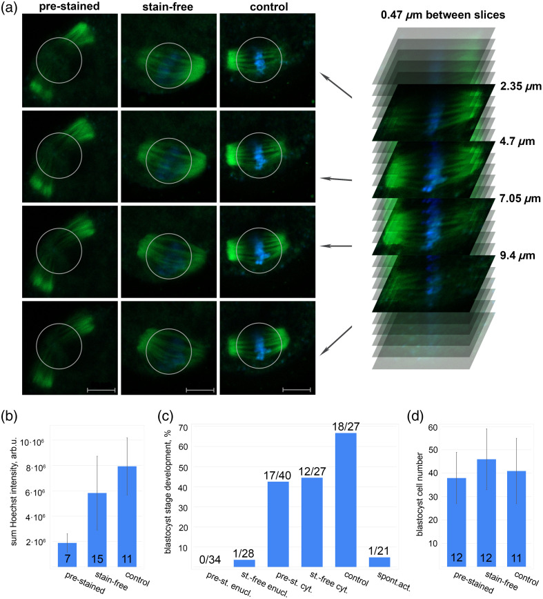
Results: We demonstrated a fundamental possibility of inactivating the metaphase plate in mouse and human oocytes by 1033 nm femtosecond laser radiation without applying a fluorescent dye. Irradiation of the spindle area, visualized by polarized light microscopy, resulted in partly or complete metaphase plate destruction but avoided the microtubules impairment. After the metaphase plate elimination, the spindle reorganized, however, it was not a complete depolymerization.
Conclusions: This method of recipient cytoplast preparation is expected to be useful for animal cloning and assisted reproductive technologies.
Keywords: enucleation; femtosecond laser; recipient cytoplast; spindle.
© 2024 The Authors.
Figures


Similar articles
-
Femtosecond laser oocyte enucleation as a low-invasive and effective method of recipient cytoplast preparation.Biomed Opt Express. 2022 Feb 14;13(3):1447-1456. doi: 10.1364/BOE.449523. eCollection 2022 Mar 1. Biomed Opt Express. 2022. PMID: 35414969 Free PMC article.
-
Comparative analysis of the metaphase II spindle of human oocytes through polarized light and high-performance confocal microscopy.Fertil Steril. 2010 Apr;93(6):2056-64. doi: 10.1016/j.fertnstert.2008.12.011. Epub 2009 Feb 24. Fertil Steril. 2010. PMID: 19243751
-
Combined multiphoton imaging and automated functional enucleation of porcine oocytes using femtosecond laser pulses.J Biomed Opt. 2010 Jul-Aug;15(4):046006. doi: 10.1117/1.3463012. J Biomed Opt. 2010. PMID: 20799808
-
Polarized light microscopy in mammalian oocytes.Reprod Domest Anim. 2010 Jun;45 Suppl 2:49-56. doi: 10.1111/j.1439-0531.2010.01621.x. Reprod Domest Anim. 2010. PMID: 20591065 Review.
-
Spindle and chromosomal alterations in metaphase II oocytes.Reprod Sci. 2013 Nov;20(11):1293-301. doi: 10.1177/1933719113483018. Epub 2013 Mar 27. Reprod Sci. 2013. PMID: 23536572 Review.
