. 2024 May 8;6(1):vdae064.
doi: 10.1093/noajnl/vdae064. eCollection 2024 Jan-Dec.

A neural tract-inspired conduit for facile, on-demand biopsy of glioblastoma

Affiliations

A neural tract-inspired conduit for facile, on-demand biopsy of glioblastoma

Martha I Betancur et al. Neurooncol Adv. .

Abstract

Background: A major hurdle to effectively treating glioblastoma (GBM) patients is the lack of longitudinal information about tumor progression, evolution, and treatment response.

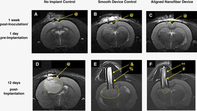
Methods: In this study, we report the use of a neural tract-inspired conduit containing aligned polymeric nanofibers (i.e., an aligned nanofiber device) to enable on-demand access to GBM tumors in 2 rodent models. Depending on the experiment, a humanized U87MG xenograft and/or F98-GFP+ syngeneic rat tumor model was chosen to test the safety and functionality of the device in providing continuous sampling access to the tumor and its microenvironment.

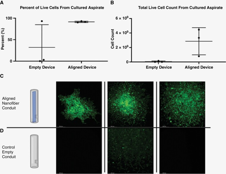
Results: The aligned nanofiber device was safe and provided a high quantity of quality genomic materials suitable for omics analyses and yielded a sufficient number of live cells for in vitro expansion and screening. Transcriptomic and genomic analyses demonstrated continuity between material extracted from the device and that of the primary, intracortical tumor (in the in vivo model).

Conclusions: The results establish the potential of this neural tract-inspired, aligned nanofiber device as an on-demand, safe, and minimally invasive access point, thus enabling rapid, high-throughput, longitudinal assessment of tumor and its microenvironment, ultimately leading to more informed clinical treatment strategies.

Keywords: glioblastoma; on-demand biopsy; tumor microenvironment.


Conflict of interest statement

S.M., B.B., R.B., and N.M. are licensing the aligned nanofiber technology discussed in this work to a private entity for translation and commercialization.

Figures

Figure 1.
Figure 1.
Aligned nanofiber topography guides cancer cells away from the tumor and toward the proximal end of the device which enables on-demand and minimally invasive access to tumor tissue, as demonstrated via histology and aspiration analyses. (A) Comparison of conduit and nanofiber materials in U87MG GBM rat model; all devices, except silicone/rolled aligned films, were attached to the tubing on both sides longitudinally to the cylindrical conduit. The aligned nanofiber film devices had significantly larger amounts of cells and tissue migrating toward the proximal end of device relative to the smooth film controls. (B) Histological evidence that cancer cells migrated to fill the entire length of implanted aligned device: immunofluorescent images of a transverse cross-sections of tissue at 4 mm from the proximal end of the devices showing tumor cells filling the aligned nanofiber devices. (C) Illustration of position of device relative to tumor and skull as well as the biopsy aspirate procedure. (D) Comparison of total aspirate volumes in the F98-GFP+ rat model between aligned nanofiber device and control empty device. ****P < .0001.
Figure 2.
Figure 2.
Representative T2-weighted MR images of U87MG model pre- and post- device implantation showing no adverse response to implant while confirming the possibility of noninvasive monitoring of tissue migration inside the device. (@: tumor, &: fluid, %: edge, **: flow voids, #: gradient). One week post-inoculation: (A) no implant control, (B) smooth film device control, (C) aligned nanofiber device. 12 days post-device implantation, (D) no implant control, (E) smooth film device control, and (F) aligned nanofiber device. In D, E, and F, tumor is surrounded by dotted white lines, bright white content inside device is fluid, and tissue inside device appears in gray tones. In (E), separation between tissue (gray) and fluid (white) is outlined by a dark edge between the two (%). Smooth film appears as a dark line at the center. In (F), inside the aligned nanofiber device, gray strips indicative of tissue appears along almost the entire length of device, and there is no well-defined edge separating the fluid inside the device from tumor. Flow voids (**) indicating interactions between tissue and fluid inside the aligned nanofiber device are visible.
Figure 3.
Figure 3.
Tumor volumes in U87MG xenograft and F98 models. (A) No significant difference in U87MG tumor volumes post-implantation was observed between different groups. When initial volume is compared to final volume for each group, only the 2 control groups show significant growth of tumors in the U87MG model. (B) The significantly lower rate of change in tumor growth in aligned-implanted tumors indicates that, when U87MG cells migrate, intracortical growth is halted to an extent. (C) and (D) All 3 groups experience a significant growth with relatively similar rate of growth in F98 model. No Implant: No device implanted; Aligned: silicone/PU conduit with PU aligned nanofiber device implanted; Smooth: silicone/PU conduit with PU smooth film device implanted; Empty: empty silicone/PU conduit device implanted.
Figure 4.
Figure 4.
CIBERSORTx and IHC analyses of devices in F98-GFP+ tumor model. (A) and (B) CIBERSORTx analysis of bulk RNA-sequencing data for samples collected from: aspirated material from inside the aligned nanofiber device, tissue attached to the aligned nanofiber film within the device conduit, the intracortical tumor in the implant and control groups, and healthy brain tissue in the F98-GFP+ tumor model. (C)–(E). Aligned nanofiber devices retrieved after aspirate biopsy, sectioned transversely, stained, and imaged showing residual tissue that remained attached to the nanofiber. Within this adherent tissue, immune cells are present throughout the length of device. (C) (proximal end of device), (D) (midpoint of device), (E) (primary tumor mass) (scale bar: 50 μm). Blue: DAPI, Red: Immune Cell Marker.
Figure 5.
Figure 5.
Cell culture from biopsy of F98-GFP+ tumor model. (A) Percentage of live cells in the culture expanded from biopsy aspirates of aligned nanofiber and control groups. (B) Absolute number of live cells in culture expanded for 7 days from biopsy aspirates of aligned nanofiber and control groups. (C) and (D) Representative images of the expanded culture after 1 week showing F98-GFP+ cells in all 3 of aspirates from aligned nanofiber devices and only a few GFP+ cells in one of the control empty devices.
Figure 6.
Figure 6.
Genomic analysis of F98-GFP+ model. (A) and (B) Aspirate DNA yield and adherent material DNA yield for aligned nanofiber device and control empty device. (C) and (D) Aspirate RNA yield and adherent material RNA yield for aligned nanofiber device and control empty device.

Similar articles

Cited by

References

    1. Ambady P, Bettegowda C, Holdhoff M.. Emerging methods for disease monitoring in malignant gliomas. CNS Oncol. 2013;2(6):511–522. - PMC - PubMed
    1. Neagu MR, Huang RY, Reardon DA, Wen PY.. How treatment monitoring is influencing treatment decisions in glioblastomas. Curr Treat Options Neurol. 2015;17(4):15. - PubMed
    1. Weller M, Cloughesy T, Perry JR, Wick W.. Standards of care for treatment of recurrent glioblastoma—are we there yet? Neuro-Oncol. 2013;15(1):4–27. - PMC - PubMed
    1. Verduin M, Compter I, Steijvers D, et al.. Noninvasive glioblastoma testing: Multimodal approach to monitoring and predicting treatment response. Dis Markers. 2018;2018:1–11. - PMC - PubMed
    1. Boonzaier NR, Piccirillo SG, Watts C, Price SJ.. Assessing and monitoring intratumor heterogeneity in glioblastoma: how far has multimodal imaging come? CNS Oncol. 2015;4(6):399–410. - PMC - PubMed

LinkOut - more resources