

Ultrasound accuracy in evaluating renal calculi in Maysan province
- PMID: 38813369
- PMCID: PMC11131636
- DOI: 10.25122/jml-2023-0477
Ultrasound accuracy in evaluating renal calculi in Maysan province
Abstract
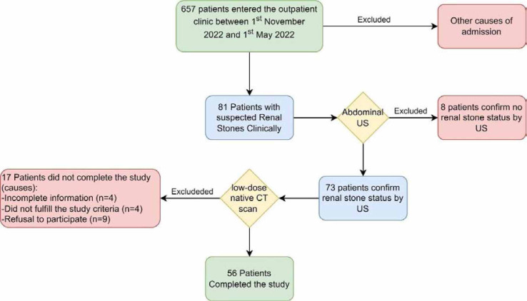
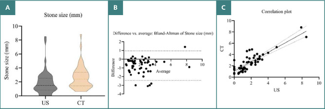
Renal calculi are a common clinical presentation. While ultrasound (US) is a widely used imaging modality for kidney stone diagnosis due to its accessibility and lower cost, its accuracy compared to computerized tomography (CT), the gold standard, remains understudied. This cross-sectional study evaluated the diagnostic accuracy of ultrasound for detecting and characterizing kidney stones compared to computed tomography (CT). Fifty-six patients with suspected kidney stones based on flank pain underwent abdominal ultrasound to assess stone presence, size, location, and the severity of any hydronephrosis (kidney swelling). These findings were then confirmed with a subsequent non-contrast CT scan. There was a fair agreement between US and CT (Kappa = 0.368) for detecting the stone location. The US could not detect 7 (12.5%) stones, being less sensitive in the middle and upper calyx compared to CT. There was a fair agreement between the US and CT (Kappa = 0.394) for detecting the severity of hydronephrosis. The US was less sensitive to moderate and severe hydronephrosis compared to CT. The abdominal ultrasound demonstrated excellent reliability for stone size measurement (intraclass correlation = 0.924), with CT measurements only slightly larger on average (mean difference 0.9 mm). Although abdominal ultrasound provides reliable stone size assessment, its capacity to accurately localize stones and assess hydronephrosis severity is limited.
Keywords: CI, Confidence Interval; CT, Computed Tomography; ICC, Intraclass Correlation; IVU, Intravenous Urography; KUB, Kidney-Ureter-Bladder X-ray; US, Ultrasound; hydronephrosis; nephrolithiasis; reliability; stone location; stone size.
© 2024 by the authors.
Conflict of interest statement
The authors declare no conflict of interest.
Figures
Similar articles
-
Sensitivity of emergency bedside ultrasound to detect hydronephrosis in patients with computed tomography-proven stones.West J Emerg Med. 2014 Feb;15(1):96-100. doi: 10.5811/westjem.2013.9.15874. West J Emerg Med. 2014. PMID: 24578772 Free PMC article.
-
Ultra-Low-Dose CT: An Effective Follow-Up Imaging Modality for Ureterolithiasis.J Endourol. 2020 Feb;34(2):139-144. doi: 10.1089/end.2019.0574. Epub 2020 Jan 10. J Endourol. 2020. PMID: 31663371
-
Ultrasound vs. Computed Tomography for Severity of Hydronephrosis and Its Importance in Renal Colic.West J Emerg Med. 2017 Jun;18(4):559-568. doi: 10.5811/westjem.2017.04.33119. Epub 2017 May 15. West J Emerg Med. 2017. PMID: 28611874 Free PMC article.
-
The Accuracy and Prognostic Value of Point-of-care Ultrasound for Nephrolithiasis in the Emergency Department: A Systematic Review and Meta-analysis.Acad Emerg Med. 2018 Jun;25(6):684-698. doi: 10.1111/acem.13388. Epub 2018 Mar 25. Acad Emerg Med. 2018. PMID: 29427476
-
Imaging in diagnosis, treatment, and follow-up of stone patients.Adv Chronic Kidney Dis. 2009 Jan;16(1):39-47. doi: 10.1053/j.ackd.2008.10.005. Adv Chronic Kidney Dis. 2009. PMID: 19095204 Review.
References
-
- Nojaba L, Guzman N. Treasure Island (FL): StatPearls Publishing; 2023. Nephrolithiasis. - PubMed
MeSH terms
LinkOut - more resources
Full Text Sources
Medical