Musculoskeletal ultrasonography in rheumatic diseases
- PMID: 38813491
- PMCID: PMC10760546
- DOI: 10.55730/1300-0144.5723
Musculoskeletal ultrasonography in rheumatic diseases
Abstract
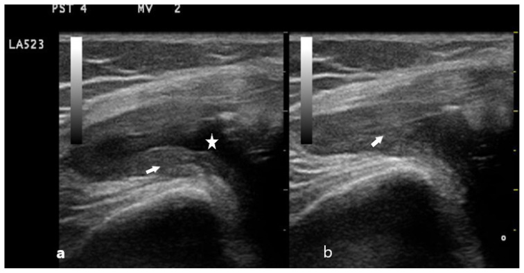
Ultrasonography is an imaging technique based on sound waves used for the evaluation of soft tissues. Sound waves have been used for a long time in nonmedical fields, including military defense systems, radar systems, and detection of icebergs. Technological advances resulted in new techniques becoming available for medical imaging, including ultrasonography, magnetic resonance imaging, and computed tomography. Nowadays, the use of imaging has become a gold standard protocol in the diagnosis of many diseases, and recently developed diagnosis and therapy options provide more efficient treatment of rheumatic diseases. Thus, it has become possible to prevent structural damage and disability in patients with rheumatic disease. Musculoskeletal ultrasonography is becoming a preferred imaging technique for rheumatic diseases, as it has many advantages. Among its advantages are being inexpensive, being radiation-free, having a dynamic image capacity, helping to detect disease activity, and helping with early detection and diagnosis of structural damage. This review summarizes the use of ultrasonography in rheumatic diseases.
Keywords: Musculoskeletal ultrasonography; crystal arthritis; rheumatoid arthritis; spondyloarthropathies.
© TÜBİTAK.
Conflict of interest statement
Disclaimer and conflict of interest statement: None of the authors of this paper has any financial or personal relationship with other people or organizations that could inappropriately influence or bias the content of the paper.
Figures


Similar articles
-
[Roles of Musculoskeletal Ultrasonography in the Management of Rheumatic Diseases].Rinsho Byori. 2015 May;63(5):580-9. Rinsho Byori. 2015. PMID: 26524897 Review. Japanese.
-
Musculoskeletal ultrasound in the diagnosis of rheumatic disease.Bull NYU Hosp Jt Dis. 2010;68(3):183-90. Bull NYU Hosp Jt Dis. 2010. PMID: 20969550 Review.
-
Clinical utility of ultrasonography in spondyloarthropathies.Curr Rheumatol Rep. 2009 Oct;11(5):317-20. doi: 10.1007/s11926-009-0045-x. Curr Rheumatol Rep. 2009. PMID: 19772825 Review.
-
Current status and recent advances on the use of ultrasonography in pediatric rheumatic diseases.World J Pediatr. 2020 Feb;16(1):52-59. doi: 10.1007/s12519-019-00312-9. Epub 2019 Sep 13. World J Pediatr. 2020. PMID: 31515696 Review.
-
[Ultrasonographic semiology--correlation between anatomy and sonography of musculoskeletal tissue].Reumatizam. 2011;58(2):85-93. Reumatizam. 2011. PMID: 22232954 Review. Croatian.
Cited by
-
Subclinical Enthesopathy in Psoriasis-An Ultrasonographic Study.Med Sci (Basel). 2024 Aug 16;12(3):40. doi: 10.3390/medsci12030040. Med Sci (Basel). 2024. PMID: 39189203 Free PMC article.
-
Ultrasonographic evaluation of hand and wrist joints in patients with systemic sclerosis.J Scleroderma Relat Disord. 2025 May 15:23971983251337227. doi: 10.1177/23971983251337227. Online ahead of print. J Scleroderma Relat Disord. 2025. PMID: 40385094 Free PMC article.
References
-
- Bruyn GAW, Schmidt WA. Introductory Guide to Musculoskeletal Ultrasound for Rheumatologists. 2nd ed. Netherlands: Bohn Stafle van Loghum Houten; 2011.
-
- Wakefield RJ, Balint PV, Szkudlarek M, Filippucci E, Backhaus M, et al. OMERACT 7 Special Interest Group. Musculoskeletal ultrasound including definitions for ultrasonographic pathology. Journal of Rheumatology. 2005;32(12):2485–2487. - PubMed
-
- D’Agostino MA, Terslev L, Aegerter P, Backhaus M, Balint P, et al. Scoring ultrasound synovitis in rheumatoid arthritis: a EULAR–OMERACT ultrasound task force – Part 1: definition and development of a standardised, consensus-based scoring system. Rheumatic and Musculoskeletal Diseases Open. 2017 Nov;3(1):e000428. doi: 10.1136/rmdopen-2016-000428. - DOI - PMC - PubMed
Publication types
MeSH terms
LinkOut - more resources
Full Text Sources