Microstructural evaluation of the brain with advanced magnetic resonance imaging techniques in cases of electrical status epilepticus during sleep (ESES)
- PMID: 38813507
- PMCID: PMC10760578
- DOI: 10.55730/1300-0144.5754
Microstructural evaluation of the brain with advanced magnetic resonance imaging techniques in cases of electrical status epilepticus during sleep (ESES)
Abstract
Background/aim: The cause and treatment of electrical status epilepticus during sleep (ESES), one of the epileptic encephalopathies of childhood, is unclear. The aim of this study was to evaluate possible microstructural abnormalities in the brain using advanced magnetic resonance imaging (MRI) techniques in ESES patients with and without genetic mutations.
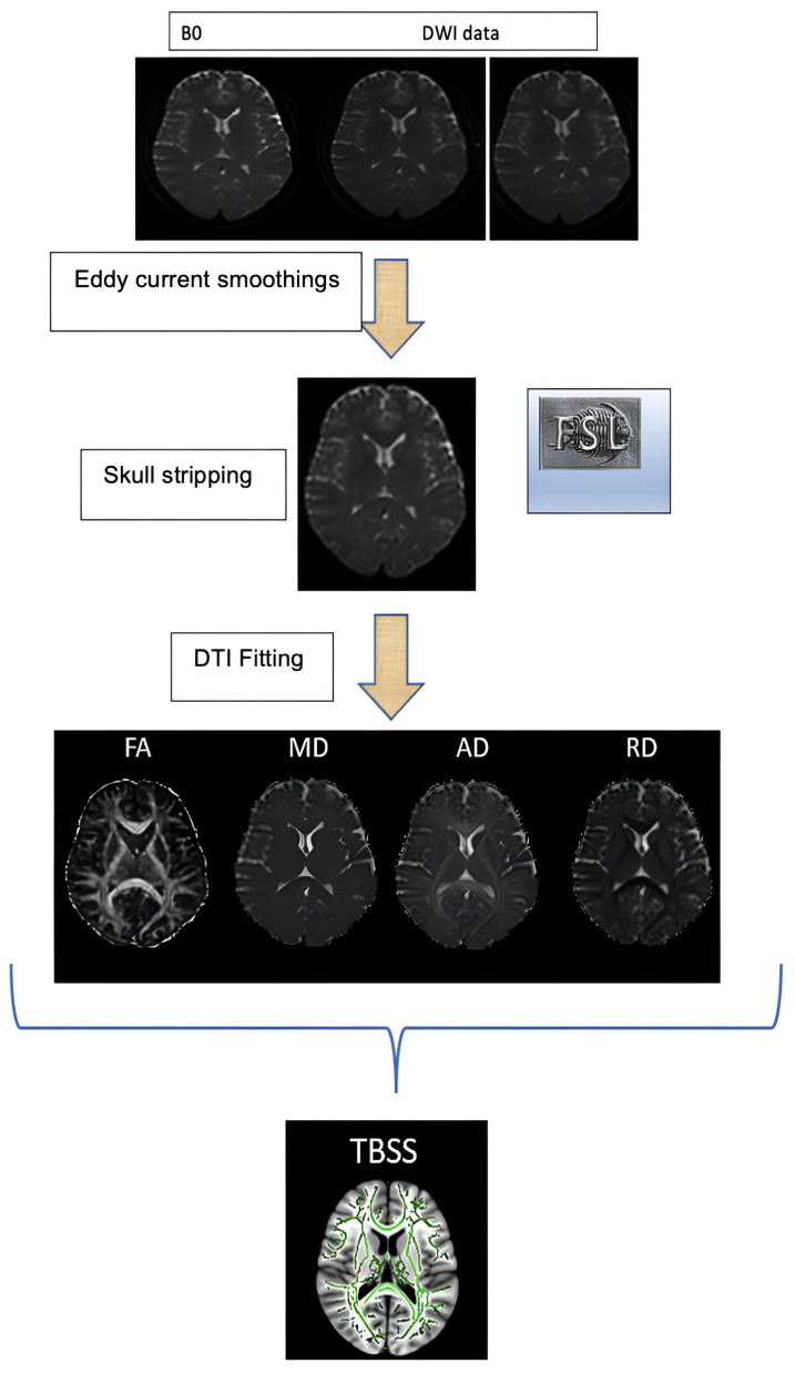
Materials and methods: This research comprised 12 ESES patients without structural thalamic lesions (6 with genetic abnormalities and 6 without) and 12 healthy children. Whole-exome sequencing was used for the genetic mutation analysis. Brain MRI data were evaluated using tractus-based spatial statistics, voxel-based morphometry, a local gyrification index, subcortical shape analysis, FreeSurfer volume, and cortical thickness. The data of the groups were compared.
Results: The mean age in the control group was 9.05 ± 1.85 years, whereas that in the ESES group was 9.45 ± 2.72 years. Compared to the control group, the ESES patients showed higher mean thalamus diffusivity (p < 0.05). ESES patients with genetic mutations had lower axial diffusivity in the superior longitudinal fasciculus and gray matter volume in the entorhinal region, accumbens area, caudate, putamen, cerebral white matter, and outer cerebellar areas. The superior and middle temporal cortical thickness increased in the ESES patients.
Conclusion: This study is important in terms of presenting the microstructural evaluation of the brain in ESES patients with advanced MRI analysis methods as well as comparing patients with and without genetic mutations. These findings may be associated with corticostriatal transmission, ictogenesis, epileptogenesis, neuropsychiatric symptoms, cognitive impairment, and cerebellar involvement in ESES. Expanded case-group studies may help to understand the physiology of the corticothalamic circuitry in its etiopathogenesis and develop secondary therapeutic targets for ESES.
Keywords: Electrical status epilepticus during sleep; genetic mutation; microstructural analysis; morphometry; tractus-based spatial statistics.
© TÜBİTAK.
Conflict of interest statement
Conflict of interest: The authors declare that they have no conflicts of interest.
Figures


Similar articles
-
Reduced thalamic volume is strongly associated with electrical status epilepticus in sleep.Acta Neurol Belg. 2021 Feb;121(1):211-217. doi: 10.1007/s13760-019-01202-7. Epub 2019 Aug 27. Acta Neurol Belg. 2021. PMID: 31456121
-
Evaluation of Thalamic Volume in Patients Diagnosed with BECTS and ESES Using the MRI-Cloud Method.Niger J Clin Pract. 2024 Dec 1;27(12):1473-1478. doi: 10.4103/njcp.njcp_556_24. Epub 2025 Mar 4. Niger J Clin Pract. 2024. PMID: 40033543
-
Electrical status epilepticus in sleep: The role of thalamus in etiopathogenesis.Seizure. 2021 Dec;93:44-50. doi: 10.1016/j.seizure.2021.10.010. Epub 2021 Oct 17. Seizure. 2021. PMID: 34687985
-
Encephalopathy related to status epilepticus during slow sleep (ESES). Pathophysiological insights and nosological considerations.Epilepsy Behav. 2023 Mar;140:109105. doi: 10.1016/j.yebeh.2023.109105. Epub 2023 Feb 7. Epilepsy Behav. 2023. PMID: 36758358 Review.
-
Encephalopathy Associated with Electrical Status Epilepticus of Sleep (ESES): A Practical Approach.Indian J Pediatr. 2020 Dec;87(12):1057-1061. doi: 10.1007/s12098-020-03422-9. Epub 2020 Jul 6. Indian J Pediatr. 2020. PMID: 32632569 Review.
References
-
- Bernhardt BC, Worsley KJ, Besson P, Concha L, Lerch JP, et al. Mapping limbic network organization in temporal lobe epilepsy using morphometric correlations: insights on the relation between mesiotemporal connectivity and cortical atrophy. Neuroimage. 2008;42(2):515–524. doi: 10.1016/j.Neuroimage.2008.04.261. - DOI - PubMed
MeSH terms
LinkOut - more resources
Full Text Sources
Medical