

Pervasive nuclear envelope ruptures precede ECM signaling and disease onset without activating cGAS-STING in Lamin-cardiomyopathy mice
- PMID: 38814785
- PMCID: PMC11290591
- DOI: 10.1016/j.celrep.2024.114284
Pervasive nuclear envelope ruptures precede ECM signaling and disease onset without activating cGAS-STING in Lamin-cardiomyopathy mice
Abstract
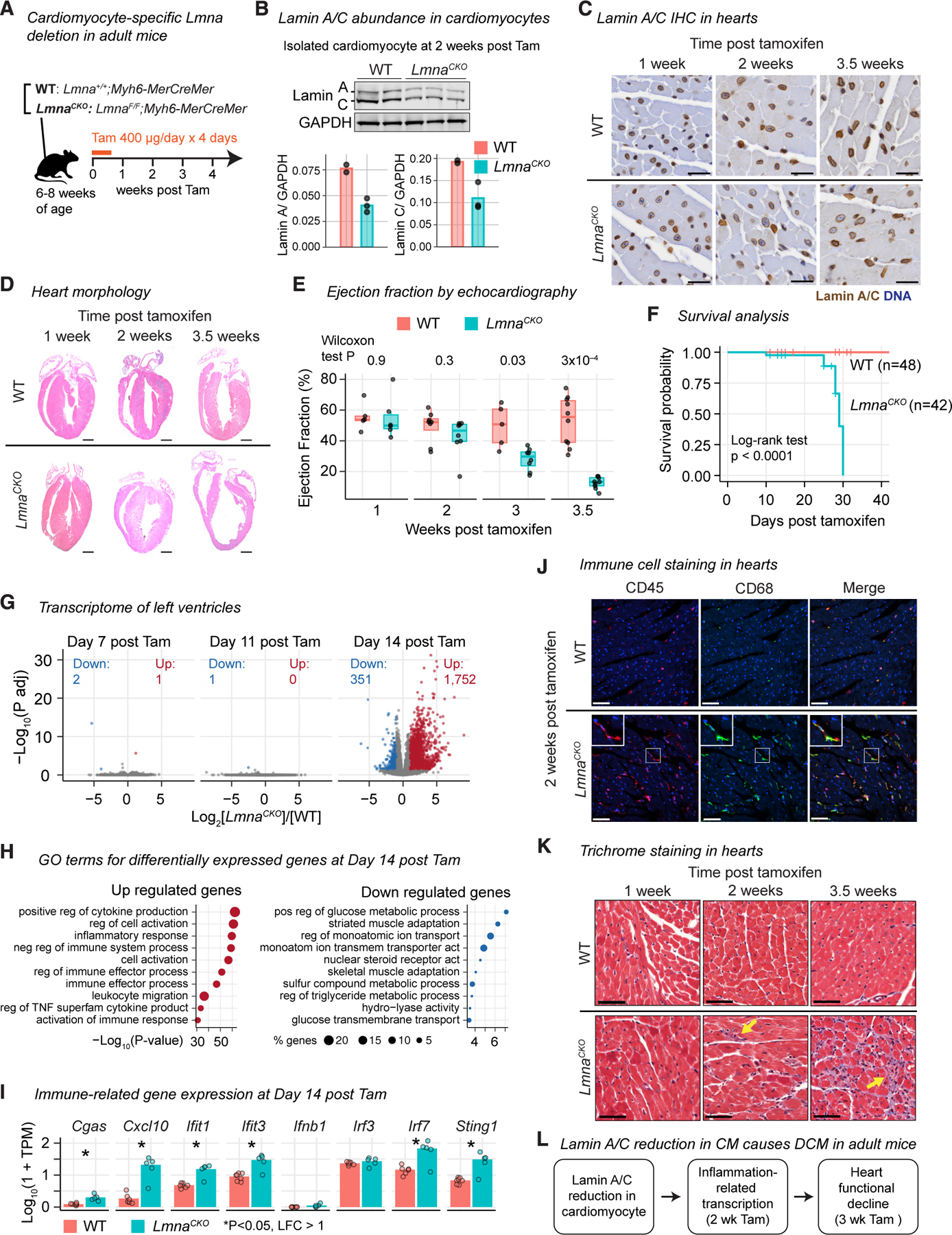
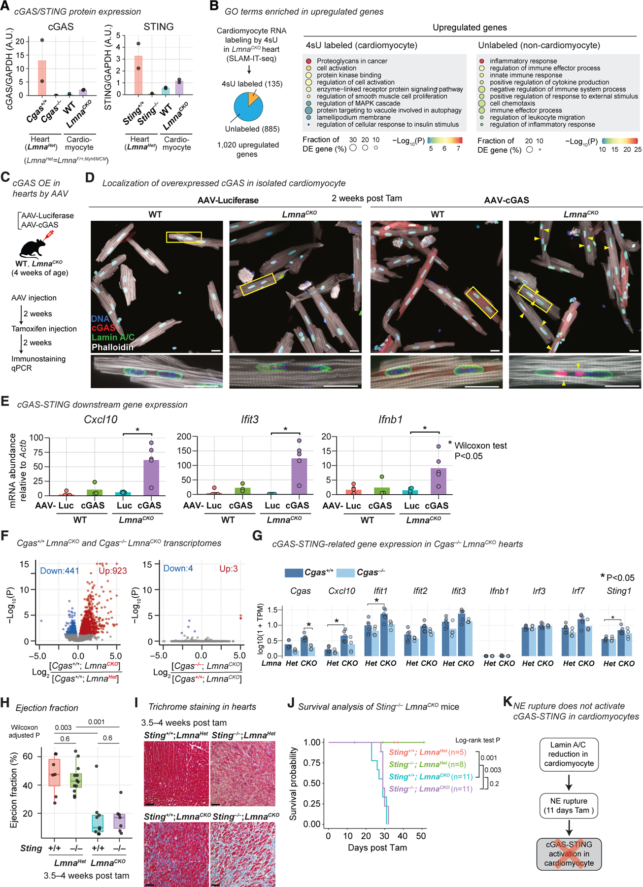
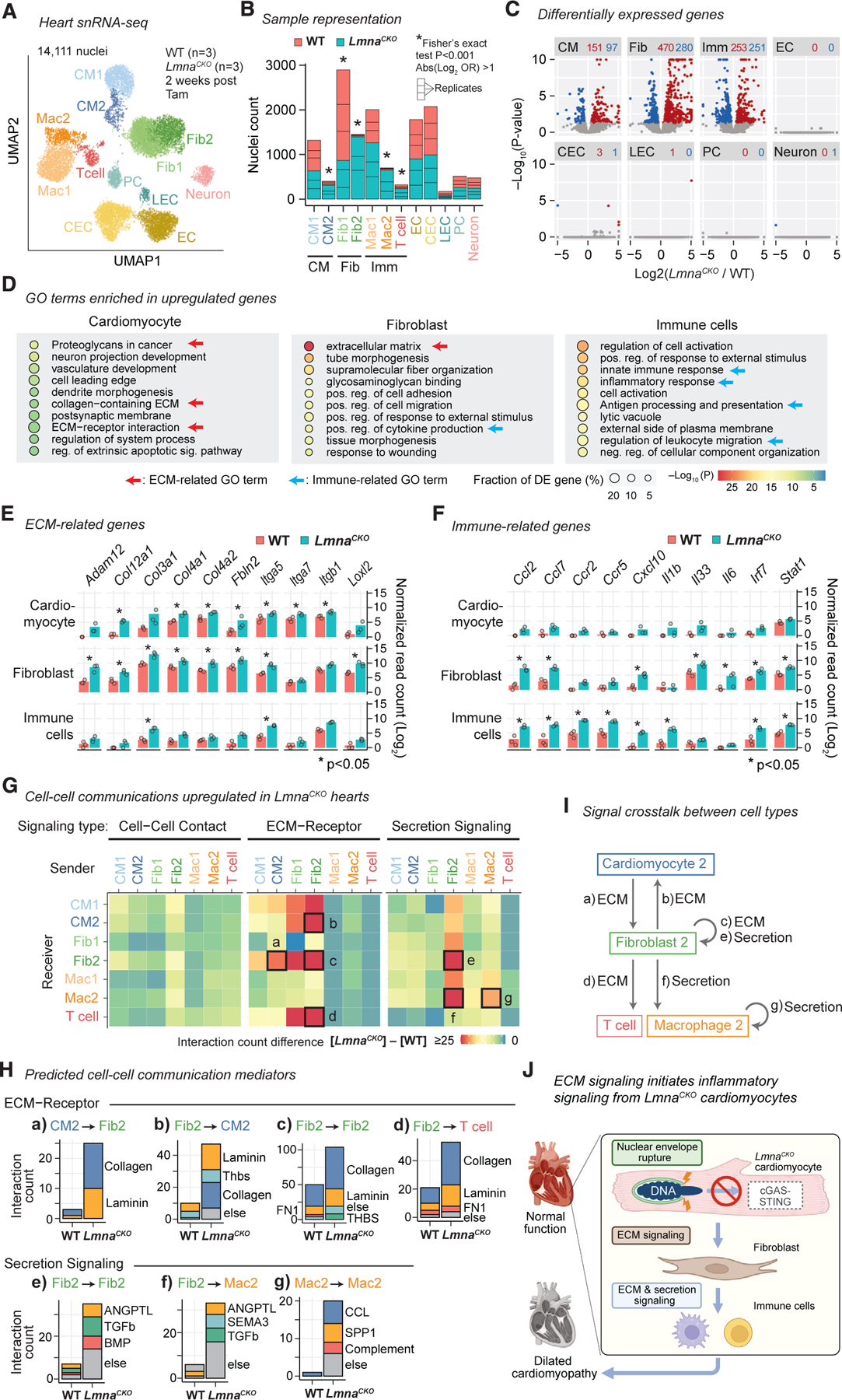
Nuclear envelope (NE) ruptures are emerging observations in Lamin-related dilated cardiomyopathy, an adult-onset disease caused by loss-of-function mutations in Lamin A/C, a nuclear lamina component. Here, we test a prevailing hypothesis that NE ruptures trigger the pathological cGAS-STING cytosolic DNA-sensing pathway using a mouse model of Lamin cardiomyopathy. The reduction of Lamin A/C in cardio-myocyte of adult mice causes pervasive NE ruptures in cardiomyocytes, preceding inflammatory transcription, fibrosis, and fatal dilated cardiomyopathy. NE ruptures are followed by DNA damage accumulation without causing immediate cardiomyocyte death. However, cGAS-STING-dependent inflammatory signaling remains inactive. Deleting cGas or Sting does not rescue cardiomyopathy in the mouse model. The lack of cGAS-STING activation is likely due to the near absence of cGAS expression in adult cardiomyocytes at baseline. Instead, extracellular matrix (ECM) signaling is activated and predicted to initiate pro-inflammatory communication from Lamin-reduced cardiomyocytes to fibroblasts. Our work nominates ECM signaling, not cGAS-STING, as a potential inflammatory contributor in Lamin cardiomyopathy.
Keywords: CP: Cell biology; ECM; Lamin A/C; Lmna; cGAS STING; dilated cardiomyopathy; extracellular matrix; laminopathy; nuclear envelope rupture; nuclear lamina.
Copyright © 2024 The Author(s). Published by Elsevier Inc. All rights reserved.
Conflict of interest statement
Declaration of interests The authors declare no competing interests.
Figures

Update of
-
Pervasive nuclear envelope ruptures precede ECM signaling and disease onset without activating cGAS-STING in Lamin-cardiomyopathy mice.bioRxiv [Preprint]. 2024 Apr 18:2023.08.28.555134. doi: 10.1101/2023.08.28.555134. bioRxiv. 2024. Update in: Cell Rep. 2024 Jun 25;43(6):114284. doi: 10.1016/j.celrep.2024.114284. PMID: 37693381 Free PMC article. Updated. Preprint.
Similar articles
-
The cGAS/STING Pathway: Friend or Foe in Regulating Cardiomyopathy.Cells. 2025 May 25;14(11):778. doi: 10.3390/cells14110778. Cells. 2025. PMID: 40497954 Free PMC article. Review.
-
Perinuclear organelle trauma at the nexus of cardiomyopathy pathogenesis arising from loss of function LMNA mutation.Nucleus. 2025 Dec;16(1):2449500. doi: 10.1080/19491034.2024.2449500. Epub 2025 Jan 9. Nucleus. 2025. PMID: 39789731 Free PMC article. Review.
-
Pervasive nuclear envelope ruptures precede ECM signaling and disease onset without activating cGAS-STING in Lamin-cardiomyopathy mice.bioRxiv [Preprint]. 2024 Apr 18:2023.08.28.555134. doi: 10.1101/2023.08.28.555134. bioRxiv. 2024. Update in: Cell Rep. 2024 Jun 25;43(6):114284. doi: 10.1016/j.celrep.2024.114284. PMID: 37693381 Free PMC article. Updated. Preprint.
-
Nucleoplasmic lamin C rapidly accumulates at sites of nuclear envelope rupture with BAF and cGAS.J Cell Biol. 2022 Dec 5;221(12):e202201024. doi: 10.1083/jcb.202201024. Epub 2022 Oct 27. J Cell Biol. 2022. PMID: 36301259 Free PMC article.
-
BRG1 Deficiency Promotes Cardiomyocyte Inflammation and Apoptosis by Activating the cGAS-STING Signaling in Diabetic Cardiomyopathy.Inflammation. 2025 Feb;48(1):299-315. doi: 10.1007/s10753-024-02058-7. Epub 2024 Jun 13. Inflammation. 2025. PMID: 38867118 Free PMC article.
Cited by
-
The cGAS/STING Pathway: Friend or Foe in Regulating Cardiomyopathy.Cells. 2025 May 25;14(11):778. doi: 10.3390/cells14110778. Cells. 2025. PMID: 40497954 Free PMC article. Review.
-
Nanoscale Curvature Regulates YAP/TAZ Nuclear Localization Through Nuclear Deformation and Rupture.Adv Sci (Weinh). 2025 Jul;12(28):e2415029. doi: 10.1002/advs.202415029. Epub 2025 Jun 3. Adv Sci (Weinh). 2025. PMID: 40462347 Free PMC article.
-
Perinuclear organelle trauma at the nexus of cardiomyopathy pathogenesis arising from loss of function LMNA mutation.Nucleus. 2025 Dec;16(1):2449500. doi: 10.1080/19491034.2024.2449500. Epub 2025 Jan 9. Nucleus. 2025. PMID: 39789731 Free PMC article. Review.
-
Microtubule forces drive nuclear damage in LMNA cardiomyopathy.bioRxiv [Preprint]. 2025 Jun 2:2024.02.10.579774. doi: 10.1101/2024.02.10.579774. bioRxiv. 2025. PMID: 38948795 Free PMC article. Preprint.
References
-
- Aebi U, Cohn J, Buhle L, and Gerace L (1986). The nuclear lamina is a meshwork of intermediate-type filaments. Nature 323, 560–564. - PubMed
-
- Röber RA, Weber K, and Osborn M (1989). Differential timing of nuclear lamin A/C expression in the various organs of the mouse embryo and the young animal: a developmental study. Development 105, 365–378. - PubMed
-
- Fatkin D, MacRae C, Sasaki T, Wolff MR, Porcu M, Frenneaux M, Atherton J, Vidaillet HJ Jr., Spudich S, De Girolami U, et al. (1999). Missense mutations in the rod domain of the lamin A/C gene as causes of dilated cardiomyopathy and conduction-system disease. N. Engl. J. Med 341, 1715–1724. - PubMed
MeSH terms
Substances
Grants and funding
LinkOut - more resources
Full Text Sources
Molecular Biology Databases
Research Materials
Miscellaneous
