

Transmission of viable Haemophilus ducreyi by Musca domestica
- PMID: 38814945
- PMCID: PMC11139276
- DOI: 10.1371/journal.pntd.0012194
Transmission of viable Haemophilus ducreyi by Musca domestica
Abstract
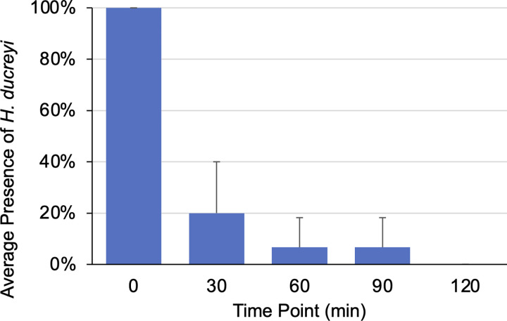
Haemophilus ducreyi was historically known as the causative agent of chancroid, a sexually-transmitted disease causing painful genital ulcers endemic in many low/middle-income nations. In recent years the species has been implicated as the causative agent of nongenital cutaneous ulcers affecting children of the South Pacific Islands and West African countries. Much is still unknown about the mechanism of H. ducreyi transmission in these areas, and recent studies have identified local insect species, namely flies, as potential transmission vectors. H. ducreyi DNA has been detected on the surface and in homogenates of fly species sampled from Lihir Island, Papua New Guinea. The current study develops a model system using Musca domestica, the common house fly, as a model organism to demonstrate proof of concept that flies are a potential vector for the transmission of viable H. ducreyi. Utilizing a green fluorescent protein (GFP)-tagged strain of H. ducreyi and three separate exposure methods, we detected the transmission of viable H. ducreyi by 86.11% ± 22.53% of flies sampled. Additionally, the duration of H. ducreyi viability was found to be directly related to the bacterial concentration, and transmission of H. ducreyi was largely undetectable within one hour of initial exposure. Push testing, Gram staining, and PCR were used to confirm the identity and presence of GFP colonies as H. ducreyi. This study confirms that flies are capable of mechanically transmitting viable H. ducreyi, illuminating the importance of investigating insects as vectors of cutaneous ulcerative diseases.
Copyright: © 2024 Stabile et al. This is an open access article distributed under the terms of the Creative Commons Attribution License, which permits unrestricted use, distribution, and reproduction in any medium, provided the original author and source are credited.
Conflict of interest statement
The authors have declared that no competing interests exist.
Figures


Similar articles
-
Haemophilus ducreyi DNA is detectable on the skin of asymptomatic children, flies and fomites in villages of Papua New Guinea.PLoS Negl Trop Dis. 2017 May 10;11(5):e0004958. doi: 10.1371/journal.pntd.0004958. eCollection 2017 May. PLoS Negl Trop Dis. 2017. PMID: 28489855 Free PMC article.
-
Chronic cutaneous ulcers secondary to Haemophilus ducreyi infection.Med J Aust. 2010 Mar 15;192(6):348-50. doi: 10.5694/j.1326-5377.2010.tb03537.x. Med J Aust. 2010. PMID: 20230355
-
Multiple Class I and Class II Haemophilus ducreyi Strains Cause Cutaneous Ulcers in Children on an Endemic Island.Clin Infect Dis. 2018 Nov 13;67(11):1768-1774. doi: 10.1093/cid/ciy343. Clin Infect Dis. 2018. PMID: 29897409 Free PMC article.
-
Chancroid and Haemophilus ducreyi.Clin Microbiol Rev. 1989 Apr;2(2):137-57. doi: 10.1128/CMR.2.2.137. Clin Microbiol Rev. 1989. PMID: 2650859 Free PMC article. Review.
-
Haemophilus ducreyi: from sexually transmitted infection to skin ulcer pathogen.Curr Opin Infect Dis. 2016 Feb;29(1):52-7. doi: 10.1097/QCO.0000000000000226. Curr Opin Infect Dis. 2016. PMID: 26658654 Review.
Cited by
-
Detection of Haemophilus ducreyi from environmental and animal samples in Cameroon.PLoS Negl Trop Dis. 2025 May 8;19(5):e0013091. doi: 10.1371/journal.pntd.0013091. eCollection 2025 May. PLoS Negl Trop Dis. 2025. PMID: 40338972 Free PMC article.
-
Infections caused by Haemophilus ducreyi: one organism, two stories.Clin Microbiol Rev. 2024 Dec 10;37(4):e0013524. doi: 10.1128/cmr.00135-24. Epub 2024 Sep 17. Clin Microbiol Rev. 2024. PMID: 39287406 Review.
References
-
- Mitjà O, Lukehart SA, Pokowas G, Moses P, Kapa A, Godornes C, et al.. Haemophilus ducreyi as a cause of skin ulcers in children from a yaws-endemic area of Papua New Guinea: a prospective cohort study. Lancet Glob Health. 2014;2(4):e235–e241. - PubMed
MeSH terms
LinkOut - more resources
Full Text Sources
