. 2024 May 30;20(5):e1012269.
doi: 10.1371/journal.ppat.1012269. eCollection 2024 May.

The Toxoplasma gondii F-Box Protein L2 Functions as a Repressor of Stage Specific Gene Expression

Affiliations

The Toxoplasma gondii F-Box Protein L2 Functions as a Repressor of Stage Specific Gene Expression

Carlos G Baptista et al. PLoS Pathog. .

Abstract

Toxoplasma gondii is a foodborne pathogen that can cause severe and life-threatening infections in fetuses and immunocompromised patients. Felids are its only definitive hosts, and a wide range of animals, including humans, serve as intermediate hosts. When the transmissible bradyzoite stage is orally ingested by felids, they transform into merozoites that expand asexually, ultimately generating millions of gametes for the parasite sexual cycle. However, bradyzoites in intermediate hosts differentiate exclusively to disease-causing tachyzoites, which rapidly disseminate throughout the host. Though tachyzoites are well-studied, the molecular mechanisms governing transitioning between developmental stages are poorly understood. Each parasite stage can be distinguished by a characteristic transcriptional signature, with one signature being repressed during the other stages. Switching between stages require substantial changes in the proteome, which is achieved in part by ubiquitination. F-box proteins mediate protein poly-ubiquitination by recruiting substrates to SKP1, Cullin-1, F-Box protein E3 ubiquitin ligase (SCF-E3) complexes. We have identified an F-box protein named Toxoplasma gondii F-Box Protein L2 (TgFBXL2), which localizes to distinct perinucleolar sites. TgFBXL2 is stably engaged in an SCF-E3 complex that is surprisingly also associated with a COP9 signalosome complex that negatively regulates SCF-E3 function. At the cellular level, TgFBXL2-depleted parasites are severely defective in centrosome replication and daughter cell development. Most remarkable, RNAseq data show that TgFBXL2 conditional depletion induces the expression of stage-specific genes including a large cohort of genes necessary for sexual commitment. Together, these data suggest that TgFBXL2 is a latent guardian of stage specific gene expression in Toxoplasma and poised to remove conflicting proteins in response to an unknown trigger of development.


Conflict of interest statement

The authors have declared that no competing interests exist.

Figures

Fig 1
Fig 1. TgFBXL2 is a Toxoplasma F-box protein.
(A). Schematic illustration of HATgFBXL2 anhydrotetracycline (ATC)-mediated gene expression. The endogenous promoter of TgFBXL2 was replaced with a SAG4 promoter construct in which a tetracycline transactivator (tTA) binding element was cloned upstream. Addition of ATC reduces transcription by preventing tTA binding to the tetracycline responsive promoter. (B). PCR of HATgFBXL2 genomic DNA showing correct integration of anhydrotetracycline responsive promoter using primers F2 and R1 as show in A. (C). Lysates prepared from HATgFBXL2 and parental TATiΔKu80 were either Western blotted (Input) or incubated with anti-HA antibodies (IP) that were captured by Protein G Sepharose. HATgFBXL2 and TgSKP1 were detected in lysates (input), flow through (FT), and immunoprecipitates (IP) by Western blotting with anti-HA and TgSKP1 antibodies. (D). Volcano plot of identified proteins in large-scale lysates immunoprecipitated with anti-HA beads and analyzed by MS. Proteins in red were 10-fold enriched from the HATgFBXL2 strain vs. parental with P-value ≤0.01 and therefore are considered bona-fide TgFBXL2 interactors. Relaxing the p-value to ≤0.05 resulted in 3 additional proteins that are likely to be false positives. See S1 Table for complete dataset. Protein candidates were selected from a list of protein hits that were detected in multiple replicates and minimally recovered with parental strain. Proteins known or predicted to interact with TgSKP1 are underlined. See S1 Table for origin of protein labels and more information about control data. (E) Protein abundances based on quantification of detected peptides for TgFBXL2 and its interactors, normalized to TgFBXL2 abundance. Values represent results from 3 biological replicates, with 3 technical replicates each. The box represents 75% of the range, and the internal bars represent the median. See S1 Table for complete dataset. Protein candidates were selected from a list of protein hits that were detected in multiple replicates and minimally recovered with parental strain. Proteins known or predicted to interact with TgSKP1 are underlined. See S1 Table for origin of protein labels and more information about control data.
Fig 2
Fig 2. TgFBXL2 is important for Toxoplasma growth.
(A). Lysates from HATgFBXL2 or parental TATiΔKu80 tachyzoites grown for 24 h ± 1μg/mL ATC were Western blotted to detect HATgFBXL2 or Histone H3 as a loading control. (B). HATgFBXL2 or TATiΔKu80 parasites were grown for 7 days on HFF monolayers in the absence or presence of 1μg/ml ATC. Representative images from 3 independent experiments performed in triplicate. (C and E). Invasion was assessed by determining numbers of vacuoles detected per 100 host cell nuclei after 24 h or 48 h ATC pre-treatment respectively. N = 3. Significance was analyzed using 2-way ANOVA. (D and F). Replication was determined by counting the number of parasites per vacuole after 24 h or 48 h ATC pre-treatment respectively.
Fig 3
Fig 3. Loss of TgFBXL2 affects tachyzoite cell cycle progression.
(A). HATgFBXL2 parasites were grown for 24 h ± 1 μg/mL ATC. Cells were fixed and stained to detect IMC3, parasite’s centrosomes (TgCentrin-1) and DNA. (*) highlight HATgFBXL2-expressing parasites showing increased number of centrosome staining. (B). HATgFBXL2 parasites were grown for either 24 h or 48 h on HFF monolayers ± ATC, then fixed and stained to detect subpellicular microtubules (Ac. tubulin), plasma membrane (SAG1) and DNA. (*) highlight HATgFBXL2 parasites showing more than 2 daughter parasites. Bars = 1 μm in top and middle panel and 2 μm in bottom panel. Quantification of vacuoles with increased number of centrosomes (C) or with asynchronous replication (D) at the indicated times. Data represents averages ± standard deviations of 3 independent experiments with at least 50 parasites examined/experiment. *, P < 0.001, One-Way ANOVA.
Fig 4
Fig 4. Loss of TgFBXL2 causes apicoplast biogenesis defect.
(A). HATgFBXL2 parasites were grown for 24, 48, or 72 h on HFF monolayers ± 1 μg/mL ATC. Cells were fixed and stained to detect plasma membrane (SAG1), apicoplast (TgAtrx1) and DNA. Shown is representative image from parasites infected for 24 h. Arrows indicate HATgFBXL2 parasites lacking apicoplast staining. Bars = 2 μm. (B). Quantification of vacuoles with parasites showing apicoplast segregation defects at the indicated times. *, p < 0.001, Two-Way ANOVA. (C). qPCR was used to quantify apicoplast genome in HATgFBXL2 parasites grown for 24, 36, or 48 h on HFF monolayers ± ATC. Actinonin and clindamycin were used as positive inhibitors of apicoplast genome replication. Shown are means and standard deviations from 4 independent experiments. (P <0.05, one-way ANOVA). (D). HATgFBXL2 parasites were grown for either 24 h or 48 h on HFF monolayers ± ATC, then fixed and stained to detect parasite’s centrosomes (TgCentrin-1), apicoplast (TgAtrx1) and DNA. Arrowheads indicate HATgFBXL2 parasites in which TgCentrin-1 is not properly associated with apicoplast during cell division. Bars = 1 μm. (E). Quantification of vacuoles with parasites showing increased number of centrosomes that lack apicoplast interaction at the indicated times. Data represents averages ± standard deviations of 3 independent experiments with at least 50 parasites examined/experiment. *, P < 0.001, One-Way ANOVA
Fig 5
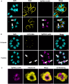
Fig 5. TgFBXL2 localizes to a perinucleolar compartment.
(A). HATgFBXL2-expressing parasites and parental TATiΔKu80 were fixed and stained to detect HATgFBXL2 (αHA), plasma membrane (SAG1) and DNA. Arrows indicate HATgFBXL2 staining in areas staining weakly with DAPI. Bars = 1 μm. (B). HATgFBXL2 and parental TATiΔKu80 tachyzoites were fixed and stained to detect HATgFBXL2 (αHA), nucleolus (SytoRNA) and DNA. Arrow highlights a parasite nucleus with HATgFBXL2 staining surrounding the nucleolus. Bars = 2 μm. (C). HATgFBXL2-expressing parasites were fixed and stained to detect HATgFBXL2 (αHA), and Histone H3 (αH3). Shown are still images from a movie of 3D rendering available as supplemental data. Bars = 0.5 μm.
Fig 6
Fig 6. Loss of TgFBXL2 upregulates pre-sexual specific genes involved in sexual commitment.
(A). Volcano plot displaying gene expression variations between the HATgFBXL2 parasites grown ± ATC for 24 h or 48 h (n = 1549, S2 Table). The red and green dots indicate those transcripts whose abundances are significantly down- and up-regulated genes, respectively, at 48 hpi, using adjusted P <0.01 (Bonferroni-corrected) and ± 2-fold change as the cut-off threshold. (B). Gene ontology for cellular component (CC) annotation of up-regulated genes in HATgFBXL2-depleted parasites. (C). Venn diagram comparing genes modulated by HATgFBXL2 to MORC or expressed by enteroepithelial stages parasites (EES). (D & E). Venn diagrams illustrating overlap between HATgFBXL2-upregulated genes (n = 355) and the RNAs expressed by merozoites, sporozoites, and bradyzoites.
Fig 7
Fig 7. Merozoite specific proteins GRA80 and ROP26 are expressed in TgFBXL2-depleted parasites.
(A). HATgFBXL2 parasites grown for 24 h or 48 h ± 1μg/ml ATC were fixed and stained to detect GRA80, IMC1 and DNA. (B). HATgFBXL2 parasites grown for 24 h or 48 h ± 1μg/ml ATC were fixed and stained to detect ROP26, IMC1 and DNA. Bars = 1 μm. (C and D). Quantification of HATgFBXL2 parasites expressing either GRA80 or ROP26 respectively. *, P < 0.001 One-Way ANOVA.
Fig 8
Fig 8. TgFBXL2 and MORC/HDAC3 do not interact.
(A). HATgFBXL2-expressing parasites were fixed and stained to detect HATgFBXL2 (αHA), and MORC. (B). HATgFBXL2-expressing parasites were fixed and stained to detect HATgFBXL2 (αHA) and HDAC3. Bars = 0.5 μm. (C). HATgFBXL2 parasites were grown for 24 h in the presence of either 100 nM FR235222 or DMSO as vehicle control. Cells were fixed and stained to detect HATgFBXL2 (aHA), SAG1 and DNA. (D&E). HATgFBXL2 parasites were grown for 48 h ± 1 μg/mL ATC. Cells were fixed and stained to detect DNA, SAG1 or either HDAC3 (D) or MORC (E).

Similar articles

Cited by

References

    1. Pappas G, Roussos N, Falagas ME. Toxoplasmosis snapshots: global status of Toxoplasma gondii seroprevalence and implications for pregnancy and congenital toxoplasmosis. Int J Parasitol. 2009;39(12):1385–94. Epub 2009/05/13. doi: 10.1016/j.ijpara.2009.04.003 . - DOI - PubMed
    1. Dubey JP. History of the discovery of the life cycle of Toxoplasma gondii. Int J Parasitol. 2009;39(8):877–82. Epub 2009/07/25. doi: 10.1016/j.ijpara.2009.01.005 . - DOI - PubMed
    1. Jones JL, Dubey JP. Foodborne toxoplasmosis. Clin Infect Dis. 2012;55(6):845–51. Epub 20120522. doi: 10.1093/cid/cis508 . - DOI - PubMed
    1. Blader IJ, Coleman BI, Chen CT, Gubbels MJ. Lytic Cycle of Toxoplasma gondii: 15 Years Later. Annu Rev Microbiol. 2015;69:463–85. doi: 10.1146/annurev-micro-091014-104100 . - DOI - PMC - PubMed
    1. Baum J, Papenfuss AT, Baum B, Speed TP, Cowman AF. Regulation of apicomplexan actin-based motility. Nat Rev Microbiol. 2006;4(8):621–8. doi: 10.1038/nrmicro1465 . - DOI - PubMed