. 2024 May 31;384(6699):eadd6260.
doi: 10.1126/science.add6260. Epub 2024 May 31.

Pharmacological modulation of septins restores calcium homeostasis and is neuroprotective in models of Alzheimer's disease

Affiliations

Pharmacological modulation of septins restores calcium homeostasis and is neuroprotective in models of Alzheimer's disease

Katrien Princen et al. Science. .

Abstract

Abnormal calcium signaling is a central pathological component of Alzheimer's disease (AD). Here, we describe the identification of a class of compounds called ReS19-T, which are able to restore calcium homeostasis in cell-based models of tau pathology. Aberrant tau accumulation leads to uncontrolled activation of store-operated calcium channels (SOCCs) by remodeling septin filaments at the cell cortex. Binding of ReS19-T to septins restores filament assembly in the disease state and restrains calcium entry through SOCCs. In amyloid-β and tau-driven mouse models of disease, ReS19-T agents restored synaptic plasticity, normalized brain network activity, and attenuated the development of both amyloid-β and tau pathology. Our findings identify the septin cytoskeleton as a potential therapeutic target for the development of disease-modifying AD treatments.


Conflict of interest statement

H.Z. has served at scientific advisory boards and/or as a consultant for Abbvie, Acumen, Alector, Alzinova, ALZPath, Amylyx, Annexon, Apellis, Artery Therapeutics, AZTherapies, Cognito Therapeutics, CogRx, Denali, Eisai, Merry Life, Nervgen, Novo Nordisk, Optoceutics, Passage Bio, Pinteon Therapeutics, Prothena, Red Abbey Labs, reMYND, Roche, Samumed, Siemens Healthineers, Triplet Therapeutics, and Wave; has given lectures in symposia sponsored by Alzecure, Biogen, Cellectricon, Fujirebio, Lilly, Novo Nordisk, and Roche; and is a cofounder of Brain Biomarker Solutions in Gothenburg AB (BBS), which is a part of the GU Ventures Incubator Program (outside submitted work). J.L.C. has provided consultation to Acadia, Actinogen, Acumen, AlphaCognition, ALZpath, Aprinoia, AriBio, Artery, Biogen, Biohaven, BioVie, BioXcel, Bristol-Myers Squib, Cassava, Cerecin, Diadem, Eisai, GAP Foundation, GemVax, Janssen, Jocasta, Karuna, Lighthouse, Lilly, Lundbeck, LSP/eqt, Merck, NervGen, New Amsterdam, Novo Nordisk, Oligomerix, Optoceutics, Ono, Otsuka, Oxford Brain Diagnostics, Prothena, reMYND, Roche, Sage Therapeutics, Signant Health, Simcere, Sinaptica, Suven, TrueBinding, Vaxxinity, and Wren pharmaceutical, assessment, and investment companies. J.W. and S.W. are cofounders and shareholders of reMYND. G.G. is a consultant for reMYND and owns reMYND warrants and shares. M.Fi. owns reMYND warrants. J.d.W. is cofounder of Augustine Tx and served as scientific advisory board member for Augustine Tx. J.T. is cofounder of Orionis Biosciences. K.Pr. and G.G. are inventors on patent WO2013/004642 held by reMYND NV: “1,2,4-thiadiazol-5-ylpiperazine derivatives useful in the treatment of neurodegenerative diseases.” K.Pr., M.V., M.Fi., and G.G. are inventors on patent application EP23209465.6 submitted by reMYND NV: “Modulators of septin 6 for use in the prevention and/or treatment of neurodegenerative disorders.”

Figures

Fig. 1.
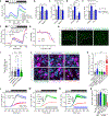
Fig. 1.. Identification of ReS19-T and its high-affinity target.
(A) Cell death and [Ca2+]cyto in ATRA-treated tauP301L-expressing BE(2)-M17 cells in presence of increasing concentrations of REM127. (B to D) Rat hippocampal neurons [22 days in vitro (DIV)] treated with vehicle or REM127 (240 nM) and exposed or not to ADDL (0.5 μM) [(B) and (C)] or ADDL (1 μM) (D) for 24 hours. (B and C) Dendritic spine density measured in dendritic segments of Marcks-GFP–transfected neurons. ADDL-/Veh, N = 39; ADDL-/REM127, N = 37; ADDL+/Veh, N = 43; and ADDL+/REM127, N = 50. Scale bar, 5 μm. Error bars indicate 95% confidence interval (CI). (D) ADDL-induced cell death in neurons exposed to the indicated treatments (N = 3). Error bars indicate SD. (E) Three-hybrid screen approach to identifying ReS19-T-interacting proteins. (F) In cellulo interaction of MFC (REM929 linked to methotrexate) with different septin isoforms (N = 3, error bars indicate SD). Inset shows phylogenetic analysis of septin isoforms probed in the three-hybrid assay. (G to J) FIDA binding isotherms of REM127 to human SEPT6, SEPT7, and SEPT2. Error bars indicate SD. (K and L) Effects of scrambled (scr) and SEPT6-lowering siRNAs on REM127-induced relief of toxicity (K) and [Ca2+]cyto overload (L) in ATRA-treated tauP301L-expressing BE(2)-M17 cells. *P < 0.05, **P < 0.01, Kruskal-Wallis with Dunn’s multiple-comparisons tests for (C) and one-way ANOVA with multiple pairwise comparisons of means (Tukey’s test) for (D). See also corresponding figs. S1 to S3.
Fig. 2.
Fig. 2.. ReS19-T specifically targets a tau-dependent pathological form of SOCE.
(A to C) SOCE measured by calcium add-back in individual Fluo-4–loaded BE(2)-M17 cells transfected with the indicated constructs and exposed to vehicle [dimethyl sulfoxide (DMSO)] or REM127 (200 nM, 24 hours). (A) Fluo-4 traces normalized to the control mRuby/vehicle condition. (B and C) Average SOCE amplitude (B) and basal [Ca2+]cyto (C) derived from time series shown in A. (D and E) Independent calcium add-back experiment showing average effect of Pyr6 (10 μM, 1 hour) on SOCE amplitude and basal [Ca2+]cyto in mRuby- or tauP301L-T2A-mRuby–expressing cells. In (A) to (E), >450 cells were analyzed for each condition. (F) SOCE measured in Fura-2–loaded BE(2)-M17 cells stably expressing tauP301L or empty vector (EV) and transfected with scrambled or SEPT6-targeting siRNAs. Shown are average calcium traces (normalized to baseline) obtained from 24 replicate wells for each condition. (G) Lactate dehydrogenase (LDH) release in ATRA-treated tauP301L cells in the presence of increasing concentrations of the SOCC inhibitors Pyr6 and BTP2. (H and I) Imaging and quantification of ER-PM contact sites in control and tauP301L cells transfected with the ER-PM reporter SPLICshort-P2AER-PM and treated with vehicle or REM127 (100 nM, 24 hours). More than 45 cells were analyzed for each condition from three independent experiments. Scale bar, 10 μm. (J and K) NFAT activity measured by nuclear translocation of mRuby-NFAT3 in mock or tauP301L-transduced cells exposed to vehicle or REM127 (20 or 100 nM, 48 hours), cyclosporin A (250 nM, 24 hours), or ionomycin (2 μM, 1 hour). (J) Confocal images showing mRuby-NFAT localization in response to indicated treatments. Scale bar, 20 μm. Yellow asterisks point to cells with clear nuclear localization of mRuby-NFAT. (K) mRuby-NFAT nucleus-to-cytoplasm intensity ratio measured for indicated treatments. N = 30 cells for each condition. (L to O) SOCE measured in Fura-2–loaded patient-derived glutamatergic neurons (tauP301L) or isogenic controls cultured for 90 DIV. N = 3 replicate wells for each condition. (O) Quantification of peak SOCE responses in control and tauP301L neurons treated with increasing concentrations of REM127 for 24 hours or Pyr6 (10 μM, 1 hour). In (A), (F), (L), (M) and (N), bars on top of time series indicate the presence (black) or absence (white) of extracellular calcium, and thapsigargin treatment (gray). Shaded error bars represent 95% CI. *P < 0.05, **P < 0.01, ***P < 0.001, one-way ANOVA with multiple pairwise comparisons of means (Tukey’s test) in (B) to (E) and (O) and Kruskal-Wallis with Dunn’s multiple-comparisons test in (I) and (K). See also corresponding figs. S4 to S6.
Fig. 3.
Fig. 3.. ReS19-T influences septin filament assembly in cell-free conditions.
(A to D) LiGaMD simulations of REM127 interaction with the SEPT2/6/7 hexamer. (A) LiGaMD identifies a high-affinity binding pocket at the NC interface between SEPT6 and SEPT7. REM127 is shown in red. (B) Blow up of the SEPT6/7 interface with amino acids predicted to interact with REM127. Hydrogen bonds formed by the compound with surrounding amino acids are shown in yellow. Amino acids are labeled according to their position in the SEPT2/6/7 hexamer. (C) Alignment showing the main cluster of amino acids in SEPT6 and SEPT7 interacting with REM127. Heatmap indicates the frequency of interactions. Amino acid conservation and type are shown at the top using the sequence logo. (D) Umbrella sampling calculations of the SEPT6/7 heterodimer showing binding free energy with and without bound ligand. (E and F) Cell-free assembly of SEPT2/6/7 filaments. (E) DLS measurements performed at different time points after mixing of purified SEPT2, SEPT6, and SEPT7 (500 nM final protein concentration) in the presence of vehicle (DMSO), GTPγS (100 μM), or REM127 (1 μM). 0′ indicates the first measurement immediately after component mixing. Distribution of particle size (y axis) is normalized across treatment and time points. (F) TEM images of SEPT2/6/7 polymerization performed in conditions identical to (E) and captured at three time points after component mixing. Arrowheads point to typical polymerized structures. Arrows point to highly ordered filaments only observed after REM127 treatment. See also corresponding figs. S7 and S8.
Fig. 4.
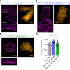
Fig. 4.. ReS19-T rescues septin filament assembly in cells expressing pathogenic tau.
(A to C) SEPT2 immunostaining in BE(2)-M17 cells transfected with mRuby or tauP301L-T2A–mRuby and treated for 24 hours with REM127 (100 nM) or vehicle. The asterisk indicates a nontransfected cell. Scale bar, 10 μm. (D) Quantification of SEPT2 circular versus filamentous structures by measuring the average minor to major axis length ratio for all SEPT2 objects segmented in individual cells (N > 13 for each group). ***P < 0.001, one-way ANOVA with multiple pairwise comparisons of means (Tukey’s test). See also corresponding fig. S9.
Fig. 5.
Fig. 5.. Rapid restoration of synaptic, network, and cognitive functions by ReS19-T in AD preclinical models.
(A) Spontaneous calcium spike activity in patient-derived tauP301L neurons and their isogenic controls exposed to vehicle or REM127 at the indicated concentrations. Mean frequency of calcium bursts was measured for 3 min over a period of 20 days from DIV 50 to 70 (three replicate wells for each condition). (B) Calcium spike activity measured 15 days after the first recording and corresponding to time point highlighted in gray in (A). (C) CA1 LTP in WT and tauP301S mice (3.5 months of age, N = 8 for each group) administered with vehicle or REM127 (20 mg/kg/day) for 2 months. Arrow indicates tetanus LTP inducing stimulus. (D) CA1 LTP in WT/Veh (N = 5), APP-Ln/Veh (N = 8), and APP-Ln/REM127 (N = 6) mice (7 months of age) administered with vehicle or REM127 (20 mg/kg/day) for 7 days. (E and F) Exposure-dependent rescue of LTP in APP-Ln mice. Cavg,u,brain: average free brain concentration of REM127 calculated from in vivo pharmacokinetic profile of the compound (table S1). Statistics were only performed within the APP-Ln group to identify exposure-dependent effects on LTP. (G) LTP in WT animals (3.5 months of age) administered with vehicle (N = 11) or REM127 (N = 6) at 20 mg/kg/day for 7 days. (H) Power analysis of EEG recordings (parietal lobe) in awake WT/Veh (N = 6), APP-Ln/Veh (N = 8), and APP-Ln/REM127 (N = 4) mice, 8 months of age, administered with vehicle or REM127 (20 mg/kg/day) for 14 days. (I and J) Morris water maze results. Shown are spatial learning (I) and memory recall (annulus crossing index) (J) in WT/Veh (N = 23), APP-Ln/Veh (N = 23), and APP-Ln/REM123 (N = 23) mice 8 months of age that were administered with vehicle or REM123 (20 mg/kg/day) for 11 weeks. Error bars indicate SEM, except for (E), where they correspond to SD. *P < 0.05, **P < 0.01, and ***P < 0.001, one-way ANOVA with multiple pairwise comparisons of means (Tukey’s test) in (A), (C) to (E), and (J); unpaired t test in (G); two-way ANOVA with repeated measures (RM) with genotype or treatment as the between-subject factor and frequency as the within subject factor in (H); and two-way ANOVA with RM and Sidak pairwise multiple-comparisons test in (I). See also corresponding fig. S10.
Fig. 6.
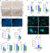
Fig. 6.. ReS19-T attenuates AD pathology.
(A to D) APP-Ln:PS1(A246E) mice (7 months old) after 3 months of vehicle (N = 16) or REM127 (N = 12) oral dosing at 20 mg/kg/day. (A to C) Aβ plaques stained with an anti-amyloid fibrils (LOC) antibody in cortex [(A) and (B)] and subiculum [(A) and (C)]. (D) CD11b immunoreactivity in cortex. (E to G) Dense core staining of Aβ plaques with Methoxy X04 in prefrontral cortex [(E) and (F)] and measurements of Aβ42 concentration in the soluble fraction of the cortex (G) of APP-SAA mice and C57Bl/6J controls orally dosed with vehicle or REM127 (25 mg/kg/day) for 3 months. N = 11 to 12 for each group. (H and I) Co-staining of Aβ fibrils (LOC) and phospho-tau (p-Ser202 and p-Thr205) using the AT8 antibody in same animal cohorts described in (E) to (G). (J to M) Five-month-old tauP301S mice treated for 3 months with vehicle or REM123 at 20 mg/kg/day. (J) Tau concentration in CSF measured by ELISA (N > 9 for each group). Pan-tau (K), pS396-tau (L), and pS199-tau (M) were measured by ELISA in cortical extracts (N = 11 for each group). Error bars indicate SD. *P < 0.05, **P < 0.01, and ***P < 0.001, unpaired t test, in (B) to (D) and (J) to (M) and one-way ANOVA with multiple pairwise comparisons of means (Tukey’s test) in (F) to (I). See also corresponding fig. S11.

Comment in

References

    1. Scheltens P. et al., Alzheimer’s disease. Lancet 397, 1577–1590 (2021). doi: 10.1016/S0140-6736(20)32205-4; - DOI - PMC - PubMed
    1. Leissring MA et al., Capacitative calcium entry deficits and elevated luminal calcium content in mutant presenilin-1 knockin mice. J. Cell Biol 149, 793–798 (2000). doi: 10.1083/jcb.149.4.793; - DOI - PMC - PubMed
    1. Etcheberrigaray R. et al., Calcium responses in fibroblasts from asymptomatic members of Alzheimer’s disease families. Neurobiol. Dis 5, 37–45 (1998). doi: 10.1006/nbdi.1998.0176; - DOI - PubMed
    1. Guo Q. et al., Increased vulnerability of hippocampal neurons to excitotoxic necrosis in presenilin-1 mutant knock-in mice. Nat. Med 5, 101–106 (1999). doi: 10.1038/4789; - DOI - PubMed
    1. Larson J, Lynch G, Games D, Seubert P, Alterations in synaptic transmission and long-term potentiation in hippocampal slices from young and aged PDAPP mice. Brain Res. 840, 23–35 (1999). doi: 10.1016/S0006-8993(99)01698-4; - DOI - PubMed

Publication types