

Relaxin-2 is a novel biomarker for differentiated thyroid carcinoma in humans
- PMID: 38815632
- PMCID: PMC11470803
- DOI: 10.1016/j.bcp.2024.116323
Relaxin-2 is a novel biomarker for differentiated thyroid carcinoma in humans
Abstract
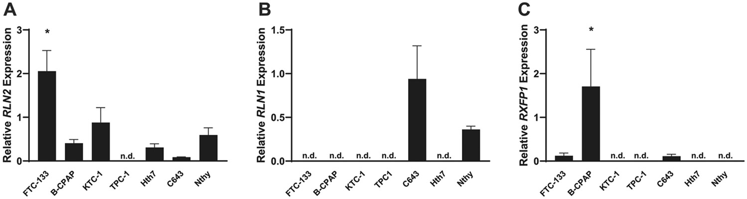
Relaxin's role in differentiated thyroid cancer (DTC) has been suggested but its characterization in a large clinical sample remains limited. We performed immunohistochemistry for relaxin-2 (RLN2), CD68 (total macrophages), CD163 (M2 macrophages) on tissue microarrays from 181 subjects with non-distant metastatic DTC, and 185 subjects with benign thyroid tissue. Mean pixels/area for each marker was compared between tumor and adjacent tissue via paired-t test and between DTC and benign subjects via t-test assuming unequal variances. RNA qPCR was performed for expression of RLN2, RLN1, and RXFP1 in cell lines. Amongst 181 cases, the mean age was 46 years, 75 % were females. Tumoral tissue amongst the DTC cases demonstrated higher mean expression of RLN2 (53.04 vs. 9.79; p < 0.0001) compared to tumor-adjacent tissue. DTC tissue also demonstrated higher mean expression of CD68 (14.46 vs. 4.79; p < 0.0001), and CD163 (23.13 vs. -0.73; p < 0.0001) than benign thyroid. These markers did not differ between tumor-adjacent and benign thyroid tissue groups; and amongst cases, did not differ by demographic or clinicopathologic features. RLN1 and RXFP1 expression was detected in a minority of the cell lines, while RLN2 was expressed by 6/7 cell lines. In conclusion, widespread RLN2 expression in DTC tissue and most cell lines demonstrates that RLN2 acts in a paracrine manner, and that RLN1 and RXFP1 are probably not involved in thyroid cancer cell signaling. RLN2 is a biomarker for thyroid carcinogenesis, being associated with but not secreted by immunosuppressive macrophages. These findings will guide further investigations for therapeutic avenues against thyroid cancer.
Keywords: Carcinogenesis; Macrophages; Relaxin; Thyroid carcinoma; Tumor microenvironment.
Published by Elsevier Inc.
Conflict of interest statement
Declaration of competing interest The authors declare that they have no known competing financial interests or personal relationships that could have appeared to influence the work reported in this paper.
Figures


Similar articles
-
Relaxin enhances the oncogenic potential of human thyroid carcinoma cells.Am J Pathol. 2006 Aug;169(2):617-32. doi: 10.2353/ajpath.2006.050876. Am J Pathol. 2006. PMID: 16877360 Free PMC article.
-
Relaxin 2/RXFP1 Signaling Induces Cell Invasion via the β-Catenin Pathway in Endometrial Cancer.Int J Mol Sci. 2018 Aug 18;19(8):2438. doi: 10.3390/ijms19082438. Int J Mol Sci. 2018. PMID: 30126180 Free PMC article.
-
Identification of a novel fusion transcript between human relaxin-1 (RLN1) and human relaxin-2 (RLN2) in prostate cancer.Mol Cell Endocrinol. 2016 Jan 15;420:159-68. doi: 10.1016/j.mce.2015.10.011. Epub 2015 Oct 21. Mol Cell Endocrinol. 2016. PMID: 26499396
-
Emerging roles for the relaxin/RXFP1 system in cancer therapy.Mol Cell Endocrinol. 2019 May 1;487:85-93. doi: 10.1016/j.mce.2019.02.001. Epub 2019 Feb 11. Mol Cell Endocrinol. 2019. PMID: 30763603 Review.
-
Role of Relaxin Signaling in Cancer: A Review.Biochem Pharmacol. 2024 Dec;230(Pt 3):116634. doi: 10.1016/j.bcp.2024.116634. Epub 2024 Nov 14. Biochem Pharmacol. 2024. PMID: 39547477 Review.
References
-
- Rahib L, Smith BD, Aizenberg R, Rosenzweig AB, Fleshman JM, Matrisian LM, Projecting cancer incidence and deaths to 2030: the unexpected burden of thyroid, liver, and pancreas cancers in the United States, Cancer Res. 74 (11) (2014) 2913–2921. - PubMed
-
- McIntyre C, Jacques T, Palazzo F, Farnell K, Tolley N, Quality of life in differentiated thyroid cancer, Int. J. Surg 50 (2018) 133–136. - PubMed
-
- Uppal N, Cunningham Nee Lubitz C, James B, The cost and financial burden of thyroid cancer on patients in the US: A review and directions for future research, JAMA Otolaryngol. Head Neck Surg 148 (6) (2022) 568–575. - PubMed
-
- Haugen BR, Alexander EK, Bible KC, Doherty GM, Mandel SJ, Nikiforov YE, Pacini F, Randolph GW, Sawka AM, Schlumberger M, Schuff KG, Sherman SI, Sosa JA, Steward DL, Tuttle RM, Wartofsky L, 2015 American Thyroid association management guidelines for adult patients with thyroid nodules and differentiated thyroid cancer: The American Thyroid Association Guidelines task force on thyroid nodules and differentiated thyroid cancer, Thyroid 26 (1) (2016) 1–133. - PMC - PubMed
Publication types
MeSH terms
Substances
Grants and funding
LinkOut - more resources
Full Text Sources
Medical
Research Materials
