Review
. 2024 Sep 9;45(9):1177-1184.
doi: 10.3174/ajnr.A8195.

Current Practice: Rationale for Screening Children with Hereditary Hemorrhagic Telangiectasia for Brain Vascular Malformations

Affiliations
Review

Current Practice: Rationale for Screening Children with Hereditary Hemorrhagic Telangiectasia for Brain Vascular Malformations

Lauren A Beslow et al. AJNR Am J Neuroradiol. .

Abstract

Background: Hereditary hemorrhagic telangiectasia is an autosomal dominant vascular dysplasia characterized by mucocutaneous telangiectasias, recurrent epistaxis, and organ vascular malformations including in the brain, which occur in about 10% of patients. These brain vascular malformations include high-flow AVMs and AVFs as well as low-flow capillary malformations. High-flow lesions can rupture, causing neurologic morbidity and mortality.

State of practice: International guidelines for the diagnosis and management of hereditary hemorrhagic telangiectasia recommend screening children for brain vascular malformations with contrast enhanced MR imaging at hereditary hemorrhagic telangiectasia diagnosis. Screening has not been uniformly adopted by some practitioners who contend that screening is not justified. Arguments against screening include application of short-term data from the adult A Randomized Trial of Unruptured Brain Arteriovenous Malformations (ARUBA) trial of unruptured sporadic brain AVMs to children with hereditary hemorrhagic telangiectasia as well as concerns about administration of sedation or IV contrast and causing patients or families increased anxiety.

Analysis: In this article, a multidisciplinary group of experts on hereditary hemorrhagic telangiectasia reviewed data that support screening guidelines and counter arguments against screening. Children with hereditary hemorrhagic telangiectasia have a preponderance of high-flow lesions including AVFs, which have the highest rupture risk. The rupture risk among children is estimated at about 0.7% per lesion per year and is additive across lesions and during a lifetime. ARUBA, an adult clinical trial of expectant medical management versus treatment of unruptured brain AVMs, favored medical management at 5 years but is not applicable to pediatric patients with hereditary hemorrhagic telangiectasia given the life expectancy of a child. Additionally, interventional, radiosurgical, and surgical techniques have improved with time. Experienced neurovascular experts can prospectively determine the best treatment for each child on the basis of local resources. The "watch and wait" approach to imaging means that children with brain vascular malformations will not be identified until a potentially life-threatening and deficit-producing intracerebral hemorrhage occurs. This expert group does not deem this to be an acceptable trade-off.


Figures

FIG 1.
FIG 1.
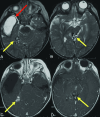
Prior brain hemorrhage in an infant with HHT. T2-weighted MR imaging (A) demonstrates a hemosiderin-lined cavity (red arrow) in the right temporal lobe of a 15-month-old girl with HHT. Additional AVFs (yellow arrows) are evident on T2 (A and B) and contrast-enhanced T1-weighted images (C and D).
FIG 2.
FIG 2.
A 13-year-old boy with HHT who presented with severe headache and altered mental status. A, Coronal noncontrast head CT shows an acute left cerebellar hemisphere hemorrhage and intraventricular hemorrhage in the bilateral lateral ventricles and fourth ventricle. Anterior-posterior (B) and lateral (C) views on digital DSA depict a left cerebellar AVM with arterial supply from the left anterior and posterior inferior cerebellar arteries, left superior cerebellar artery, and deep venous drainage through the transverse sinus.
FIG 3.
FIG 3.
An infant diagnosed with HHT due to a familial ENG variant who had screening MR imaging of the brain and MRA of the head at 2 months of life had a pial AVF with hemosiderin deposition. Sagittal reformat (A) TOF-MRA demonstrates the right fontal AVF (yellow arrow). Axial (B) and coronal (C) T2-weighted MR images confirm the location of the AVF (yellow arrows) as well as adjacent hemosiderin deposition (red arrows) suggestive of prior hemorrhage.
FIG 4.
FIG 4.
A girl diagnosed with HHT due to a familial ENG variant had screening MR imaging that revealed a punctate area of enhancement in the left posterior frontal lobe surrounded by a subtle halo of enhancement (yellow arrow) on a high-resolution 3D T1 postgadolinium image (A). This finding corresponds to a subcentimeter AVM nidus (yellow arrow) on lateral (B) and anterior-posterior (C) DSA. An additional subcentimeter AVM nidus (blue arrow) supplied by the contralateral anterior cerebral artery is also identified on DSA.

References

    1. Faughnan ME, Mager JJ, Hetts SW, et al. Second International Guidelines for The Diagnosis and Management of Hereditary Hemorrhagic Telangiectasia. Ann Intern Med 2020;173:989–1001 10.7326/M20-1443 - DOI - PubMed
    1. Eker OF, Boccardi E, Sure U, et al. European Reference Network for Rare Vascular Diseases (VASCERN) position statement on cerebral screening in adults and children with hereditary haemorrhagic telangiectasia (HHT). Orphanet J Rare Dis 2020;15:165 10.1186/s13023-020-01386-9 - DOI - PMC - PubMed
    1. Dakeishi M, Shioya T, Wada Y, et al. Genetic epidemiology of hereditary hemorrhagic telangiectasia in a local community in the northern part of Japan. Hum Mutat 2020;19:140–48 10.1002/humu.10026 - DOI - PubMed
    1. Govani FS, Shovlin CL. Hereditary haemorrhagic telangiectasia: a clinical and scientific review. Eur J Hum Genet 2009;17:860–71 10.1038/ejhg.2009.35 - DOI - PMC - PubMed
    1. McDonald J, Wooderchak-Donahue W, VanSant Webb C, et al. Hereditary hemorrhagic telangiectasia: genetics and molecular diagnostics in a new era. Front Genet 2015;6:1 10.3389/fgene.2015.00001 - DOI - PMC - PubMed

MeSH terms

LinkOut - more resources