. 2024 May 30;15(1):4617.
doi: 10.1038/s41467-024-48696-1.

The debranching enzyme Dbr1 regulates lariat turnover and intron splicing

Affiliations

The debranching enzyme Dbr1 regulates lariat turnover and intron splicing

Luke Buerer et al. Nat Commun. .

Abstract

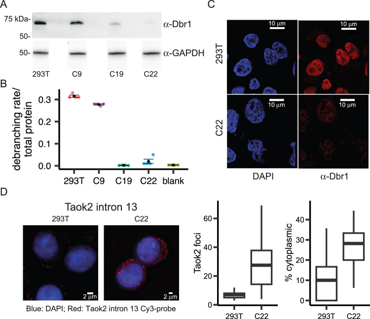
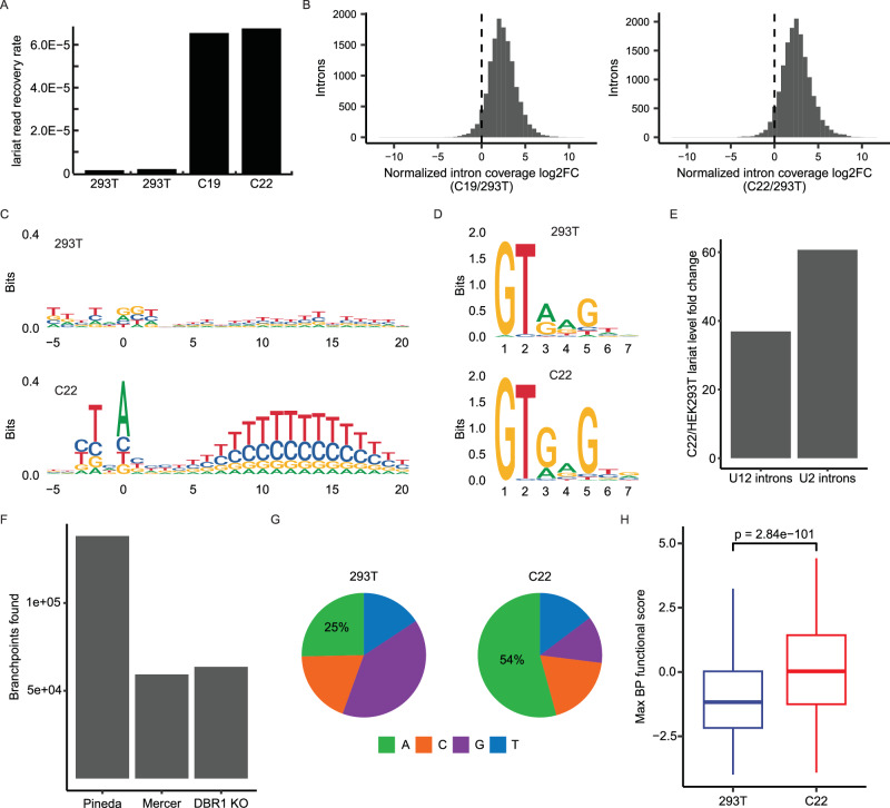
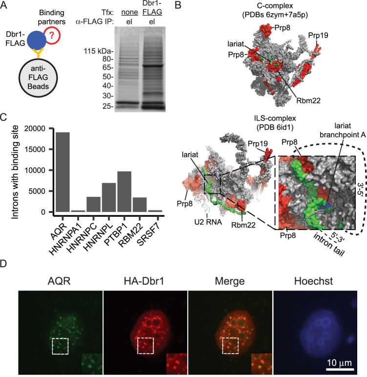
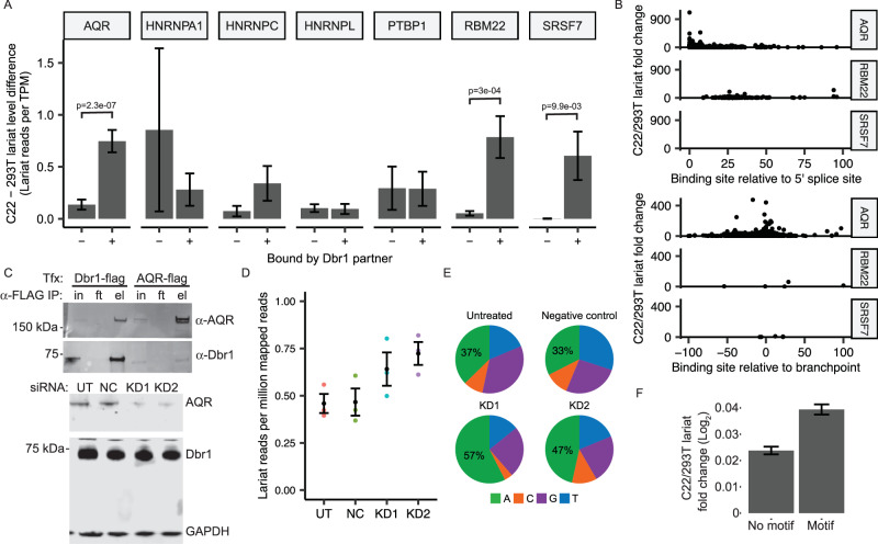
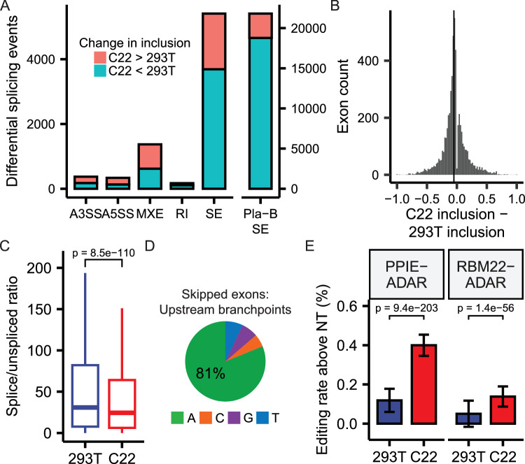
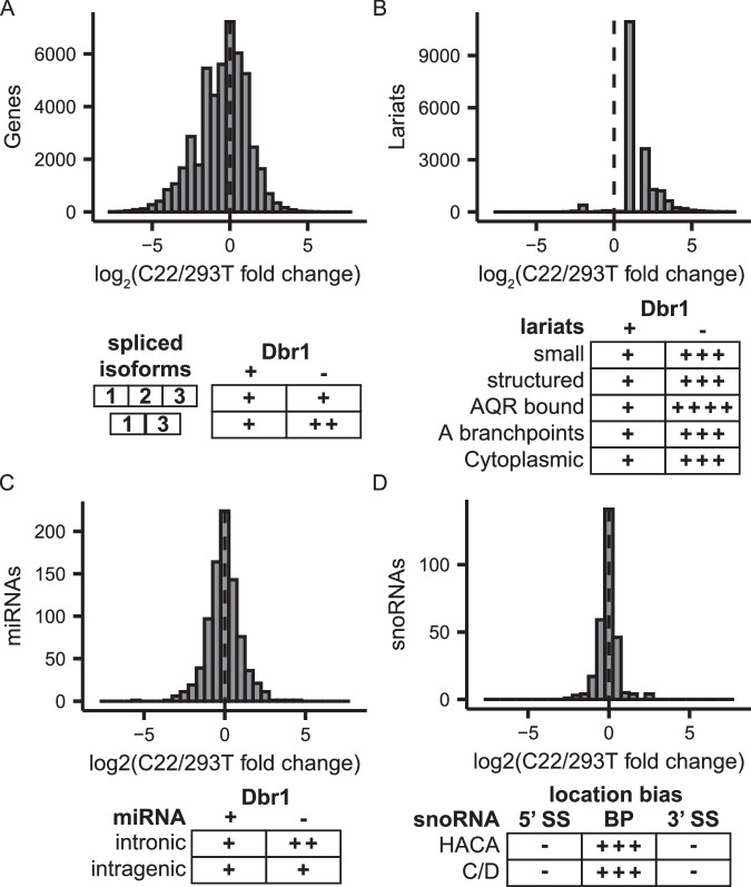
The majority of genic transcription is intronic. Introns are removed by splicing as branched lariat RNAs which require rapid recycling. The branch site is recognized during splicing catalysis and later debranched by Dbr1 in the rate-limiting step of lariat turnover. Through generation of a viable DBR1 knockout cell line, we find the predominantly nuclear Dbr1 enzyme to encode the sole debranching activity in human cells. Dbr1 preferentially debranches substrates that contain canonical U2 binding motifs, suggesting that branchsites discovered through sequencing do not necessarily represent those favored by the spliceosome. We find that Dbr1 also exhibits specificity for particular 5' splice site sequences. We identify Dbr1 interactors through co-immunoprecipitation mass spectrometry. We present a mechanistic model for Dbr1 recruitment to the branchpoint through the intron-binding protein AQR. In addition to a 20-fold increase in lariats, Dbr1 depletion increases exon skipping. Using ADAR fusions to timestamp lariats, we demonstrate a defect in spliceosome recycling. In the absence of Dbr1, spliceosomal components remain associated with the lariat for a longer period of time. As splicing is co-transcriptional, slower recycling increases the likelihood that downstream exons will be available for exon skipping.


Conflict of interest statement

We disclose W.G.F. as the founder of Walah Scientific and member of the scientific advisory board for Remix Therapeutics. All other authors declare no competing interests.

Figures

Fig. 1
Fig. 1. The predominantly nuclear Dbr1 supplies all debranching activity in 293 T cell extract.
A Dbr1 protein levels in wild type 293T and DBR1 knockout cells lines (C9, C19, C22) assayed by western blot. B Fluorogenic debranching activity assay of extracts from C9, C19, and C22 cells compared to 293T control lysates (n = 4 independent experiments for each cell line; sample values shown as colored points; mean ± SE shown in black). C Fluorescent immuno-microscopy of 293T and C22 cells, using DAPI stain and α-Dbr1 polyclonal antibodies. D FISH analysis of Taok2 intron 13 in 293T and C22 cells (left), and quantification of cytoplasmic and nuclear intron foci (right; n = 35 cells for 293 T, 58 cells for C22; center line represents median; lower and upper bounds of the box represent the 25th and 75th percentile, respectively; lower and upper whiskers extend to the smallest or largest value no further than 1.5× the inter-quartile range from the 25th or 75th percentile value, respectively; outliers not shown). Source data are provided as a Source Data file.
Fig. 2
Fig. 2. The DBR1 knockout cell line C22 is 20-fold less effective in lariat turnover.
A The lariat read recovery rate (lariat reads/total mapped reads) in C19, C22 and two 293T control samples. B Fold change between DBR1 KO and wild type samples in the coverage of individual introns that were classified by ShapeShifter as exhibiting lariat accumulation. C Sequence logo of branchsites from lariat reads recovered in 293T and C22 samples from introns with a single recovered branchpoint. D Sequence logo of the top 100 5’ splice sites ranked by lariat read counts in 293T and C22 samples. E Change in lariat levels between C22 and 293T samples for annotated U12 introns as well as U2 introns from the genes containing U12 introns. F Tally of branchpoints reported in previous mapping studies (Pineda and Mercer) and those found in DBR1 KO cell lines. G Branchpoint nucleotide composition of lariats reads recovered in 293T and C22 samples. H Distribution of the maximum branchpoint functional score in an 11 bp window centered on branchpoints from lariat reads recovered in 293T (n = 1374 branchpoints) and C22 samples (n = 15829 branchpoints; p value from two-sided t-test; center line represents median; lower and upper bounds of the box represent the 25th and 75th percentile, respectively; lower and upper whiskers extend to the smallest or largest value no further than 1.5x the inter-quartile range from the 25th or 75th percentile value, respectively; outliers not shown). Source data are provided as a Source Data file.
Fig. 3
Fig. 3. Dbr1 interacts with spliceosomal proteins and other splicing factors.
A Dbr1-FLAG and binding partners were isolated through co-immunoprecipitation with anti-FLAG magnetic beads. The eluate shows a clear band for Dbr1 on a Coomassie-stained gel (n = 3 independent replicates for both non-transfected control cells and Dbr1-FLAG transfected cells). B Significant interactions between Dbr1 and spliceosome factors Prp8, Prp19, and Cwc2 (red) were detected, and these factors are adjacent to the lariat (green) in cryo-EM structures of the C and intron lariat spliceosome (ILS) complexes. After transition to the ILS complex, the lariat becomes accessible to Dbr1. C The count of human introns with an eCLIP binding site from ENCODE for Dbr1 co-IP partners that are RNA-binding proteins. D Immunofluorescence using anti-AQR and anti-HA antibodies in U2OS cells shows Dbr1 and AQR co-localize to nuclear speckles (n = 3 independent replicates for each treatment). Source data are provided as a Source Data file.
Fig. 4
Fig. 4. AQR recruits Dbr1 to branchpoints.
A For each Dbr1-interacting RNA-binding protein, the change in normalized lariat levels between C22 and 293T samples for introns with and without reported eCLIP binding sites (mean ± SE shown; n = 15087 introns for AQR, 230 introns for HNRNPA1, 2462 introns for HNRNPC, 4322 introns for HNRNPL, 6330 introns for PTBP1, 2576 introns for RBM22 and 280 introns for SRSF7; p-values from two-sided t-test). B For the three RBPs with significant changes in (A), the change in lariat levels between C22 and 293T samples is compared to the location of RBP binding sites relative to the 5’ splice site and branchpoint. C Reciprocal co-IP of Dbr1 and AQR in 293T cells (top; n = 3 independent replicates), and Dbr1 and AQR levels in untreated (UT), non-targeted control (NC) and two AQR-targeted siRNA knockdown samples (KD1 and KD2, bottom; n = 3 independent replicates). D Lariat mapping of RNA-seq data from control and AQR knockdown samples (n = 3 independent replicates shown in color with mean ± SE in black). E Branchpoint nucleotide composition of lariat reads recovered in control and AQR knockdown samples. F Fold-change in lariat levels between C22 and 293T samples for introns containing the AQR binding motif learned from eCLIP peak sequences (mean ± SE shown; n = 17304 introns for both motif and no motif categories). Source data are provided as a Source Data file.
Fig. 5
Fig. 5. Dbr1 enhances splicing of cassette exons.
A The count of differential splicing events computed by rMATS (FDR < 0.05) between C22 and 293T samples for alternative 3’ splice site (A3SS), alternative 5’ splice site (A5SS), mutually-exclusive exon (MXE), retained intron (RI), and skipped exon (SE) events. The skipped exon counts observed in a Pladienolide B splicing inhibition experiment are shown for comparison (right panel). B Change in inclusion between C22 and 293T samples for differentially-spliced skipped exons (FDR < 0.05). Vertical line indicates the median inclusion change (−0.05). C Distribution of the ratio of spliced (exon-exon) to unspliced (exon-intron and intron-exon) reads for introns in 293T and C22 samples (n = 59723 introns; p-value from two-sided t-test; center line represents median; lower and upper bounds of the box represent the 25th and 75th percentile, respectively; lower and upper whiskers extend to the smallest or largest value no further than 1.5x the inter-quartile range from the 25th or 75th percentile value, respectively; outliers not shown). D Branchpoint nucleotide composition in introns upstream of exons that are differentially skipped in C22 samples. E The percentage of edited adenosine bases in 10 sequenced lariats from cells transfected with PPIE-ADAR or RBM22-ADAR fusion proteins after correction with the base editing rate observed in non-transfected (NT) cells (mean ± SE shown; for 293T, n = 213 bases for non-transfected sample, 212 bases for PPIE-ADAR-transfected sample, 204 bases for RBM22-ADAR-transfected sample; for C22, n = 441 bases for non-transfected sample, 407 bases for PPIE-ADAR-transfected sample, 400 bases for RBM22-ADAR-transfected sample; p-value from Pearson’s Chi-squared test). Source data are provided as a Source Data file.
Fig. 6
Fig. 6. Phenotypes related to Dbr1 loss.
AD Quantitative (top) and qualitative (bottom) changes due to DBR1 KO in transcripts (A), lariats (B), miRNAs (C) and snoRNAs (D). Source data are provided as a Source Data file.

Update of

References

    1. Tseng C-K, Cheng S-C. Both Catalytic Steps of Nuclear Pre-mRNA Splicing Are Reversible. Science. 2008;320:1782–1784. doi: 10.1126/science.1158993. - DOI - PubMed
    1. Koodathingal P, Novak T, Piccirilli JA, Staley JP. The DEAH box ATPases Prp16 and Prp43 cooperate to proofread 5’ splice site cleavage during pre-mRNA splicing. Mol. Cell. 2010;39:385–395. doi: 10.1016/j.molcel.2010.07.014. - DOI - PMC - PubMed
    1. Smith DJ, Konarska MM, Query CC. Insights into branch nucleophile positioning and activation from an orthogonal pre-mRNA splicing system in yeast. Mol. Cell. 2009;34:333–343. doi: 10.1016/j.molcel.2009.03.012. - DOI - PMC - PubMed
    1. Ruskin B, Green Michael R. An RNA Processing Activity That Debranches RNA Lariats. Science. 1985;229:135–140. doi: 10.1126/science.2990042. - DOI - PubMed
    1. Chapman KB, Boeke JD. Isolation and characterization of the gene encoding yeast debranching enzyme. Cell. 1991;65:483–492. doi: 10.1016/0092-8674(91)90466-C. - DOI - PubMed