. 2024 May 30;15(5):379.
doi: 10.1038/s41419-024-06768-6.

Biallelic variants in CSMD1 are implicated in a neurodevelopmental disorder with intellectual disability and variable cortical malformations

Affiliations

Biallelic variants in CSMD1 are implicated in a neurodevelopmental disorder with intellectual disability and variable cortical malformations

Elizabeth A Werren et al. Cell Death Dis. .

Abstract

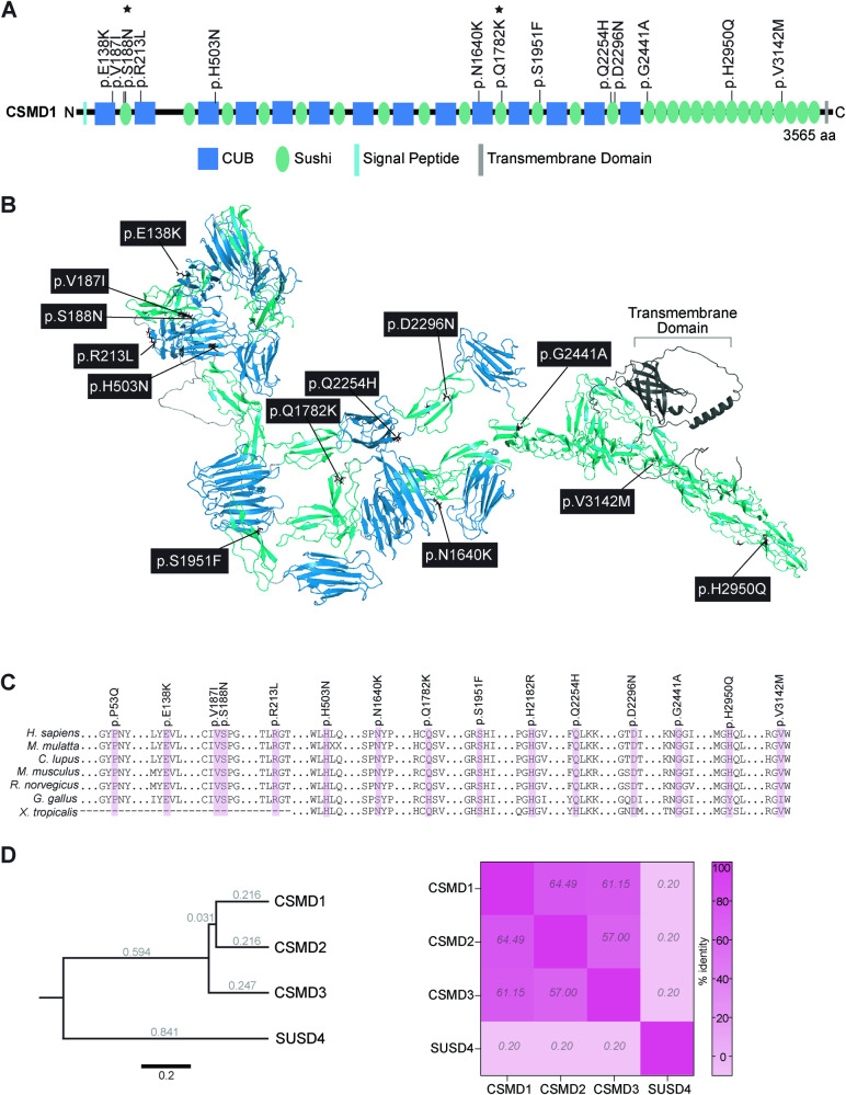
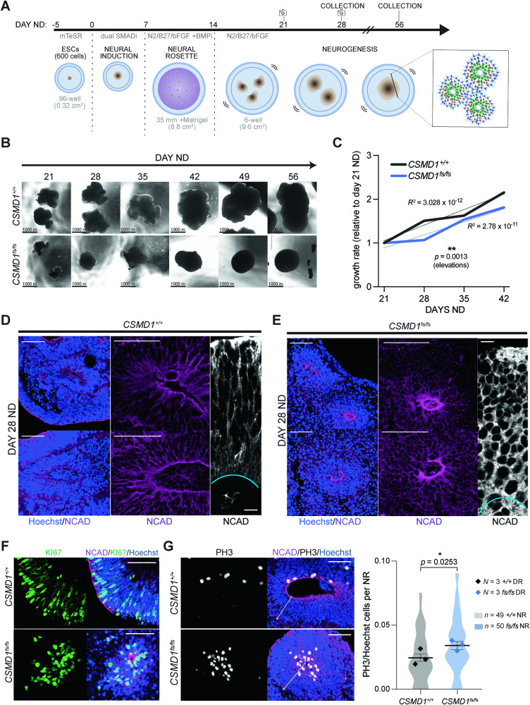
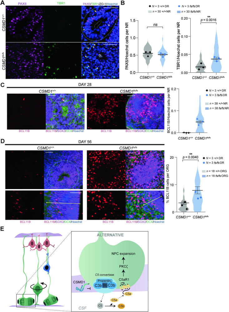
CSMD1 (Cub and Sushi Multiple Domains 1) is a well-recognized regulator of the complement cascade, an important component of the innate immune response. CSMD1 is highly expressed in the central nervous system (CNS) where emergent functions of the complement pathway modulate neural development and synaptic activity. While a genetic risk factor for neuropsychiatric disorders, the role of CSMD1 in neurodevelopmental disorders is unclear. Through international variant sharing, we identified inherited biallelic CSMD1 variants in eight individuals from six families of diverse ancestry who present with global developmental delay, intellectual disability, microcephaly, and polymicrogyria. We modeled CSMD1 loss-of-function (LOF) pathogenesis in early-stage forebrain organoids differentiated from CSMD1 knockout human embryonic stem cells (hESCs). We show that CSMD1 is necessary for neuroepithelial cytoarchitecture and synchronous differentiation. In summary, we identified a critical role for CSMD1 in brain development and biallelic CSMD1 variants as the molecular basis of a previously undefined neurodevelopmental disorder.


Conflict of interest statement

The authors declare no competing interests.

Figures

Fig. 1
Fig. 1. Families with inherited CSMD1 variants.
A Pedigree drawings of segregating NDD phenotypes in families 1-7, with generations listed on the left-hand side. Females are represented as circles and males are denoted by squares. Affected family members are indicated by solid black coloring while unaffected are unfilled. Consanguineous partnerships are represented by double lines. Sanger sequencing confirmation was performed on individuals in families 1 and 5 (chromatograms shown). B Sagittal (left) and T2 coronal (right) MRIs of P1 at 12 months old (right) relative to control (left). Arrowheads point to thin corpus callosum. Also note normal cerebellar vermis, widened lateral ventricles, and abnormal cortex, suggestive of polymicrogyria.
Fig. 2
Fig. 2. Modeling variant effect on CSMD1 protein.
A CSMD1 linear protein map with inherited missense variants identified in individuals with NDD. Stars denote inherited CSMD1 variants not included in our study cohort but previously published. All variants identified by exome and genome sequencing in affected individuals localize to the CUB (blue) and sushi (green) domains of CSMD1. B Model of CSMD1 protein structure with mapped clinical variants. Each Cub and Sushi domain was individually modeled using AlphaFold2, and then assembled using DeepAsembly to build the complete CSMD1 structure. Protein domains and variant residues were illustrated using color and licorice functions in PyMOL v2.5.2. C Species conservation of affected residues. D Rooted phylogenetic tree of CSMD family proteins and the complement regulator SUSD4 as an outgroup (left). Branches are labeled with genetic distance as measured by substitutions per site. Scale bar, 0.2 substitutions per site. Heatmap illustrating percent sequence identity of SUSD4 and CSMD family proteins to each other determined from protein alignment (right).
Fig. 3
Fig. 3. Characterization of neuroepithelium polarity and proliferation in CSMD1+/+ and CSMD1fs/fs forebrain organoids.
A Schematic of forebrain organoid differentiation, starting with 600 ESCs per well in 96-well V-bottom cell culture plates. Cross-sectional images were captured of live organoid tissue at days 21, 28, 35, and 42 ND. B Representative images across developmental time points by genotype of organoids growing in suspension, used for cross-section area growth analysis from day 21 ND to day 42 ND. C Growth rate determined by mean relative to day 21 ND, from day 21 ND to day 42 ND (right). N = 100-300 organoids from 3 independent differentiations (30–100 organoids per differentiation) per genotype per time point. Statistical significance determined using simple linear regression (elevations, p = 0.0013; slopes, p =>0.9999). Representative images of NRs across independent organoid replicates to demonstrate gross morphology (Hoescht, N-Cadherin) for CSMD1+/+ (D) and CSMD1fs/fs (E). Lower 20X magnification N-cadherin immunostaining showing organization of entire NR (left; scale bar 50 μm); higher magnification of NR (middle; scale bar 50 μm); NR radial segment (right; scale bar 10 μm). KI67 (F) and PH3 (G) immunostaining of day 28 NRs for each genotype with respective quantifications. Scale bars, 50 μm. Data are shown as points representing differentiation batch means (N = 3 per genotype) super-imposed on a violin plot of the distribution of individual NRs (CSMD1fs/fs n = 50; CSMD1+/+ n = 49). Error bars represent mean ± SEM for all NRs. Significance determined by unpaired two-tailed t test on all NR data (PH3/Hoechst, p = 0.0253). Variance, F-test (PH3/Hoechst, F = 1.328, p = 0.3268).
Fig. 4
Fig. 4. CSMD1fs/fs differentiation defects of deep layer cortical neurons in early corticogenesis.
A Immunostaining of NPCs (PAX6) and early born neurons (TBR1) with quantifications (B). Scale bars, 50 μm. Data are shown as points representing differentiation batch means (N = 3 per genotype) super-imposed on a violin plot of the distribution of individual NRs (n = 30 per genotype). Error bars represent mean ± SEM for all NRs. ns, not significant. Significance determined by: unpaired two-tailed t test for PAX6/Hoechst (p = 0.3524) and Mann-Whitney U test for TBR1/Hoechst (p = 0.0016). Variance, F-test (PAX6/Hoechst, F = 1.065, p = 0.8672; TBR1/Hoechst, F = 4.241, p = 0.0002). C Immunostaining of layer 5 excitatory neurons (BCL11B) in day 28 NR per genotype with quantifications (right). Scale bars, 50 μm. Data are shown as points representing differentiation batch means (N = 3 per genotype) super-imposed on a violin plot of the distribution of individual NRs (n = 38 per genotype). Error bars represent mean ± SEM for all NRs. Statistical significance was not assessed given the absence of BCL11B cells in control NRs. D BCL11B immunostaining in day 56 NRs (left) and percentage of BCL11B cells per whole organoids per genotype (right). Data are shown as points representing differentiation batch means (N = 3 per genotype) super-imposed on a violin plot of the distribution of individual organoids (n = 18 per genotype). Error bars represent mean ± SEM for all organoids. ORG, organoids. Statistical significance was assessed with Mann-Whitney U test (p = 0.0040) given the variance of the data (F = 4.498, p = 0.0034). E Illustration of proposed model of CSMD1 regulation of the alternative complement pathway in NPCs of the developing neuroepithelium via inhibition of C5 convertase functions. CSF, cerebrospinal fluid. Left: NPCs at the apical surface are green, migrating neurons are blue, and mature neurons at the cortical plate are red.

References

    1. Dalakas MC, Alexopoulos H, Spaeth PJ. Complement in neurological disorders and emerging complement-targeted therapeutics. Nat Rev Neurol. 2020;16:601–17. doi: 10.1038/s41582-020-0400-0. - DOI - PMC - PubMed
    1. Coulthard LG, Hawksworth OA, Woodruff TM. Complement: The Emerging Architect of the Developing Brain. Trends Neurosci. 2018;41:373–84. doi: 10.1016/j.tins.2018.03.009. - DOI - PubMed
    1. Jeanes A, Coulthard LG, Mantovani S, Markham K, Woodruff TM. Co-ordinated expression of innate immune molecules during mouse neurulation. Mol Immunol. 2015;68:253–60. doi: 10.1016/j.molimm.2015.09.004. - DOI - PubMed
    1. Costanzo F, Zanni G, Fucà E, Di Paola M, Barresi S, Travaglini L, et al. Cerebellar Agenesis and Bilateral Polimicrogyria Associated with Rare Variants of CUB and Sushi Multiple Domains 1 Gene (CSMD1): A Longitudinal Neuropsychological and Neuroradiological Case Study. Int J Environ Res Public Health. 2022;19. - PMC - PubMed
    1. Song W, Li Q, Wang T, Li Y, Fan T, Zhang J, et al. Putative complement control protein CSMD3 dysfunction impairs synaptogenesis and induces neurodevelopmental disorders. Brain Behav Immun. 2022;102:237–50. doi: 10.1016/j.bbi.2022.02.027. - DOI - PubMed

Publication types

LinkOut - more resources