. 2024 May 31;14(1):12486.
doi: 10.1038/s41598-024-63006-x.

Spectrum of MRI findings in central nervous system affection in Lyme neuroborreliosis

Affiliations

Spectrum of MRI findings in central nervous system affection in Lyme neuroborreliosis

T Volk et al. Sci Rep. .

Abstract

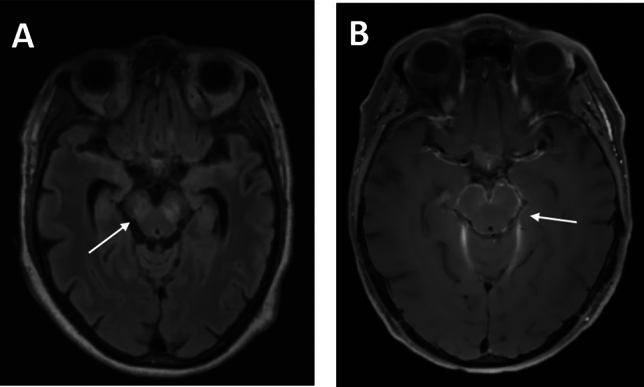
Affections of the central nervous system (CNS) rarely occur in Lyme neuroborreliosis (LNB). CNS manifestations can have residual neurological symptoms despite antibiotic treatment. We explored the spectrum of CNS affections in patients with LNB in a tertiary care center in a region endemic for Lyme borreliosis. We retrospectively included patients treated at a tertiary care center from January 2020-December 2021 fulfilling the case criteria for LNB as stated in the current German guideline on LNB. Clinical data, cerebrospinal fluid (CSF) findings and MRI imaging were collected. We included 35 patients with LNB, 24 with early manifestations and 11 with CNS-LNB. CNS-LNB patients had encephalomyelitis (n = 6) or cerebral vasculitis (n = 5). Patients with early LNB and CNS-LNB differed regarding albumin CSF/serum quotient and total protein in CSF. Duration from onset of symptoms until diagnosis was statistically significantly longer in patients with encephalomyelitis. MRI findings were heterogeneous and showed longitudinal extensive myelitis, perimedullar leptomeningeal enhancement, pontomesencephalic lesions or cerebral vasculitis. CNS-LNB can present with a variety of clinical syndromes and MRI changes. No clear pattern of MRI findings in CNS-LNB could be identified. The role of MRI consists in ruling out other causes of neurological symptoms.

Keywords: CSF; Cerebral MRI imaging; Encephalitis; Encephalomyelitis; Lyme disease; Lyme neuroborreliosis; Myelitis; Neuroinfectious diseases; Vasculitis.


Conflict of interest statement

The authors declare no competing interests.

Figures

Figure 1
Figure 1
Cerebral MRI in CNS-LNB patients with encephalitis. (A) bilateral T2-hyperintense mesencephalic lesions in a patient with LNB encephalitis (T2 FLAIR). (B) leptomeningeal contrast enhancement around the cerebral pedunculi as a sign of basal meningitis as well as signs of vessel wall inflammation (T1w with contrast agent). White arrows depict representative findings.
Figure 2
Figure 2
Cerebral MRI in CNS-LNB. (A) ‘Rounded M’-sign with T2-hyperintense pontine lesion in a patient with encephalomyelitis. (B) subtle ‘Tarsier-sign’ with T2-hypertintense mesencephalic lesion at the cerebral peduncles in the same patient. White arrows depict representative findings.
Figure 3
Figure 3
Spinal MRI in LNB patients with spinal affections. (A) longitudinal extensive transverse myelitis (LETM) of the cervical myelon (T2) (B1/2) perimedullar and radicular leptomeningeal enhancement (T1 fat sat with contrast agent) (C) LETM with hyperintense alterations of spinal grey matter (‘H’-sign) in a patient with concomitant CNS vasculitis (T2). White arrows depict representative findings.
Figure 4
Figure 4
Cerebral MRI and MRI-angiography in CNS-LNB patients with cerebral vasculitis (Table 2, patient 8) (A1) ischemic lesion in the left MCA territory of the middle cerebral artery (MCA) (diffusion-weighted imaging). (A2) concomitant stenosis of the distal M1-segment as seen in the time-of-flight (TOF) angiography. (A3/4) correspondant contrast enhancement in vessel-wall imaging showing extensive vessel wall inflammation (T1w with contrast agent). (B) sagittal reconstruction showing concentric vessel-wall enhancement of the MCA in a patient with basal meningitis and vasculitis (Figure 1, T1w with contrast agent, Table 2: patient 7). (C1) scattered ischemic lesions in the right MCA territory (diffusion weighted imaging) (C2) concomitant stenosis of the distal M1-segment as seen in the time-of-flight (TOF) angiography. (C3) correspondant contrast enhancement in vessel-wall imaging showing local vessel wall inflammation (T1w with contrast agent, Table 2: patient 10). Each letters corresponds to one patient. White arrows depict representative findings.

References

    1. Burn L, Tran TMP, Pilz A, Vyse A, Fletcher MA, Angulo FJ, et al. Incidence of lyme borreliosis in Europe from National Surveillance Systems (2005–2020) Vector Borne Zoonotic Dis. 2023;23(4):156–171. doi: 10.1089/vbz.2022.0071. - DOI - PMC - PubMed
    1. Enkelmann J, Böhmer M, Fingerle V, Siffczyk C, Werber D, Littmann M, et al. Incidence of notified Lyme borreliosis in Germany, 2013–2017. Sci. Rep. 2018;8(1):14976. doi: 10.1038/s41598-018-33136-0. - DOI - PMC - PubMed
    1. Koedel U, Fingerle V, Pfister HW. Lyme neuroborreliosis—Epidemiology, diagnosis and management. Nat. Rev. Neurol. 2015;11(8):446–456. doi: 10.1038/nrneurol.2015.121. - DOI - PubMed
    1. Mygland A, Ljøstad U, Fingerle V, Rupprecht T, Schmutzhard E, Steiner I, et al. EFNS guidelines on the diagnosis and management of European Lyme neuroborreliosis. Eur. J. Neurol. 2010;17(1):8–16. doi: 10.1111/j.1468-1331.2009.02862.x. - DOI - PubMed
    1. van Samkar A, Bruinsma RA, Vermeeren YM, Wieberdink RG, van Bemmel T, den Reijer PM, et al. Clinical characteristics of Lyme neuroborreliosis in Dutch children and adults. Eur. J. Pediatr. 2023;182(3):1183–1189. doi: 10.1007/s00431-022-04749-5. - DOI - PubMed