

Equivalence study of the resin-dentine interface of internal tunnel restorations when using an enamel infiltrant resin with ethanol-wet dentine bonding
- PMID: 38816512
- PMCID: PMC11139992
- DOI: 10.1038/s41598-024-63289-0
Equivalence study of the resin-dentine interface of internal tunnel restorations when using an enamel infiltrant resin with ethanol-wet dentine bonding
Abstract
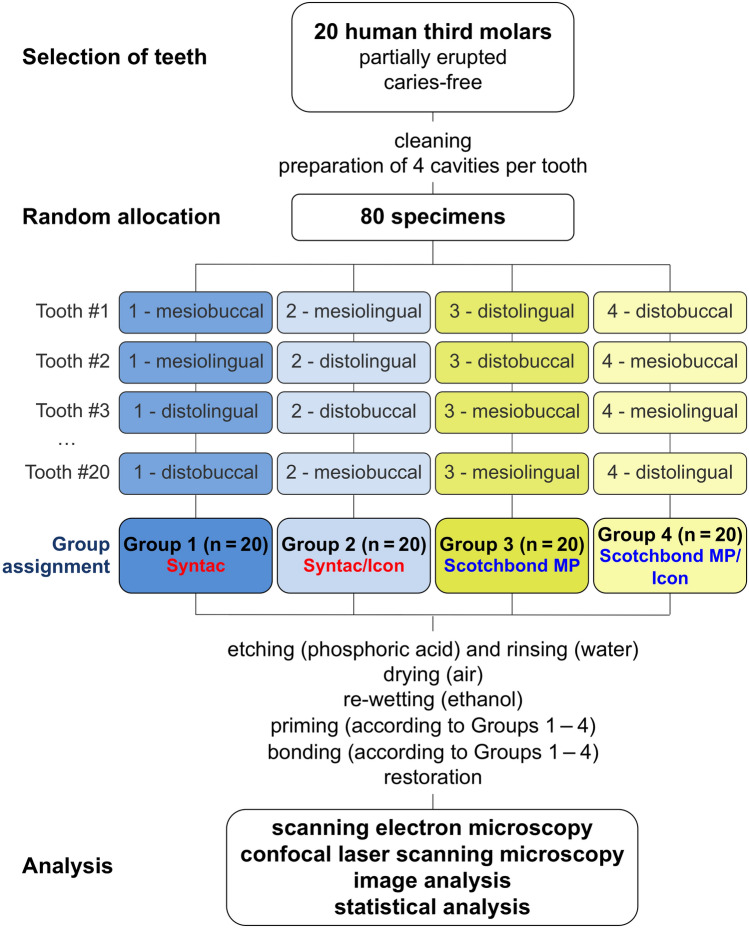
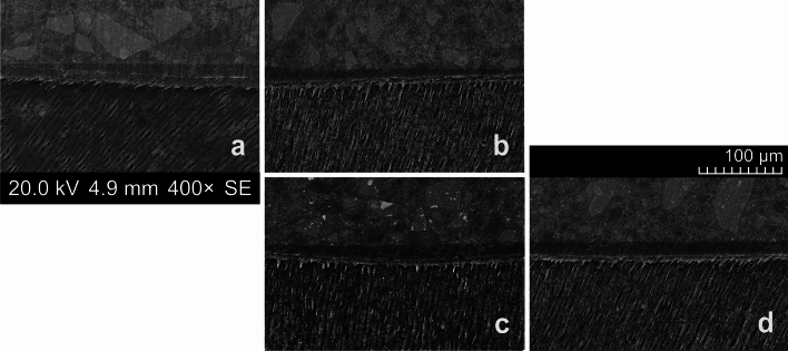
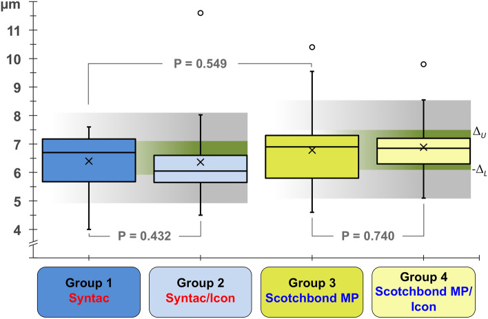
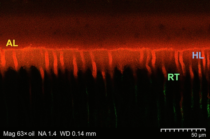
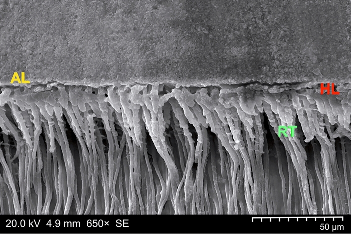
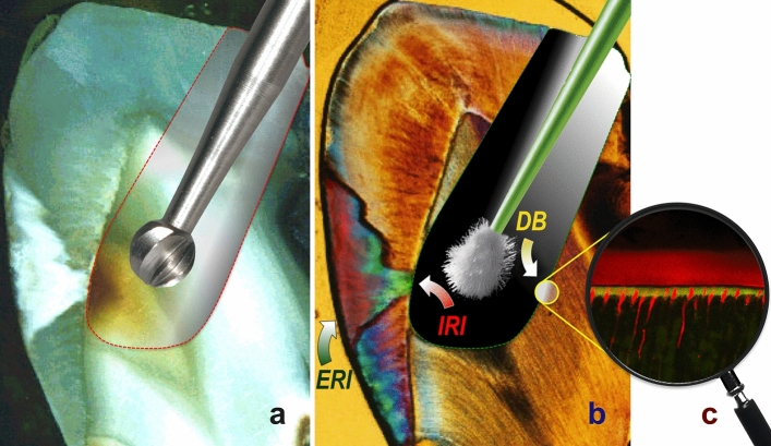
This preregistered ex vivo investigation examined the dentinal hybrid layer formation of a resinous infiltrant (Icon), with reference to both thickness (HLT) and homogeneity when combined with modified tunnel preparation (occlusal cavity only) and internal/external caries infiltration. The adhesives Syntac and Scotchbond MP were used as controls (Groups 1 and 3) or in combination with Icon (Groups 2 and 4). A split-tooth design using healthy third molars from 20 donors resulted in 20 prepared dentine cavities per experimental group. The cavity surfaces (n = 80) were etched (37% H3PO4), rinsed, and air-dried. Rewetting with ethanol was followed by application of the respective primers. After labeling with fluorescent dyes, either Syntac Adhesive/Heliobond or Scotchbond MP Adhesive was used alone or supplemented with Icon. HLT, as evaluated by scanning electron microscopy, did not significantly differ (P > 0.05), and confocal laser scanning microscopy revealed homogeneously mixed/polymerized resin-dentine interdiffusion zones in all groups. Icon can be successfully integrated into an ethanol-wet dentine bonding strategy, and will result in compact and homogeneous hybrid layers of comparable thickness considered equivalent to the non-Icon controls, thus allowing for preservation of the tooth's marginal ridge and interdental space in the case of internal/external infiltration of proximal caries.
Keywords: Confocal laser scanning microscopy; Dental adhesives; Dentine bonding; Ethanol-wet bonding; Hybrid layer; Icon; Resin infiltration; Scanning electron microscopy; Scotchbond multi-purpose; Split-tooth design; Syntac.
© 2024. The Author(s).
Conflict of interest statement
A.M.K. is named as inventor in Brazilian, Canadian, Chinese, European, Indian, Japanese, Korean, Russian, Swiss, U.K., and U.S. patents (held by Charité – Universitätsmedizin Berlin, Germany) for the resin infiltration of initially carious enamel (“Method and means for infiltrating enamel lesions”, Patent Number: EP 1854445 A1). These patents have been licensed by DMG (Hamburg, Germany), and A.M.K. receives patent royalties from this license, disbursed by Charité – Universitätsmedizin Berlin. Furthermore, A.M.K. and E.L. are Editorial Board Members and Academic Editors of Scientific Reports; according to the Journal’s recusal process, they were involved neither in the peer review nor in the editorial decision-making process for the present paper. S.S. and W.F. declare that they have no competing interests. This investigation will be part of J.-S.B.’s final examination requirements, and will serve as the basis for her diploma thesis.
Figures
Similar articles
-
Ex vivo investigation on internal tunnel approach/internal resin infiltration and external nanosilver-modified resin infiltration of proximal caries exceeding into dentin.PLoS One. 2020 Jan 28;15(1):e0228249. doi: 10.1371/journal.pone.0228249. eCollection 2020. PLoS One. 2020. PMID: 31990942 Free PMC article.
-
Adjunctive application of chlorhexidine and ethanol-wet bonding on durability of bonds to sound and caries-affected dentine.J Dent. 2014 Jun;42(6):709-19. doi: 10.1016/j.jdent.2014.04.001. Epub 2014 Apr 13. J Dent. 2014. PMID: 24732576
-
Effect of etching agent on dentinal adhesive interface in primary teeth.J Clin Pediatr Dent. 2000 Spring;24(3):205-9. J Clin Pediatr Dent. 2000. PMID: 11314144
-
Microscopic features of clinically successful dentine bonding.Dent Update. 1998 Sep;25(7):281-6. Dent Update. 1998. PMID: 10478022 Review.
-
Current perspectives on dental adhesion: (2) Concepts for operatively managing carious lesions extending into dentine using bioactive and adhesive direct restorative materials.Jpn Dent Sci Rev. 2020 Nov;56(1):208-215. doi: 10.1016/j.jdsr.2020.08.003. Epub 2020 Sep 20. Jpn Dent Sci Rev. 2020. PMID: 32983288 Free PMC article. Review.
Cited by
-
Influence of chlorhexidine dentin disinfection on universal adhesive performance: Interfacial adaptation and bond strength assessments.PLoS One. 2024 Dec 31;19(12):e0315036. doi: 10.1371/journal.pone.0315036. eCollection 2024. PLoS One. 2024. PMID: 39739701 Free PMC article.
-
Diagnostic accuracy of dental caries detection using ensemble techniques in deep learning with intraoral camera images.PLoS One. 2024 Sep 6;19(9):e0310004. doi: 10.1371/journal.pone.0310004. eCollection 2024. PLoS One. 2024. PMID: 39241044 Free PMC article.
References
-
- Kielbassa AM, Ulrich I, Treven L, Mueller J. An updated review on the resin infiltration technique of incipient proximal enamel lesions. Med. Evol. 2010;16:3–15. doi: 10.13140/RG.2.2.36646.37443. - DOI
-
- Kielbassa AM, Müller J, Gernhardt CR. Closing the gap between oral hygiene and minimally invasive dentistry: A review on the resin infiltration technique of incipient (proximal) enamel lesions. Quintessence Int. 2009;40:663–681. - PubMed
MeSH terms
Substances
LinkOut - more resources
Full Text Sources
