

Single cell RNA analysis uncovers the cell differentiation and functionalization for air breathing of frog lung
- PMID: 38816547
- PMCID: PMC11139932
- DOI: 10.1038/s42003-024-06369-1
Single cell RNA analysis uncovers the cell differentiation and functionalization for air breathing of frog lung
Abstract
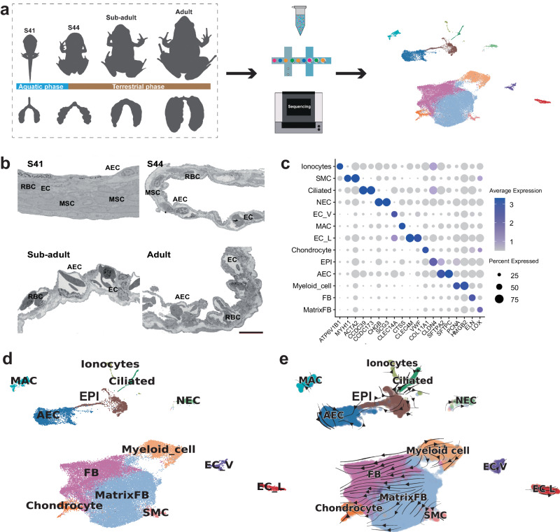
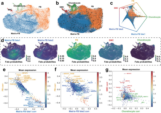
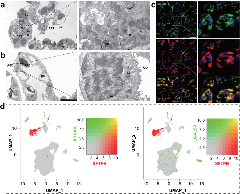
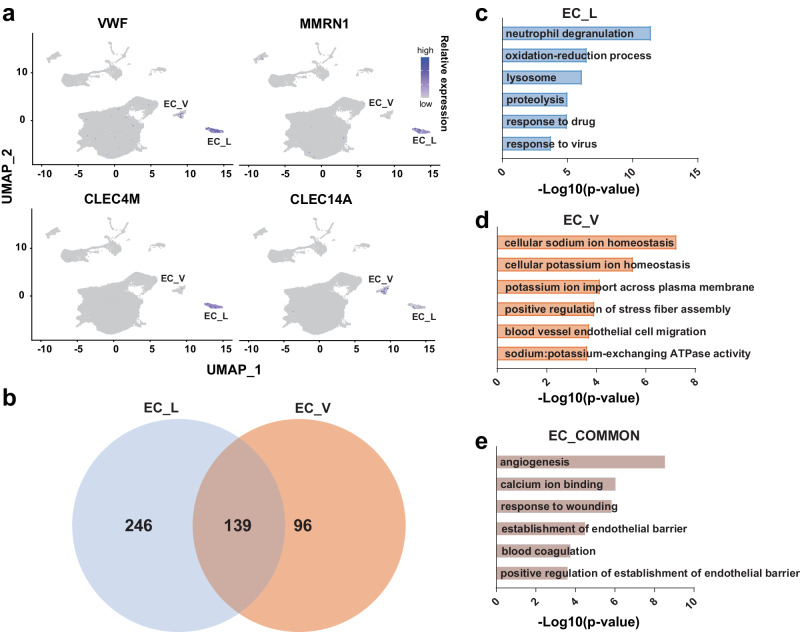
The evolution and development of vertebrate lungs have been widely studied due to their significance in terrestrial adaptation. Amphibians possess the most primitive lungs among tetrapods, underscoring their evolutionary importance in bridging the transition from aquatic to terrestrial life. However, the intricate process of cell differentiation during amphibian lung development remains poorly understood. Using single-cell RNA sequencing, we identify 13 cell types in the developing lungs of a land-dwelling frog (Microhyla fissipes). We elucidate the differentiation trajectories and mechanisms of mesenchymal cells, identifying five cell fates and their respective driver genes. Using temporal dynamics analyses, we reveal the gene expression switches of epithelial cells, which facilitate air breathing during metamorphosis. Furthermore, by integrating the published data from another amphibian and two terrestrial mammals, we illuminate both conserved and divergent cellular repertoires during the evolution of tetrapod lungs. These findings uncover the frog lung cell differentiation trajectories and functionalization for breathing in air and provide valuable insights into the cell-type evolution of vertebrate lungs.
© 2024. The Author(s).
Conflict of interest statement
The authors declare no competing interests.
Figures

Similar articles
-
Gene Expression Program Underlying Tail Resorption During Thyroid Hormone-Dependent Metamorphosis of the Ornamented Pygmy Frog Microhyla fissipes.Front Endocrinol (Lausanne). 2019 Jan 25;10:11. doi: 10.3389/fendo.2019.00011. eCollection 2019. Front Endocrinol (Lausanne). 2019. PMID: 30740088 Free PMC article.
-
From Water to Land: The Structural Construction and Molecular Switches in Lungs during Metamorphosis of Microhyla fissipes.Biology (Basel). 2022 Mar 30;11(4):528. doi: 10.3390/biology11040528. Biology (Basel). 2022. PMID: 35453728 Free PMC article.
-
Osteological development of a small and fast metamorphic frog, Microhyla fissipes (Anura, Neobatrachia, Microhylidae).J Anat. 2021 Dec;239(6):1318-1335. doi: 10.1111/joa.13510. Epub 2021 Jul 15. J Anat. 2021. PMID: 34268788 Free PMC article.
-
Molecular mechanism and evolutional significance of epithelial-mesenchymal interactions in the body- and tail-dependent metamorphic transformation of anuran larval skin.Int Rev Cytol. 2007;260:213-60. doi: 10.1016/S0074-7696(06)60005-3. Int Rev Cytol. 2007. PMID: 17482907 Review.
-
Fundamental structural aspects and features in the bioengineering of the gas exchangers: comparative perspectives.Adv Anat Embryol Cell Biol. 2002;163:III-XII, 1-108. doi: 10.1007/978-3-642-55917-4. Adv Anat Embryol Cell Biol. 2002. PMID: 11892241 Review.
Cited by
-
Evolution, Diversity, and Conservation of Herpetofauna.Animals (Basel). 2024 Jul 7;14(13):2004. doi: 10.3390/ani14132004. Animals (Basel). 2024. PMID: 38998116 Free PMC article.
-
Cellular and Molecular Basis of Environment-Induced Color Change in a Tree Frog.Animals (Basel). 2024 Dec 1;14(23):3472. doi: 10.3390/ani14233472. Animals (Basel). 2024. PMID: 39682437 Free PMC article.
References
-
- Burggren WW, Infantino RL. JR. The respiratory transition from water to air breathing during amphibian metamorphosis. Am. Zool. 2015;34:238–246. doi: 10.1093/icb/34.2.238. - DOI
Publication types
MeSH terms
Grants and funding
LinkOut - more resources
Full Text Sources
