. 2024 May 30;43(1):153.
doi: 10.1186/s13046-024-03066-z.

Placental growth factor promotes neural invasion and predicts disease prognosis in resectable pancreatic cancer

Affiliations

Placental growth factor promotes neural invasion and predicts disease prognosis in resectable pancreatic cancer

Andreas Göhrig et al. J Exp Clin Cancer Res. .

Abstract

Background: Surgery represents the only curative treatment option for pancreatic ductal adenocarcinoma (PDAC), but recurrence in more than 85% of patients limits the success of curative-intent tumor resection. Neural invasion (NI), particularly the spread of tumor cells along nerves into extratumoral regions of the pancreas, constitutes a well-recognized risk factor for recurrence. Hence, monitoring and therapeutic targeting of NI offer the potential to stratify recurrence risk and improve recurrence-free survival. Based on the evolutionary conserved dual function of axon and vessel guidance molecules, we hypothesize that the proangiogenic vessel guidance factor placental growth factor (PlGF) fosters NI. To test this hypothesis, we correlated PlGF with NI in PDAC patient samples and functionally assessed its role for the interaction of tumor cells with nerves.

Methods: Serum levels of PlGF and its soluble receptor sFlt1, and expression of PlGF mRNA transcripts in tumor tissues were determined by ELISA or qPCR in a retrospective discovery and a prospective validation cohort. Free circulating PlGF was calculated from the ratio PlGF/sFlt1. Incidence and extent of NI were quantified based on histomorphometric measurements and separately assessed for intratumoral and extratumoral nerves. PlGF function on reciprocal chemoattraction and directed neurite outgrowth was evaluated in co-cultures of PDAC cells with primary dorsal-root-ganglia neurons or Schwann cells using blocking anti-PlGF antibodies.

Results: Elevated circulating levels of free PlGF correlated with NI and shorter overall survival in patients with PDAC qualifying for curative-intent surgery. Furthermore, high tissue PlGF mRNA transcript levels in patients undergoing curative-intent surgery correlated with a higher incidence and greater extent of NI spreading to tumor-distant extratumoral nerves. In turn, more abundant extratumoral NI predicted shorter disease-free and overall survival. Experimentally, PlGF facilitated directional and dynamic changes in neurite outgrowth of primary dorsal-root-ganglia neurons upon exposure to PDAC derived guidance and growth factors and supported mutual chemoattraction of tumor cells with neurons and Schwann cells.

Conclusion: Our translational results highlight PlGF as an axon guidance factor, which fosters neurite outgrowth and attracts tumor cells towards nerves. Hence, PlGF represents a promising circulating biomarker of NI and potential therapeutic target to improve the clinical outcome for patients with resectable PDAC.

Keywords: Axon guidance molecule; Cancer neuroscience; Nerves; Neural invasion; Pancreatic cancer; Placental growth factor (PlGF).


Conflict of interest statement

CF has a patent pending related to the content of this work. All other authors disclose no potential conflicts of interest.

Figures

Fig. 1
Fig. 1
High circulating PlGF/sFlt1 serum ratio is associated with neural invasion and shorter survival in patients with PDAC undergoing curative-intent surgery. A PlGF/sFlt1circ is elevated in PDAC patients (n = 73) compared to healthy controls (ctr; n = 79). B PlGF/sFlt1circ does not differ between patients with non-resectable (locally advanced or metastatic tumors, PT; n = 37) and patients receiving curative-intent surgery (CS; n = 36). C In patients receiving curative-intent surgery, those with evidence of NI (Pn1; n = 20) exhibited higher PlGF/sFlt1circ than patients without NI (Pn0; n = 16). D and E Comparable PlGF/sFlt1circ ratios in patients with (D N1-2, n = 27) or without (N0; n = 9) lymph node metastasis and in patients with R0 (E n = 24) versus R1 resection (n = 12). F and G In Kaplan–Meier estimates PlGF/sFlt1circ above cut-off correlates with shorter OS in patients with resectable PDAC (F HR: 4.05; 95% confidence interval: 1.22 to 13.48; Log-rank p = 0.007; n = 17 below and n = 19 above cut-off), but not with palliative disease (G HR: 0.845; 95% confidence interval: 0.33 to 2.19; p = 0.696; n = 20 below and n = 17 above cut-off). H Tumor-related pain was quantified using visual analogue scales (VAS 0–10) and grouped into no or low (0–3, n = 20), or moderate to strong pain (4–10, n = 16). Shown are PlGF/sFlt1circ for the curative surgery cohort of patients. A-E and H scatter dot plots with median and interquartile range. *, P < 0.05; ****, P < 0.0001; ns, not significant
Fig. 2
Fig. 2
Circulating PlGF/sFlt1 ratio predicts neural invasion in a prospective cohort of PDAC patients undergoing curative-intent surgery. A PlGF/sFlt1circ is elevated in patients with NI (n = 32) compared to patients without NI (n = 9). B-D PlGF/sFlt1circ does not correlate with either incidence (B) or extent (C) of lymph node metastasis (B n = 12 for N0 and n = 29 for N1-2; C n = 22 for below and n = 19 for above median); or with R0 versus R1 resection margins (D n = 28 for R0, n = 13 for R1). **, P < 0.01; ns, not significant
Fig. 3
Fig. 3
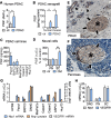
PlGF and its receptors are expressed at the tumor-nerve interface. A PlGF mRNA-transcript expression in human PDAC (n = 13) and healthy pancreas (ctr, n = 9). B Human (representing tumor cell derived) and murine (representing host derived) PlGF proteins in DANG orthotopic xenograft tumors (PDAC) and paired pancreas (ctr) determined using species-specific ELISA. C ELISA-based quantification of PlGF in supernatants of human PDAC cell line cultures (n = 3). D Primary neurons (PN) and Schwann cells (SC) from newborn mice were cultivated with control media (ctr) or conditioned media (CM) from MiaPaCa tumor cell cultures and PlGF mRNA expression determined (n = 3). Tumor cell conditioned supernatants induce PlGF expression in Schwann cells. E and F Representative IHC for the PlGF receptor Nrp1 in tissues of PDAC (E) and healthy pancreas (F). Intratumoral (in E) and intrapancreatic (in F) nerves are indicated by asterisks. Nrp1 expression in ductal epithelial cancer cells (arrowheads) and nerves (arrows). G and H mRNA transcripts and protein expression of Nrp1 and VEGFR1, respectively, in (G) human PDAC cell lines, the immortalized human pancreatic ductal epithelial cell line HPDE, as well as in (H) dorsal root ganglia (DRG), primary neurons (PN) and Schwann cells (SC; n = 3). *, P < 0.05; **, P < 0.01
Fig. 4
Fig. 4
Morphologic parameters of neural invasion and plasticity: Neural invasion of extratumoral nerves predicts early disease recurrence and overall survival. A Representative IHC images and magnifications of intratumoral (A1 and A2) and extratumoral NI (A3-A5; scale bars, 200 μm). Shown are perineural invasion (PNI; open arrows) and intraneural invasion (INI; filled arrows) of intratumoral (in A1 and A2) and extratumoral (intrapancreatic in A3; extrapancreatic in A4 and A5) nerves (asterisks). B Illustration summarizing morphologic parameters of neural plasticity and invasion (created with BioRender.com). Neural plasticity is quantified by number, caliber and area of intra-/extratumoral nerves (middle panel). Neural invasion is assessed by presence, localization (perineural vs. intraneural) and extent (circumference; score 1–4) of tumor cells within the neural space (right panel). C Incidence of tumors with (NI +) or without (NI-) neural invasion (n = 20; p = 0.19, Fisher’s exact test) in PDAC specimens (intratumoral) and corresponding adjacent healthy pancreas (extratumoral). D and E Data on DSF and OS was available for n = 17 patients. A high neural dissemination score of extratumoral nerves is associated with shorter DFS (D) and OS (E) in patients receiving curative-intent surgery. Kaplan–Meier estimates depict DSF and OS of patients with extensive neural invasion of extratumoral nerves (score 2; n = 6) versus absent or focal neural invasion (score 0 and 1, respectively; n = 11; HR: 4.57; 95% confidence interval: 1.09 to 19.22; Log-rank p = 0.0038 in D HR: 4.63; 95% confidence interval: 1.00 to 21.52; Log-rank p = 0.0004 in E). F Percentage of tumor-invaded nerves per total nerves in PDAC specimens (intratumoral) and adjacent healthy pancreas (extratumoral). G and H High fraction of invaded extratumoral nerves is associated with shorter DFS (G) and OS (H) in patients receiving curative-intent surgery. Kaplan–Meier estimates depict DSF and OS of patients with extratumoral nerve fractions above (n = 8) or below (n = 9) median (HR: 3.29; 95% confidence interval: 0.95 to 11.39; Log-rank p = 0.0210 in G; HR: 3.93; 95% confidence interval: 1.06 to 14.58; Log-rank p = 0.0013 in H). **, P < 0.01
Fig. 5
Fig. 5
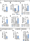
PlGF mRNA transcript levels correlate with the extent of neural invasion of extratumoral nerves. A-I Analyses refer to n = 20 PDAC samples that allowed for examination of extratumoral nerves. A PlGF mRNA transcript levels dependent on presence (NI +) or absence of neural invasion (NI-). B Incidence of NI in tumors with PlGF mRNA transcripts < median and > median. C Semiquantitative assessment of the NI dissemination as extensive (score 2), focal (score 1) or absent (score 0) in tumors with PlGF mRNA transcripts < median and > median. D PlGF mRNA transcript levels in tumors without (PNI absent) or with perineural invasion (PNI present). E Circumferential range of PNI was morphometrically determined and scored as 0 (absent), 1 (1/4 circumference), 2 (1/2 circumference), 3 (3/4 circumference), and 4 (whole circumference). Shown are PNI scores of affected nerves in tumors with PlGF mRNA transcripts < median and > median. F The PNI area fraction was determined by calculating the ratio of the PNI tumor cell area and the area of the corresponding nerve. G PlGF mRNA transcripts in tumors without (L0) and with (L1) lymphangioinvasion. H and I, Comparable PlGF mRNA expression in tumors with (N1-2) and without (N0) lymphatic metastasis (H) and, conversely, similar fractions of tumor infiltrated lymph nodes per total lymph nodes in tumors with PlGF mRNA transcripts < median and > median (I). *, P < 0.05; **, P < 0.01; ns, not significant
Fig. 6
Fig. 6
PlGF mediates mutual chemoattraction between tumor cells and Schwann cells and stimulates neurite outgrowth. A Neutralizing anti-PlGF antibodies inhibit directed migration of Schwann cells from the upper transwell chamber towards conditioned media from DANG or Panc1 monolayers (lower chamber) as compared to IgG1 control (n = 3). B Anti-PlGF inhibits directed migration of Panc1 and Capan-2 cells towards chemoattractant stimuli from conditioned Schwann cell supernatants placed in the lower chamber (n = 3). C and D Whole primary DRGs (containing neurons and Schwann cells) from newborn mice were incubated with supernatants from various PDAC cells (C) or medium containing recombinant nerve growth factor (NGF), glial-derived nerve growth factor (GDNF) and PlGF (D). Overall neurite length was determined using NeuroQuant® software based on selective staining of primary neurons for neuron-specific β3-tubulin (n = 3–5). PlGF stimulates neurite outgrowth, whereas neutralizing antibodies to PlGF secreted by DANG cells inhibit neurite outgrowth. EG Representative images of β3-tubulin stained primary neurons cultured with control media (E), DANG supernatant (F) and DANG supernatant with anti-PlGF (G). *, P < 0.05
Fig. 7
Fig. 7
Chemotherapy induces PlGF expression within the neural compartment of PDAC. A Treatment of mice bearing orthotopic DANG tumors with the chemotherapeutic agent gemcitabine induces PlGF production by tumor epithelial cells (human) and stromal cells (mouse) as determined using species-specific ELISA. B Conditioned tumor cell supernatant (CM) and gemcitabine dose-dependently induce PlGF expression in Schwann cells (n = 3–5). *,P < 0.05
Fig. 8
Fig. 8
Cartoon summarizing the proposed model for the role of PlGF in neural invasion in PDAC. PlGF is expressed in various stromal cell types within the desmoplastic microenvironment and in tumor cells. Released PlGF then binds to and activates its corresponding receptors VEGFR1 and NRP1, which exhibit differential expression: NRP1 receptors are present on tumor cells, neurons and Schwann cells. Neurons and Schwann cells additionally express VEGFR1. PlGF-mediated activation of VEGFR1 and NRP1 stimulates the mutual attraction between tumor cells and neurons as well as Schwann cells, thereby supporting directed neurite outgrowth towards tumor cells and neural invasion. By directed migration tumor cells move along nerves from intratumoral into extratumoral regions of the adjacent normal pancreas thereby escaping curative-intent surgery. Chemotherapy induces PlGF production within the tumor microenvironment, thus creating a PlGF-rich tumor supportive niche which in turn may support neural invasion. Free circulating PlGF levels correlate with the presence of neural invasion and with the fraction of invaded nerves, providing a non-invasive biomarker of neural invasion. In addition, therapeutic targeting of PlGF is feasible and might be exploited to counteract neural invasion, and thereby reduce recurrence rates following curative-intent surgery. Created with BioRender

References

    1. Rawla P, Sunkara T, Gaduputi V. Epidemiology of pancreatic cancer: global trends, etiology and risk factors. World J Oncol. 2019;10(1):10–27. doi: 10.14740/wjon1166. - DOI - PMC - PubMed
    1. Collisson EA, Bailey P, Chang DK, Biankin AV. Molecular subtypes of pancreatic cancer. Nat Rev Gastroenterol Hepatol. 2019;16(4):207–220. doi: 10.1038/s41575-019-0109-y. - DOI - PubMed
    1. Mizrahi JD, Surana R, Valle JW, Shroff RT. Pancreatic cancer. Lancet. 2020;395(10242):2008–2020. doi: 10.1016/S0140-6736(20)30974-0. - DOI - PubMed
    1. Wood LD, Canto MI, Jaffee EM, Simeone DM. Pancreatic cancer: pathogenesis, screening, diagnosis, and treatment. Gastroenterology. 2022;163(2):386–402. doi: 10.1053/j.gastro.2022.03.056. - DOI - PMC - PubMed
    1. Bapat AA, Hostetter G, Von Hoff DD, Han H. Perineural invasion and associated pain in pancreatic cancer. Nat Rev Cancer. 2011;11(10):695–707. doi: 10.1038/nrc3131. - DOI - PubMed