Review
. 2024 May 30;22(1):520.
doi: 10.1186/s12967-024-05347-9.

Boosting antitumor efficacy using docetaxel-loaded nanoplatforms: from cancer therapy to regenerative medicine approaches

Affiliations
Review

Boosting antitumor efficacy using docetaxel-loaded nanoplatforms: from cancer therapy to regenerative medicine approaches

Nima Beheshtizadeh et al. J Transl Med. .

Abstract

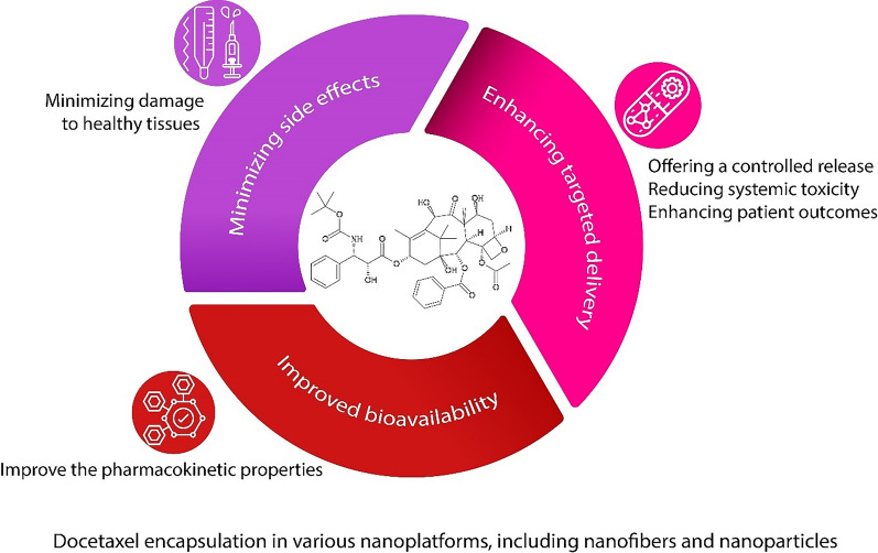
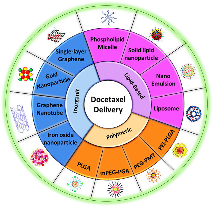
The intersection of nanotechnology and pharmacology has revolutionized the delivery and efficacy of chemotherapeutic agents, notably docetaxel, a key drug in cancer treatment. Traditionally limited by poor solubility and significant side effects, docetaxel's therapeutic potential has been significantly enhanced through its incorporation into nanoplatforms, such as nanofibers and nanoparticles. This advancement offers targeted delivery, controlled release, and improved bioavailability, dramatically reducing systemic toxicity and enhancing patient outcomes. Nanofibers provide a versatile scaffold for the controlled release of docetaxel, utilizing techniques like electrospinning to tailor drug release profiles. Nanoparticles, on the other hand, enable precise drug delivery to tumor cells, minimizing damage to healthy tissues through sophisticated encapsulation methods such as nanoprecipitation and emulsion. These nanotechnologies not only improve the pharmacokinetic properties of docetaxel but also open new avenues in regenerative medicine by facilitating targeted therapy and cellular regeneration. This narrative review highlights the transformative impact of docetaxel-loaded nanoplatforms in oncology and beyond, showcasing the potential of nanotechnology to overcome the limitations of traditional chemotherapy and pave the way for future innovations in drug delivery and regenerative therapies. Through these advancements, nanotechnology promises a new era of precision medicine, enhancing the efficacy of cancer treatments while minimizing adverse effects.

Keywords: Antitumor activity; Docetaxel-loaded nanoplatforms; Regenerative medicine; Tissue engineering.


Conflict of interest statement

The authors declare that they have no competing interests.

Figures

Fig. 1
Fig. 1
A schematic diagram of encapsulation of DTX in nanoplatforms, including nanoparticles and nanofibers, and their advantages
Fig. 2
Fig. 2
Schematic illustration of drug loaded Electrospun fibers. A Blend electrospinning involves co-solving pharmaceuticals and polymers in solutions before spinning, B dual concentric nozzles are used in co-axial electrospinning to spin distinct medicament and polymer mixtures, C Emulsion electrospinning involves the emulsification of medication solutions into insoluble polymer solutions, which is subsequently followed by the spinning process, D Following the immobilization process, drugs are attached to artificially created nanofiber matrices through either physical or chemical interactions
Fig. 3
Fig. 3
Depiction of RGD_PLGA Nanoparticles with DTX. A Schematic of PLGA-NPs. B In vivo effect of PLGA-NP on tumor diameter at the end of the experiment. *p < 0.05; **p < 0.01; two-way ANOVA. A study of the MRI and histological characteristics of 4T1 tumors obtained from mice. C Representative axial T2w-MR images of tumor region in mice with RGD_PLGA, Ctrl_ PLGA, free DTX, or physiological saline solution as a control (N = 6 per group). D Representative tumors from four treatment groups were histologically stained with hematoxylin/eosin. Necrotic areas are indicated by red arrows (Reprinted with permission from [77])
Fig. 4
Fig. 4
Folate-PEG-DTX on a Nanoscale. A Depicts the structure of the DTX-PEG micelle. B Illustrates the release behavior of DTX at 37 °C using DTX encapsulated in FA-PEG-DTX and PEG-DTX formulations in both citrate buffer (pH = 5.4) and phosphate buffer (pH = 7.4). C Shows the cytotoxic impact on the 4T1 cell line following 48 h of exposure to DTX encapsulated in FA-PEG-DTX, DTX encapsulated in PEG-DTX, and unencapsulated DTX. D Details the comparative in vivo efficacy of various formulations on tumor size reduction in BALB/c mice implanted with 4T1 tumors (n = 4) (Reprinted with permission [78])
Fig. 5
Fig. 5
DTX-loaded solid lipid nanoparticles Solid lipid nanoparticles encapsulating DTX. A The composition of SLN-DTX. B Release pattern of SLN-DTX at neutral (pH 7.4) and acidic (pH 5.0) conditions in PBS over a period of 10 days, presented as average ± standard error (****p < 0.0001). C Changes in tumor size in Balb/c mice bearing 4T1 tumors treated with SLN-DTX, DTX, Blank-SLN, and PBS, with significant distinctions noted against the PBS group (***p < 0.001; ****p < 0.0001). D Lung image. E Statistical evaluation of lung tumor nodules. F Microscopic examination of lung tissue, highlighting metastatic locations with black arrows and red dotted lines. Averages ± standard error are provided (**p < 0.01; ****p < 0.0001) (Reprinted with permission from [80])
Fig. 6
Fig. 6
DTX encapsulated in trastuzumab-coated nanoparticles. A Depicts eTmab-PPLNs and their uptake by cells. B Demonstrates the cytotoxic effects on BT474 cells of various treatments: DTX solution, Tmab, PPLNs, pTmab-PPLNs, and eTmab-PPLNs. C Shows CLSM (Confocal Laser Scanning Microscopy) visuals of BT474 cells after incubation with fluorescently tagged PPLNs, eTmab-PPLNs, or a combination of Tmab and eTmab-PPLNs. Here, the mixture of eTmab-PPLN with Tmab is shown, where Tmab without fluorescence labels was introduced prior to the double fluorescence-tagged eTmab-PPLNs (Reprinted with permission from [87])
Fig. 7
Fig. 7
DTX-loaded NPs within liposomes. A Assessment of liposomal penetration into MCF-7 and MCF-7/ADR cells using confocal laser scanning microscopy (CLSM) after a 2-h exposure to different liposomal formulations, including free coumarin-6, TPGS-coumarin-6 liposome, PEG-coumarin-6 liposome, and DSPC-coumarin-6 liposome. The scale bar is set at 50 µm. The capacity of the various liposomal configurations to induce apoptosis in B MCF-7 cells and C MCF-7/ADR cells, with statistical significance denoted by *P < 0.05 when compared to TPGS-chol-liposome (n = 3) (Reprinted with permission from [89])
Fig. 8
Fig. 8
DTX within Silver Nanoclusters. A Creation of fluorescent AgNCs embedded in Chitosan. B The oral bioavailability of DTX suspension versus DTX-Ag-NCPs was evaluated in rabbits (n = 5) by measuring blood levels at set times (via HPLC) following the administration of 10 mg/kg of each formulation. C The comparative in vitro effectiveness of DTX suspension, DTX-Ag-NCPs, and Ag-NCPs on the human breast cancer cell line MDA-MB-231 was assessed. D The impact on the organ to body weight ratio in Swiss albino mice was examined following the OECD 425 guidelines for acute oral toxicity, with tests conducted on DTX, DTX-Ag-NCPs, and Ag-NCPs. The error bar denotes the Mean ± S.D. from three separate trials (Reprinted with permission from [97])
Fig. 9
Fig. 9
HSA nanoparticles loaded with DTX targeting non-small cell lung cancer. A Evaluation of DNP's effect against metastasis in vitro. (I) Assessment of PC9 cell adhesion following treatment with DNPs (DTX INJ) or DTX. (II) The rate of cell migration and (III) invasiveness derived from (IV). (IV) Photographic evidence of cells migrating or invading through matrigel-coated transwell barriers post-treatment with DNPs or DTX, stained with crystal violet and captured using a fluorescent microscope. Scale bar = 200 µm. B Assessment of DNP's antitumor activity in a live mouse model. (I) Tumor site count averages. (II) Survival rate monitoring of mice under treatment. (III) Quantification of lung H&E staining and tumor dimensions utilizing OLYMPUS OlyVIA software, with (IV) tumor areas highlighted by dotted black lines. Scale bar = 200 µm. Data represent means ± SD. Significance levels are indicated as *p < 0.05, **p < 0.01, and ***p < 0.001 between the groups indicated (Reprinted with permission from [101])
Fig. 10
Fig. 10
DTX-loaded mPEG-b-PLA-Phe(Boc) micelle studies. A Comparative release patterns of DTX from Taxotere®, DTX/mPEG-b-PLA micelles, and DTX-PMs. Examination of the distribution of Taxotere® and DTX-PMs in B the entire blood volume and C plasma of Sprague–Dawley rats. D Assessment of the A549 xenograft model’s reaction to treatment in vivo, including (I) a graph tracking tumor growth over time, (II) analysis of the tumor suppression rate, and (III) photographs of mice bearing A549 tumors (Reprinted with permission from [102])
Fig. 11
Fig. 11
Biomimetic nanoparticles encapsulating DTX for lung cancer treatment. A Schematic representation of in vivo delivery using PM/PLGA/DTX nanoparticles. B Graph showing the release of DTX in vitro in PBS (pH 7.4) at 37 °C for PLGA/DTX, PM/PLGA/DTX, and free DTX, with averages shown as mean ± SD (n = 3). C The volume of tumors in mice and D the rates of tumor suppression following administration of free DTX, PLGA/DTX, and PM/PLGA/DTX, reported as mean ± SD (n = 8), ***P < 0.001. E Examination of tissue pathology; H&E-stained tumor tissue images from treatments with free DTX, PLGA/DTX, and PM/PLGA/DTX. Scale bar = 50 μm. F Quantitative assessment of the necrotic areas within tumors for each treatment group (Reprinted with permission from [105])
Fig. 12
Fig. 12
Nanoparticles delivering DTX for melanoma metastasis and growth suppression. A DTX-CSCD nanoparticles self-assemble, target tumors, and dispense medication in reaction to redox/enzymatic triggers. B Live tracking of DTX distribution in melanoma-bearing mice post intravenous delivery via the tail with Taxotere®, DTX-CSAD3, and DTX-CSCD3, averaged data as mean ± SD (n = 3). C Comparative anti-tumor effectiveness of normal saline (NS), Taxotere®, DTX-CSAD3, and DTX-CSCD3 nanoparticles in mice with B16F10 tumors, results expressed as mean ± SD (n = 5), with statistical significance indicated by *p < 0.05, ***p < 0.001, compared to the DTX-CSCD3 group. D Examination of lung tissue via H&E staining from B16F10 melanoma mice treated with various compounds, where yellow circles highlight areas of lung metastasis (Reprinted with permission from [106])
Fig. 13
Fig. 13
Biomimetic nanoparticles mirroring cancer cells for improved prostate cancer treatment. A Illustration of a biomimetic platform enveloped by a cancer cell membrane, enhancing immune evasion and targeted delivery of TCM/SCM/DTX analogs for better antitumor effects. B Release profile of DTX in vitro from TCM/SCM/DTX and SCM/DTX at pH 7.4 and 37 °C, with results shown as mean ± SD (n = 3). Observations of antitumor activity in a living organism. C Alterations in tumor size, D tumor suppression rates, and E microscopic examination of tumor tissue sections stained with H&E in mice treated with DTX, SCM/DTX, and TCM/SCM/DTX (Reprinted with permission from [108])
Fig. 14
Fig. 14
Enhanced efficacy and safety of DTX in prostate cancer treatment through the synergistic use of lactoferrin. A Observations on how DTX and DTX-LfNPs impact the growth of Mat-LyLu prostate cancer cells over 48 h. Following 48 h post-administration, a reduction in the IC50 value for DTXLfNPs was noted in comparison to that of soluble DTX. B The bioavailability of DTX within the prostate cancer tissues of male Wistar rats over a 24-h period. The results are presented as mean ± SD for six subjects, with **P < 0.01 (using Student’s t-test). C Evaluation of tumor suppression: Measurements of tumor volumes and (D) weights were taken at the three-week mark. The data are displayed as mean ± SD for six subjects, and ****P < 0.0001 (according to ANOVA’s post-test). E Histological examination of prostate tissues treated with either DTX or DTX-LfNPs at 20× magnification, using saline as a baseline control. Areas of necrosis are marked with red arrows (Reprinted with permission from [109])
Fig. 15
Fig. 15
Titanate Nanotubes Engineered with Gold Nanoparticles DTX. A Illustration of TiONts-AuNPs-PEG3000-DTX. B The therapeutic effect of TiONts-DTX and TiONts-AuNPs-PEG3000-DTX injection into PC-3 xenografted tumors with or without radiotherapy administered in three daily doses of 4 Gy in three groups of 6–7 animals each. * p = 0.035, the nonparametric Mann–Whitney test was utilized to perform the analysis (Reprinted with permission from [111])
Fig. 16
Fig. 16
Nanoparticles of albumin for combined therapeutic strategies in prostate cancer. A Near-infrared (NIR) fluorescence imaging of prostate cancer in mice using HSA@IR780@DTX nanoparticles. (I) Fluorescence imagery following nanoparticle injection into prostate cancer-afflicted mice, with tumor locations marked by white arrows. (II) Fluorescence imagery of dissected major organs from mice 48 h post-injection. (III) Semiquantitative analysis of nanoparticle distribution across major organs, presented as mean ± SD (n = 3). B Employing a combination of PTT and chemotherapy in a subcutaneous tumor model. (I) Comparative analysis of thermal response in tumor-bearing mice treated with PBS, HSA@IR780 nanoparticles, and HSA@IR780@DTX nanoparticles, with results shown as mean ± SD (n = 3). (II) Tumor growth trajectories in different mouse cohorts subjected to varied treatments, with error bars denoting standard errors of the mean (n = 5). C In vivo assessment of nanoparticle therapeutic impact. (I) Comparative imagery of mice with prostate cancer pre- and post-treatment. (II) Microscopic examination of tumor tissues stained with H&E following nanoparticle treatment (Reprinted with permission from [114])
Fig. 17
Fig. 17
Schematic illustration of various nanoparticle-based DTX delivery systems

Similar articles

Cited by

References

    1. Siegel RL, Giaquinto AN, Jemal A. Cancer statistics, 2024. CA Cancer J Clinrnal. 2024;74(1):12–49. - PubMed
    1. Malvezzi M, Santucci C, Boffetta P, Collatuzzo G, Levi F, La Vecchia C, Negri E. European cancer mortality predictions for the year 2023 with focus on lung cancer. Ann Oncol. 2023;34(4):410–419. - PubMed
    1. Xu P, Li C, Yuan J, Bao Z, Liu W. Predict lncRNA-drug associations based on graph neural network. Front Genet. 2024;15:1388015. - PMC - PubMed
    1. Misra R, Acharya S, Sahoo SK. Cancer nanotechnology: application of nanotechnology in cancer therapy. Drug Discov Today. 2010;15(19–20):842–850. - PubMed
    1. Zhang Y, Li M, Gao X, Chen Y, Liu T. Nanotechnology in cancer diagnosis: progress, challenges and opportunities. J Hematol Oncol. 2019;12(1):1–13. - PMC - PubMed