

Clinical, histopathological characteristics and malignant transformation of proliferative verrucous leukoplakia with 36 patients: a retrospective longitudinal study
- PMID: 38816724
- PMCID: PMC11138006
- DOI: 10.1186/s12903-024-04360-0
Clinical, histopathological characteristics and malignant transformation of proliferative verrucous leukoplakia with 36 patients: a retrospective longitudinal study
Abstract
Background: Proliferative verrucous leukoplakia (PVL), distinguished by its malignant transformation rate of 43.87% to 65.8%, stands as the oral potentially malignant disorder with the highest propensity for malignancy. PVL is marked by distinctive heterogeneity regarding the clinical or histopathological characteristics as well as prognostic factors pertinent to this condition. The purpose of this study is to compile and assess the clinicopathological features, malignant transformation, and associated risk factors in patients diagnosed with PVL.
Methods: This study is a hospital-based retrospective longitudinal study of 36 patients diagnosed with PVL from 2013 to 2023. We conducted complete clinical and histopathological evaluations of the patients.
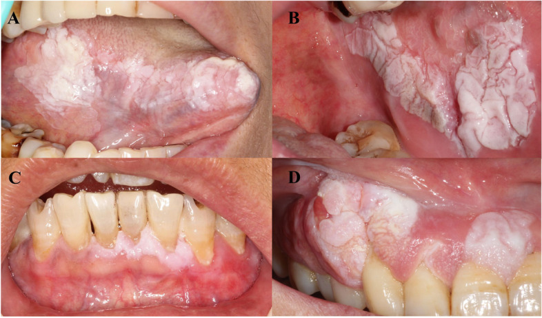
Results: The cohort comprised 16 males and 20 females, yielding a male-to-female ratio of 1:1.25. The follow-up period ranged from 8 to 125 months, with an average of 47.50 months. The most common clinical type of lesion was the verrucous form (58.33%), and the gingiva was the most common site (44.44%). Each patient had between 2 to 7 lesions, averaging 3.36 per patient. During the follow-up period, twelve patients (33.3%) developed oral cancer, with an average time to malignant transformation of 35.75 months. Kaplan-Meier survival analysis indicated that patients with complaints of pain, roughness, or a rough sensation, with diabetes, and the presence of cytologic atypia histologically showed a higher risk of malignant transformation (p < 0.05). In this study, the rate of malignant transformation in the treatment group (5/23) was lower than that in the untreated group (7/13), however, no statistically significant difference (p = 0.05).
Conclusion: The main complaints of pain, roughness, or foreign body sensation, coupled with cytologic atypia histologically are indicative of an increased risk of malignant transformation in PVL. Further research is needed to elucidate the influence of these clinicopathological parameters on the malignant progression of PVL.
Keywords: Malignant transformation; Oral cavity; Oral epithelial dysplasia; Proliferative verrucous leukoplakia.
© 2024. The Author(s).
Conflict of interest statement
The authors declare that they have no competing interests.
Figures


Similar articles
-
Potentially malignant disorders revisited-The lichenoid lesion/proliferative verrucous leukoplakia conundrum.J Oral Pathol Med. 2018 Jul;47(6):557-565. doi: 10.1111/jop.12716. Epub 2018 Apr 29. J Oral Pathol Med. 2018. PMID: 29663518
-
[Proliferative verrucous leukoplakia. Report of five cases].Mund Kiefer Gesichtschir. 2003 May;7(3):164-70. doi: 10.1007/s10006-003-0472-1. Epub 2003 May 1. Mund Kiefer Gesichtschir. 2003. PMID: 12764683 German.
-
Malignant transformation of proliferative Verrucous Leukoplakia-systematic review & meta-analysis.BMC Oral Health. 2025 Feb 1;25(1):175. doi: 10.1186/s12903-025-05565-7. BMC Oral Health. 2025. PMID: 39893387 Free PMC article.
-
Lichenoid areas may arise in early stages of proliferative verrucous leukoplakia: A long-term study of 34 patients.J Oral Pathol Med. 2022 Jul;51(6):573-581. doi: 10.1111/jop.13317. Epub 2022 Jun 3. J Oral Pathol Med. 2022. PMID: 35596256 Free PMC article.
-
Clinicopathologic analysis of verrucous hyperplasia, verrucous carcinoma and squamous cell carcinoma as part of the clinicopathologic spectrum of oral proliferative verrucous leukoplakia: A literature review and analysis.Pathol Res Pract. 2019 Dec;215(12):152670. doi: 10.1016/j.prp.2019.152670. Epub 2019 Sep 25. Pathol Res Pract. 2019. PMID: 31630872 Review.
Cited by
-
Malignant Transformation of Proliferative Verrucous Leukoplakia: A Description of the Clinical Oral Characteristics of These Squamous Cell Carcinomas.Cancers (Basel). 2025 Apr 1;17(7):1199. doi: 10.3390/cancers17071199. Cancers (Basel). 2025. PMID: 40227760 Free PMC article.
-
Cell-mediated mucositis of the oral cavity: narrative review on etiology, clinico-pathological aspects and malignant transformation.Pathologica. 2025 Apr;117(2):84-100. doi: 10.32074/1591-951X-1093. Pathologica. 2025. PMID: 40474707 Free PMC article. Review.
-
Squamous Cell Carcinoma with Prominent Eosinophils.Head Neck Pathol. 2024 Oct 28;18(1):115. doi: 10.1007/s12105-024-01718-2. Head Neck Pathol. 2024. PMID: 39466476 Review.
References
MeSH terms
LinkOut - more resources
Full Text Sources
