. 2024 May 30;22(1):517.
doi: 10.1186/s12967-024-05233-4.

CircDCAF8 promotes the progression of hepatocellular carcinoma through miR-217/NAP1L1 Axis, and induces angiogenesis and regorafenib resistance via exosome-mediated transfer

Affiliations

CircDCAF8 promotes the progression of hepatocellular carcinoma through miR-217/NAP1L1 Axis, and induces angiogenesis and regorafenib resistance via exosome-mediated transfer

Jiahao Gong et al. J Transl Med. .

Abstract

Background: Circular RNAs (circRNAs), which are a new type of single-stranded circular RNA, have significant involvement in progression of many diseases, including tumors. Currently, multiple circRNAs have been identified in hepatocellular carcinoma (HCC). Our study aims to investigate the function and mechanism of circDCAF8 in HCC.

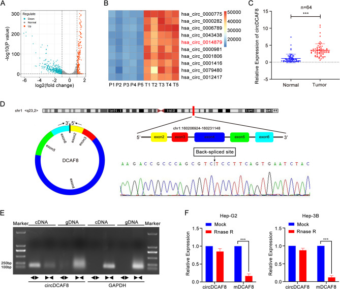
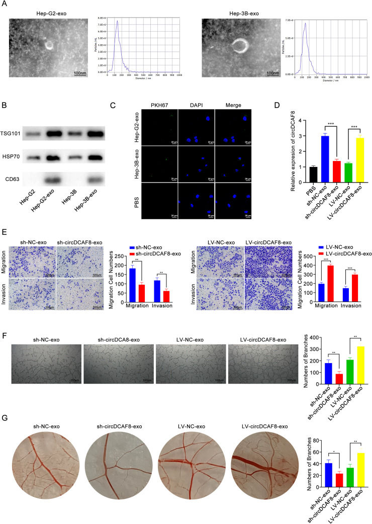
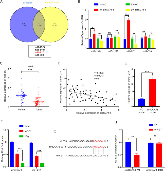
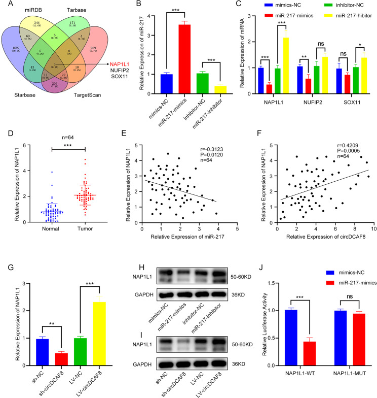
Methods: The expression of circDCAF8 (hsa_circ_0014879) in HCC and para-carcinoma tissue samples was determined using quantitative real-time polymerase chain reaction (qRT-PCR). The biological function of circDCAF8 in HCC was confirmed by experiments conducted both in vitro and in vivo. And the relationship between circDCAF8, miR-217 and NAP1L1 was predicted by database and verified using qRT-PCR, RNA-binding protein immunoprecipitation (RIP) and dual-luciferase reporter assays. Exosomes isolated from HCC cells were utilized to assess the connection of exosomal circDCAF8 with HCC angiogenesis and regorafenib resistance.

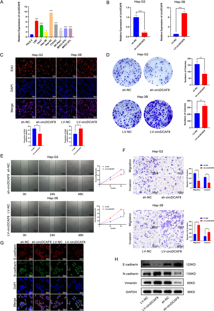
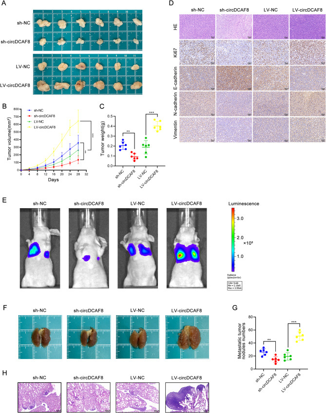
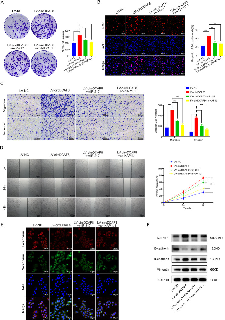
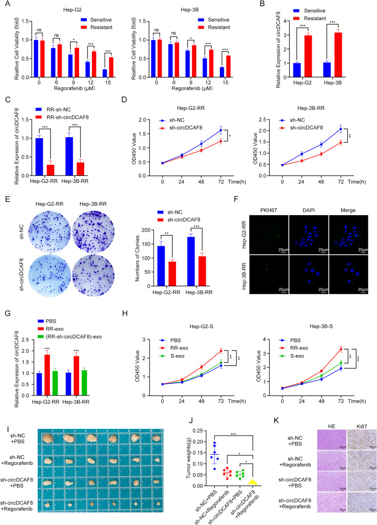
Results: CircDCAF8 is upregulated in HCC tissues and cell lines, and is linked to an unfavourable prognosis for HCC patients. Functionally, circDCAF8 was proved to facilitate proliferation, migration, invasion and Epithelial-Mesenchymal Transformation (EMT) in HCC cells. Animal examinations also validated the tumor-promoting characteristics of circDCAF8 on HCC. Besides, exosomal circDCAF8 promoted angiogenesis in HUVECs. Mechanistically, circDCAF8 interacted with miR-217 and NAP1L1 was a downstream protein of miR-217. CircDCAF8 promoted NAP1L1 expression by sponging miR-217. In addition, exosomes may transfer circDCAF8 from regorafenib-resistant HCC cells to sensitive cells, where it would confer a resistant phenotype.

Conclusion: CircDCAF8 facilitates HCC proliferation and metastasis via the miR-217/NAP1L1 axis. Meanwhile, circDCAF8 can promote angiogenesis and drive resistance to regorafenib, making it a viable therapeutic target for HCC patients.


Conflict of interest statement

The authors declare no conflicts of interests.

Figures

Fig. 1
Fig. 1
Selection and identification of circDCAF8. A The volcano plot of DECs in GSE94508. B A heatmap of the top 10 upregulated circRNAs in 5 paired samples of HCC. C Relative expression of circDCAF8 in human HCC tissues and paired adjacent nontumor tissues of 64 patients was determined by qRT-PCR. D Sanger sequencing of the annotated genomic region of circDCAF8 was performed to confirm the Back-spliced site of circDCAF8. E The divergent primers detected circDCAF8 in cDNA but not in gDNA by agarose gel electrophoresis. GAPDH was used as a negative control. F qRT–PCR analysis for the expression of circDCAF8 and mDCAF8 after treatment with RNase R in Hep-G2 and Hep-3B cells. Data are representative of three independent experiments and are presented as means ± SDs. (*p < 0.05; **p < 0.01; ***p < 0.001)
Fig. 2
Fig. 2
CircDCAF8 promotes proliferation, migration, invasion and EMT in HCC cells. A Relative expression of circDCAF8 in HCC cell lines and THLE-2 (human immortalized hepatocytes). B The knockdown efficiency of circCAF8 in Hep-G2 cells and overexpression efficiency in Hep-3B cells were determined by qRT-PCR. C, D EdU and colony formation assays evaluated the proliferation of sh-circDCAF8 cells and LV-circDCAF8 cells. Scale bar = 50 μm. E Wound healing assay determined cell migration ability. Scale bar = 100 μm. F Transwell assay measured invasion and migration ability with or without matrix. Scale bar = 500 μm. G, H Immunofluorescence and western blot detected the expression of EMT-related proteins. Scale bar = 50 μm. Data are representative of three independent experiments and are presented as means ± SDs. (*p < 0.05; **p < 0.01; ***p < 0.001)
Fig. 3
Fig. 3
CircDCAF8 promoted HCC proliferation and metastasis in vivo. A Tumors formed in nude mice by subcutaneous injection of circDCAF8 stable knockdown or overexpression cells. B, C The volume and weight of the subcutaneous tumor. D H&E, Ki67, E-cadherin, N-cadherin and Vimentin staining of xenograft tumors. Scale bar = 50 μm. E Fluorescence intensity alterations in pulmonary metastasis models. F Lung metastasis induced by tail vein injection of circDCAF8 knockdown or overexpression cells in nude mice. G Lung metastatic nodules were counted. H H&E stain of lung metastases. Scale bar = 200 μm. n = 6 mice per group. Data are presented as means ± SDs. (*p < 0.05; **p < 0.01; ***p < 0.001)
Fig. 4
Fig. 4
Exosomal circDCAF8 promoted the migration, invasion and tube formation of HUVECs. A TEM and NTA of exosomes isolated from Hep-G2 and Hep-3B. Scale bar = 100 nm. B Exosomal protein positive markers (Tsg101, HSP70 and CD63) were detected by western blot from purified exosomes and exosome-depleted cell extracts. C Laser confocal microscopy showed that the exosomes secreted by HCC cells were ingested by HUVECs. Scale bar = 20 μm. D qRT-PCR was performed to detect circDCAF8 expression in HUVECs after coculture with circDCAF8 knockdown or overexpression exosomes. E Migration and invasion of exosomes ingested by HUVECs was detected using the transwell assay. Scale bar = 500 μm. F Tube formation assay measured the tube formation ability of HUVECs ingested exosomes. Scale bar = 100 μm. G Chick chorioallantoic membrane assay showed that exosomal circDCAFB promoted the angiogenesis of chick embryo chorioallantoic membrane. Data are presented as means ± SDs. (*p < 0.05; **p < 0.01; ***p < 0.001)
Fig. 5
Fig. 5
CircDCAF8 acts as a sponge of miR-217. A Downstream miRNAs of circDCAF8 predicted by circbank and circinteractome databases. B Relative expression of downstream miRNAs was determined by qRT-PCR in circDCAF8 knockdown and overexpression cells. C Relative expression of miR-217 in human HCC tissues and paired adjacent nontumor tissues of 64 patients was determined by qRT-PCR. D Spearman correlation analysis showed circDCAF8 expression was negatively correlated with the miR-217 expression. E Pull down assays showed that miR-217 was enriched by the circDCAF8 probe. F RIP assay confirmed circDCAF8 and miR-217 could bind with RNA-induced silencing complex (RISC). G A schematic of wild-type (WT) and mutant (MUT) circDCAF8 luciferase reporter vectors. H Luciferase reporter assay unveiled the molecular combination of miR-217 with circDCAF8 wild type in HEK-293T cells. Data are presented as means ± SDs. (ns, not significant; *p < 0.05; **p < 0.01; ***p < 0.001)
Fig. 6
Fig. 6
NAP1L1 is a target gene of miR-217. A Downstream genes of miR-217 predicted by miRDB, Tarbase, Starbase and TargetScan databases. B The overexpression and knockdown efficiency of miR-217 were determined by qRT-PCR. C Relative expression of downstream genes was determined by qRT-PCR in miR-217 overexpression and knockdown cells. D Relative expression of NAP1L1 in human HCC tissues and paired adjacent nontumor tissues of 64 patients was determined by qRT-PCR. E, F Spearman correlation analysis showed NAP1L1 expression was negatively correlated with the miR-217 expression(E) and positively correlated with circDCAF8 expression(F). G Relative expression of NAP1L1 was determined by qRT-PCR in circDCAF8 knockdown and overexpression cells. H, I Western blot was performed to detect protein expression levels of NAP1L1 in miR-217 and circDCAF8 knockdown or overexpression cells. J Luciferase reporter assay unveiled the molecular combination of miR-217 with circDCAF8 wild type in HEK-293T cells. Data are presented as means ± SDs. (ns, not significant; *p < 0.05; **p < 0.01; ***p < 0.001)
Fig. 7
Fig. 7
CircDCAF8 promoted HCC progression through the miR-217/NAP1L1 axis. A, B Colony formation and EdU assays evaluated the proliferation ability in LV-circDCAF8 Hep-3B cells and LV-circDCAF8 Hep-3B cells transfected with miR-217 mimics or sh-NAP1L1 vectors. Scale bar = 50 μm. C Transwell assay measured invasion and migration ability with or without matrix. Scale bar = 500 μm. D Wound healing assay determined cell migration ability. Scale bar = 100 μm. E, F Immunofluorescence and western blot detected the expression of NAP1L1 and EMT-related proteins. Scale bar = 50 μm. Data are representative of three independent experiments and are presented as means ± SDs. (*p < 0.05; **p < 0.01; ***p < 0.001)
Fig. 8
Fig. 8
CircDCAF8 transmits regorafenib resistance by exosomes. A Cell viability of cells treated with different doses of regorafenib for 48 h. B Relative expression of circDCAF8 in regorafenib sensitive and resistant cells. C The knockdown efficiency of circCAF8 in regorafenib resistant cells were determined by qRT-PCR. D, E CCK8 and colony formation assays evaluated the proliferation of sh-circDCAF8 regorafenib resistant cells. F Exosomes secreted by regorafenib resistant cells were ingested by sensitive cells. Scale bar = 20 μm. G qRT-PCR was used to detect the relative expression of circDCAF8 in sensitive cells after incubation with exosomes from different sources. H CCK8 assay detected proliferation capacity in exosome-treated sensitive cells. I Regorafenib therapy or circDCAF8 knockdown could both inhibit the proliferation of the tumors formed in nude mice. J The weight of the subcutaneous tumor. K H&E and Ki67 staining of xenograft tumors. Scale bar = 50 μm. Data are presented as means ± SDs. In A-E, G, H, n = 3; in I-K, n = 6. (ns, not significant; *p < 0.05; **p < 0.01; ***p < 0.001)

Similar articles

Cited by

References

    1. SUNG H, FERLAY J, SIEGEL RL, et al. Global Cancer statistics 2020: GLOBOCAN estimates of incidence and Mortality Worldwide for 36 cancers in 185 Countries[J] CA Cancer J Clin. 2021;71(3):209–49. doi: 10.3322/caac.21660. - DOI - PubMed
    1. MCGLYNN K A, PETRICK J L, LONDON W T. Global epidemiology of hepatocellular carcinoma: an emphasis on demographic and regional variability[J] Clin Liver Dis. 2015;19(2):223–38. doi: 10.1016/j.cld.2015.01.001. - DOI - PMC - PubMed
    1. FORNER A, REIG M, BRUIX J. Hepatocellular carcinoma[J] Lancet. 2018;391(10127):1301–14. doi: 10.1016/S0140-6736(18)30010-2. - DOI - PubMed
    1. MARRERO J A, KULIK L M, SIRLIN C B, et al. Diagnosis, staging, and management of Hepatocellular Carcinoma: 2018 Practice Guidance by the American Association for the study of Liver Diseases[J] Hepatology. 2018;68(2):723–50. doi: 10.1002/hep.29913. - DOI - PubMed
    1. VICENS Q. WESTHOF E Biogenesis Circular RNAs[J] Cell. 2014;159(1):13–4. - PubMed

Publication types

MeSH terms