. 2024 May 30;25(1):534.
doi: 10.1186/s12864-024-10448-2.

Differential gene expression and immune cell infiltration in maedi-visna virus-infected lung tissues

Affiliations

Differential gene expression and immune cell infiltration in maedi-visna virus-infected lung tissues

Xiaona Shi et al. BMC Genomics. .

Abstract

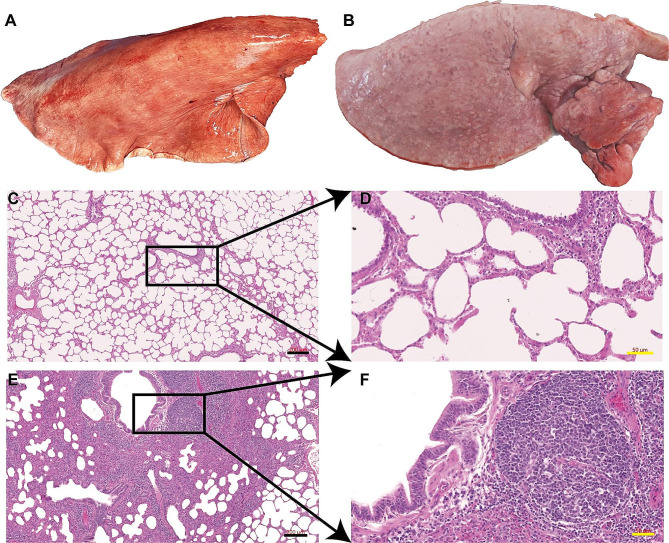
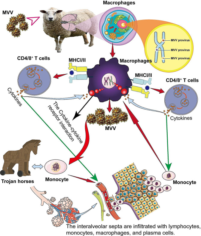
Background: Maedi-visna virus (MVV) is a lentivirus that infects monocyte/macrophage lineage cells in sheep, goats, and wild ruminants and causes pneumonia, mastitis, arthritis, and encephalitis. The immune response to MVV infection is complex, and a complete understanding of its infection and pathogenesis is lacking. This study investigated the in vivo transcriptomic patterns of lung tissues in sheep exposed to MVV using the RNA sequencing technology.

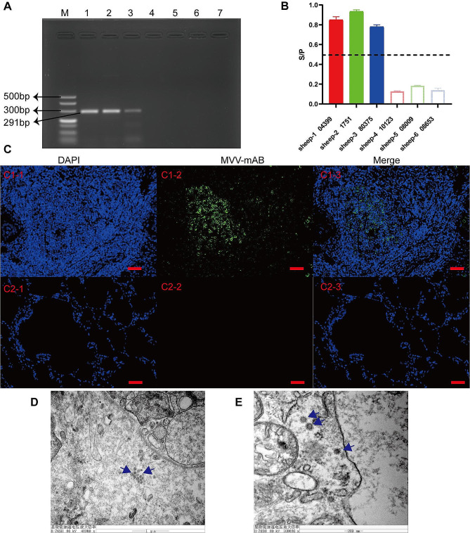
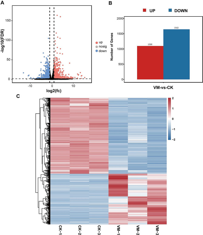
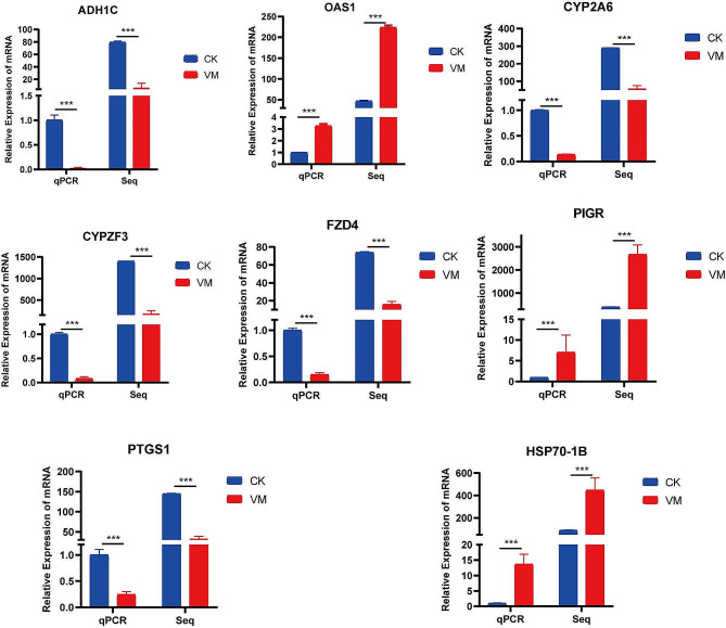
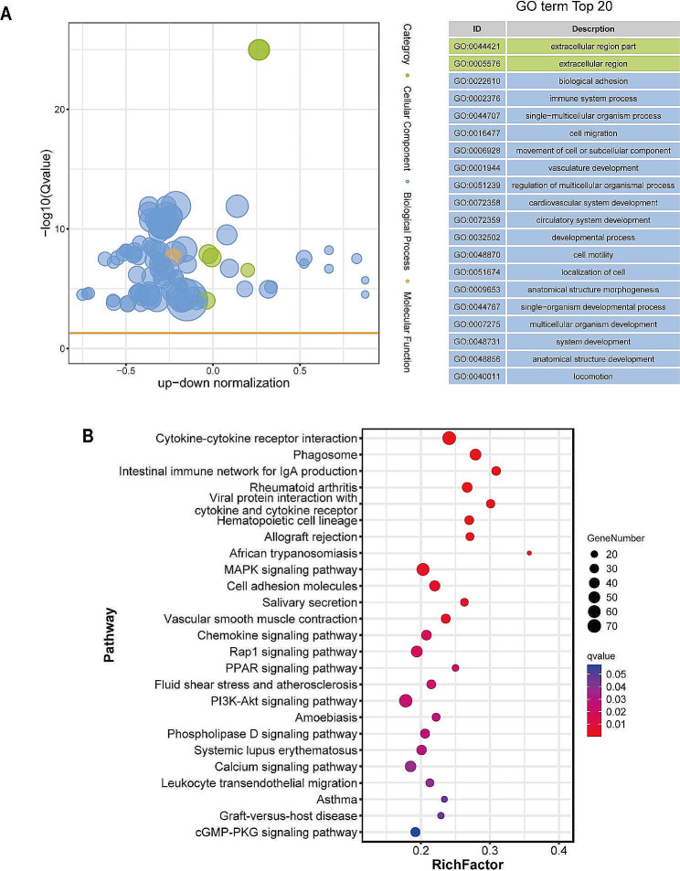
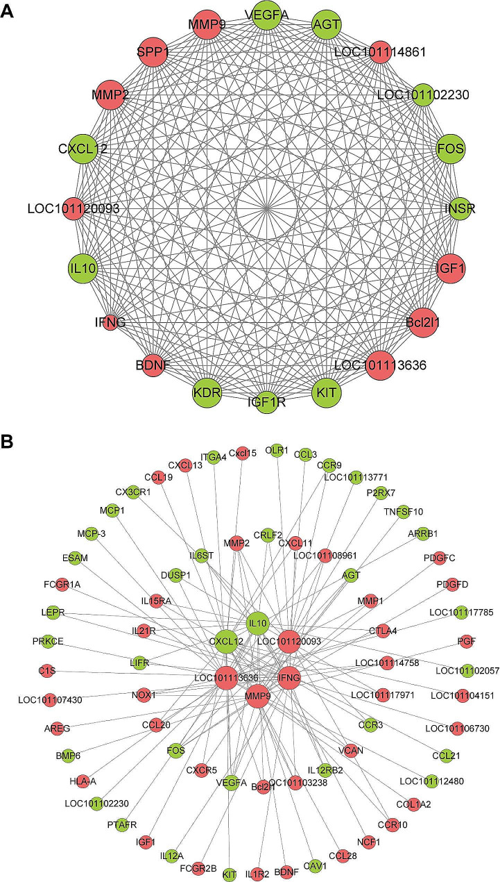
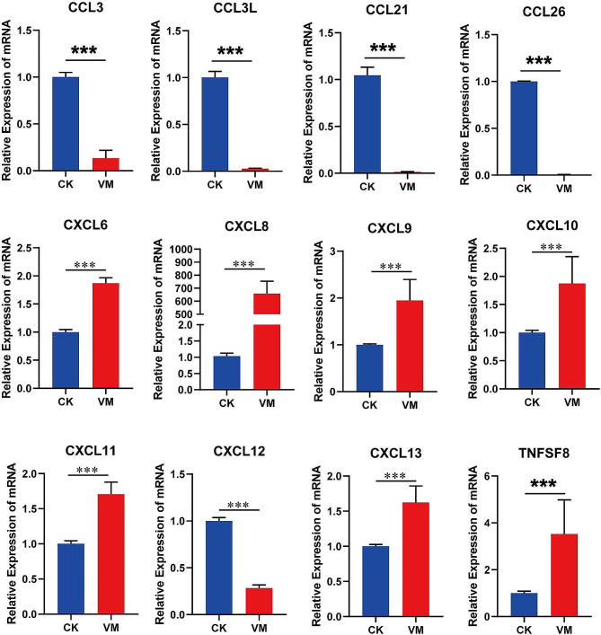
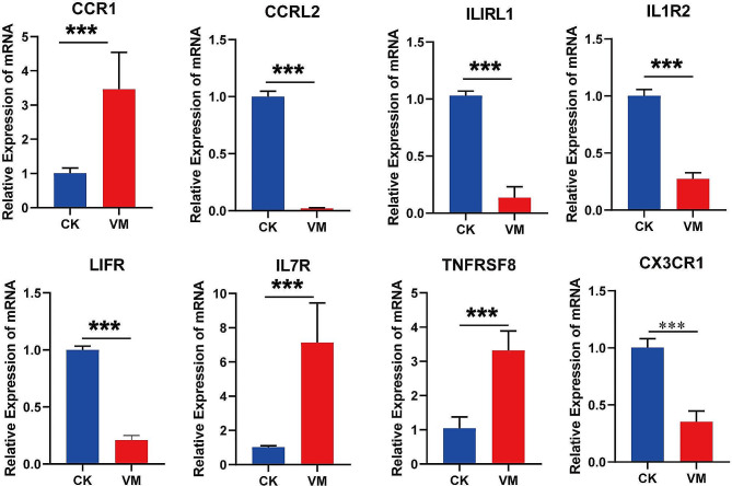
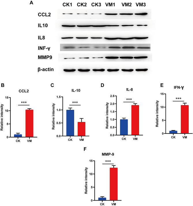
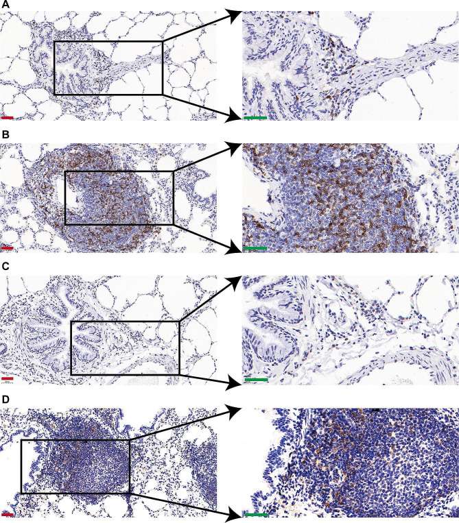
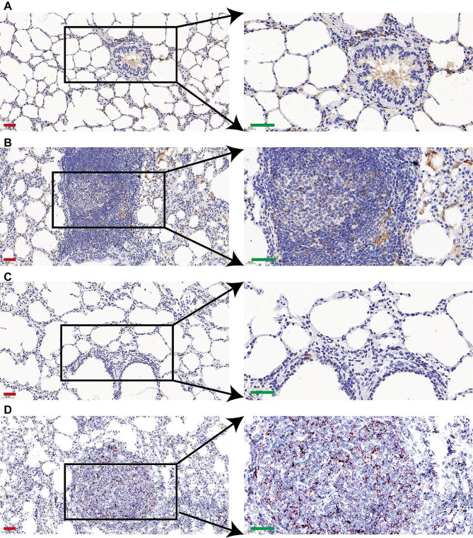
Result: The results indicated that 2,739 genes were significantly differentially expressed, with 1,643 downregulated genes and 1,096 upregulated genes. Many variables that could be unique to MVV infections were discovered. Gene Ontology analysis revealed that a significant proportion of genes was enriched in terms directly related to the immune system and biological responses to viral infections. Kyoto Encyclopedia of Genes and Genomes analysis revealed that the most enriched pathways were related to virus-host cell interactions and inflammatory responses. Numerous immune-related genes, including those encoding several cytokines and interferon regulatory factors, were identified in the protein-protein interaction network of differentially expressed genes (DEGs). The expression of DEGs was evaluated using real-time polymerase chain reaction and western blot analysis. CXCL13, CXCL6, CXCL11, CCR1, CXCL8, CXCL9, CXCL10, TNFSF8, TNFRSF8, IL7R, IFN-γ, CCL2, and MMP9 were upregulated. Immunohistochemical analysis was performed to identify the types of immune cells that infiltrated MVV-infected tissues. B cells, CD4+ and CD8+ T cells, and macrophages were the most prevalent immune cells correlated with MVV infection in the lungs.

Conclusion: Overall, the findings of this study provide a comprehensive understanding of the in vivo host response to MVV infection and offer new perspectives on the gene regulatory networks that underlie pathogenesis in natural hosts.

Keywords: Differential expression; Immunopathogenesis; Maedi-Visna virus; Ovine progressive pneumonia; Pathogenesis; RNA-seq; Sheep.


Conflict of interest statement

The authors declare no competing interests.

Figures

Fig. 1
Fig. 1
Histopathologic characteristics of Maedi-visna virus (MVV)-infected lungs. (A) Overall appearance of the lung from a healthy sheep; (B) appearance of Maedi-visna disease in an infected sheep lung sample; (C and D) microscopic examination of the lung from a healthy sheep (black bar, 200 μm; yellow bar, 50 μm); (E and F) microscopic view of MVV-infected lungs (black bar, 200 μm; yellow bar, 50 μm)
Fig. 2
Fig. 2
Identification of Maedi-visna virus (MVV) within the lungs of naturally infected sheep. (A) Polymerase chain reaction products were subjected to agarose-gel electrophoresis. Lane M presents 500 bp molecular weight markers; Lanes 1–3 present lung tissue samples from three MVV-infected sheep; Lanes 4–6 present lung tissue samples from three healthy sheep; and Lane 7 presents the negative control. (B) Enzyme-Linked ImmunoSorbent Assay for MVV antibody production. (C) Immunofluorescence of the capsid protein in the lungs of MVV-infected or -uninfected sheep (scale bar, 50 μm). (D and E) Transmission electron micrographs of MVV-infected lungs. (D) Viral buds can be observed on the outer membrane of macrophages infected with MVV (scale bar, 1,000 nm), whereas (E) intracytoplasmic particles are visible within the macrophages (scale bar, 200 nm)
Fig. 3
Fig. 3
Phylogenetic trees inferred from gag and env regions. (A) Neighbor-Joining method phylogenetic trees based on the gag region of 79 sequences: 1 analyzed in this study (labeled by a red pentagram) and 78 whole genome sequences of SRLVs available in GenBank. (B) Neighbor-Joining method phylogenetic trees based on the env region of 79 sequences: 1 analyzed in this study (labeled by a red pentagram) and 78 whole genome sequences of SRLVs available in GenBank. The numbers on the nodes indicatethe percentage of bootstrap values obtained from 1000 replicates. The tree was generated using the neighbor-joining method coupled with the p-distance model and bootstrap analysis of 1,000 replicates
Fig. 4
Fig. 4
Quantitative assessment of DEGs. (A) Visualization of differentially expressed genes (DEGs) using volcanic plots. The log2FoldChange value is plotted on the x-axis, whereas the –log10padj value is plotted on the y-axis. Differential gene screening criteria are indicated by the black dotted line, which serves as the threshold line. (B) CK, control. Upregulated and downregulated DEGs are presented in the red and blue columns, respectively. Visualization of DEGs using a heatmap
Fig. 5
Fig. 5
Comparison of fold-changes in differentially expressed genes between RNA-seq and quantitative reverse transcription-polymerase chain reaction. Data are presented as the mean ± standard deviation of values from three replicates. Statistical analysis was conducted using Student’s t-test, with significance indicated at p < 0.001
Fig. 6
Fig. 6
Gene ontology and Kyoto Encyclopedia of Genes and Genomes enrichment analyses of differentially expressed genes. (A) Gene ontology functional classification of differentially expressed genes (DEGs) according to diverse biological processes was performed. The results are visualized using a bubble plot, where the size of the bubbles represents the number of associated genes, and significantly enriched categories are labeled. (B) Kyoto Encyclopedia of Genes and Genomes (KEGG) enrichment analysis of the DEGs was performed. The horizontal axis of the graph indicates the proportion of DEGs annotated to a particular KEGG pathway relative to the overall number of DEGs. The y-axis represents the KEGG pathway. The number of genes annotated to the KEGG pathway is indicated by the size of the dots, and the significance of enrichment is represented by the color gradient, which ranges from red to blue
Fig. 7
Fig. 7
Protein-protein interaction (PPI) network analysis. (A) Analysis of differentially expressed genes using the STRING database. Hub genes were identified by visualizing the modules in the PPI network. The amplification of various core factors revealed significant aggregation and interaction components in the PPI network. (B) Cluster network of the key genes LOC101113636, LOC101120093, MMP9, IFN-γ, CXCL12, and IL10
Fig. 8
Fig. 8
Expression of chemokine genes in the cytokine-cytokine receptor interaction pathway
Fig. 9
Fig. 9
Expression of chemokine ligand and receptor genes in the cytokine-cytokine receptor interaction pathway
Fig. 10
Fig. 10
Relative protein expression of significantly altered inflammatory cytokines. (A) CCL2, IFN-γ, IL-8, IL-10, and MMP9 expression in the lungs of MVV-infected sheep confirmed through western blotting. (BF) Quantitative analysis of CCL2, IFN, IL-8, IL-10, and MMP9 expression
Fig. 11
Fig. 11
CD4 and CD8 markers used for immunostaining of immune cells in lung sections. (A) Control samples for CD4 immunostaining. (B) CD4 immunostaining of lungs from Maedi-visna virus (MVV)-infected sheep. (C) Control samples for CD8 immunostaining. (D) CD8 immunostaining of lungs from MVV-infected sheep (red bar, 50 μm and green bar, 40 μm)
Fig. 12
Fig. 12
Immunostaining of CD68-positive macrophages and CD19‐positive B cells infiltrating the lung. (A) Control samples for CD68 immunostaining. (B) CD68 immunostaining of lungs from Maedi-visna virus (MVV)-infected sheep. (C) Control samples for CD19 immunostaining. (D) CD19 immunostaining of lungs from MVV-infected sheep (red bar, 50 μm; green bar, 40 μm)
Fig. 13
Fig. 13
Schematic diagram of the pathogenic mechanism of ovine progressive pneumonia caused by Maedi-visna virus (MVV). MVV infection begins with the presence of macrophages in the lung mucosa. Antigen presenting cells, also known as macrophages, process viral proteins into antigens. T lymphocytes recognize the viral protein-major histocompatibility complex, triggering the synthesis of interferons I and II. This attracts additional inflammatory cells to the core, thereby supporting ongoing viral replication and persistent inflammation. CD4/8 + T cells undergo activation and differentiation into Th1 and Th2 effector cells and various subpopulations, such as Tfh cells. Cytotoxic T cells secrete cytokines such as INF-γ and chemokines to recruit immune cells. Continuous immune activation occurs in MVV-induced lesions, releasing cytokines that stimulate the transformation of monocytes into macrophages. The distinction between macrophages enables the ongoing display of viral antigens to T cells. Consequently, the stimulated T cells generate additional cytokines, thereby establishing a detrimental cycle

Similar articles

Cited by

References

    1. Borquez Cuevas MY, Hernández Chávez JF, Armenta Leyva B, Cedillo Cobián JR, Molina Barrios RM. Ovine Progressive Pneumonia: Diagnosis and Seroprevalence in the South of Sonora, Mexico. Case Reports In Veterinary Medicine 2021, 2021:6623888. - PMC - PubMed
    1. Kalogianni AI, Bossis I, Ekateriniadou LV, Gelasakis AI. Etiology, epizootiology and Control of Maedi-Visna in dairy Sheep: a review. Animals: Open Access J MDPI 2020, 10(4). - PMC - PubMed
    1. De Boer GF. Zwoegerziekte virus, the causative agent for progressive interstitial pneumonia (maedi) and meningo-leucoencephalitis (visna) in sheep. Res Vet Sci. 1975;18(1):15–25. doi: 10.1016/S0034-5288(18)33625-7. - DOI - PubMed
    1. Torsteinsdottir S, Andresdottir V, Arnarson H, Petursson G. Immune response to maedi-visna virus. Front Bioscience: J Virtual Libr. 2007;12:1532–43. doi: 10.2741/2166. - DOI - PubMed
    1. Pálsson PA. Maedi and Visna in sheep. Front Biology. 1976;44:17–43. - PubMed

MeSH terms