. 2024 May 30;19(1):75.
doi: 10.1186/s13020-024-00933-x.

A simplified herbal decoction attenuates myocardial infarction by regulating macrophage metabolic reprogramming and phenotypic differentiation via modulation of the HIF-1α/PDK1 axis

Affiliations

A simplified herbal decoction attenuates myocardial infarction by regulating macrophage metabolic reprogramming and phenotypic differentiation via modulation of the HIF-1α/PDK1 axis

Zhi-Jun Lin et al. Chin Med. .

Abstract

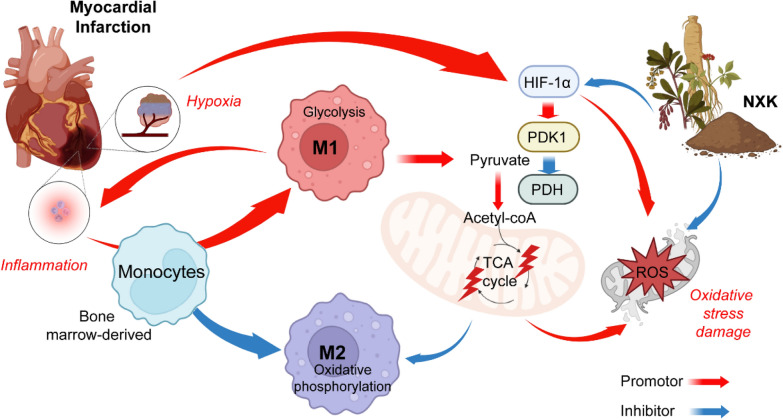
Background: Myocardial infarction (MI) poses a global public health challenge, often associated with elevated mortality rates and a grim prognosis. A crucial aspect of the inflammatory injury and healing process post-MI involves the dynamic differentiation of macrophages. A promising strategy to alleviate myocardial damage after MI is by modulating the inflammatory response and orchestrating the shift from pro-inflammatory (M1) to anti-inflammatory (M2) macrophages, aiming to achieve a reduced M1/M2 ratio. Nuanxinkang (NXK), a simplified herbal decoction, has demonstrated noteworthy cardioprotective, inflammation-regulating, and myocardial energy metabolism-regulating properties.

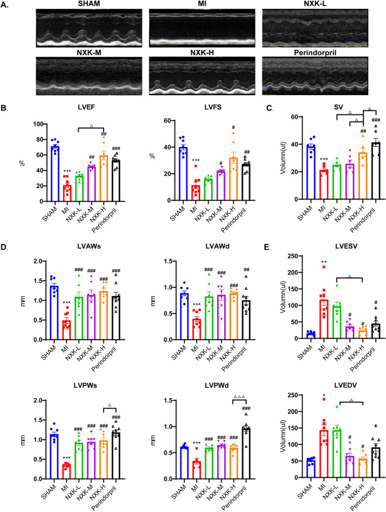
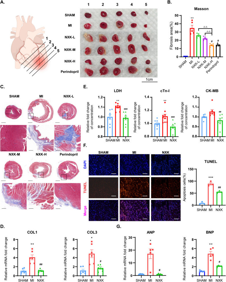
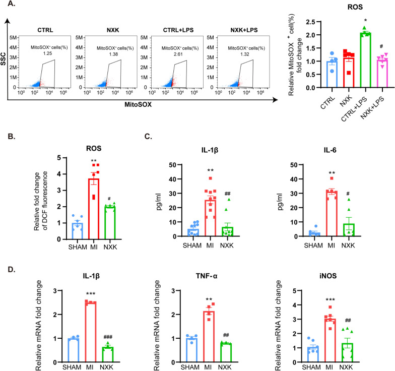
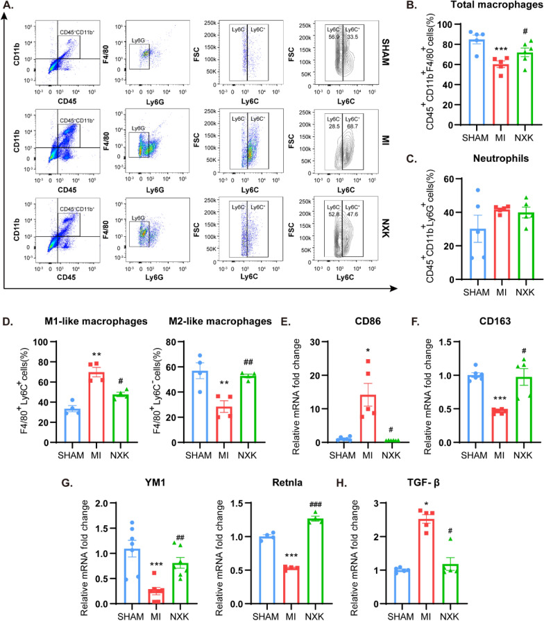
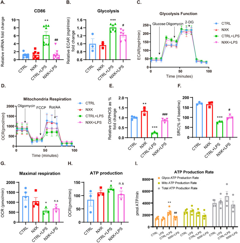
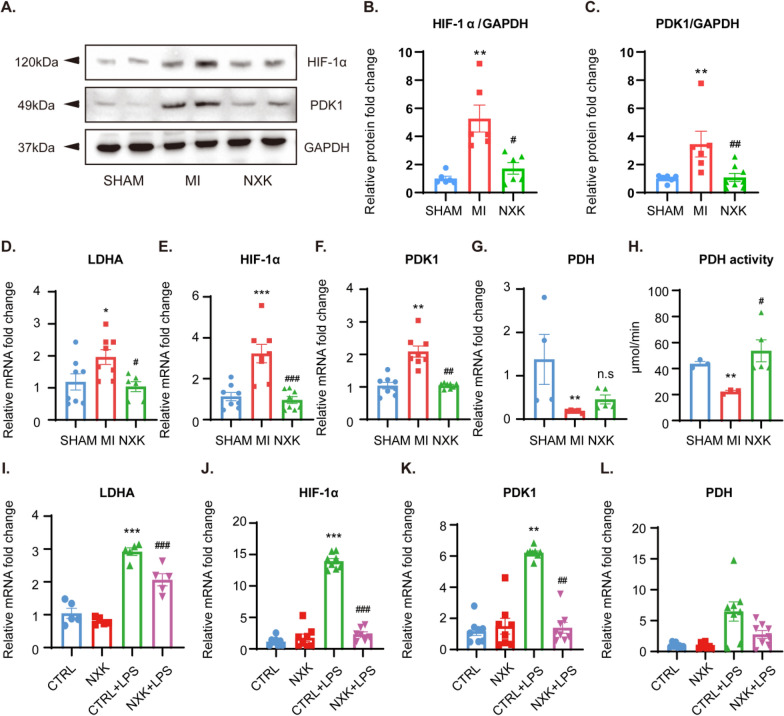
Methods: In this study, we constructed an MI model by ligating coronary arteries to investigate the efficacy of NXK in improving ventricular remodeling and cardiac function. Mice were administered NXK (1.65 g/kg/d) or an equivalent volume of regular saline via gavage for 28 consecutive days, commencing the day after surgery. Then, we conducted echocardiography to assess the cardiac function, Masson staining to illustrate the extent of myocardial fibrosis, TUNEL staining to reveal myocardial apoptosis, and flow cytometry to analyze the polarization of M1 and M2 macrophages in the hearts. Besides, a lipopolysaccharide (LPS)-induced pro-inflammatory macrophage (M1) polarization model was implemented in RAW264.7 cells to elucidate the underlying mechanism of NXK in regulating macrophage polarization. RAW264.7 cells were pre-treated with or without NXK-containing serum. Oxidative stress was detected by MitoSox staining, followed by Seahorse energy metabolism assay to evaluate alterations in mitochondrial metabolic patterns and ATP production. Both In vivo and in vitro, HIF-1α and PDK1 were detected by fluorescent quantitative PCR and Western blotting.

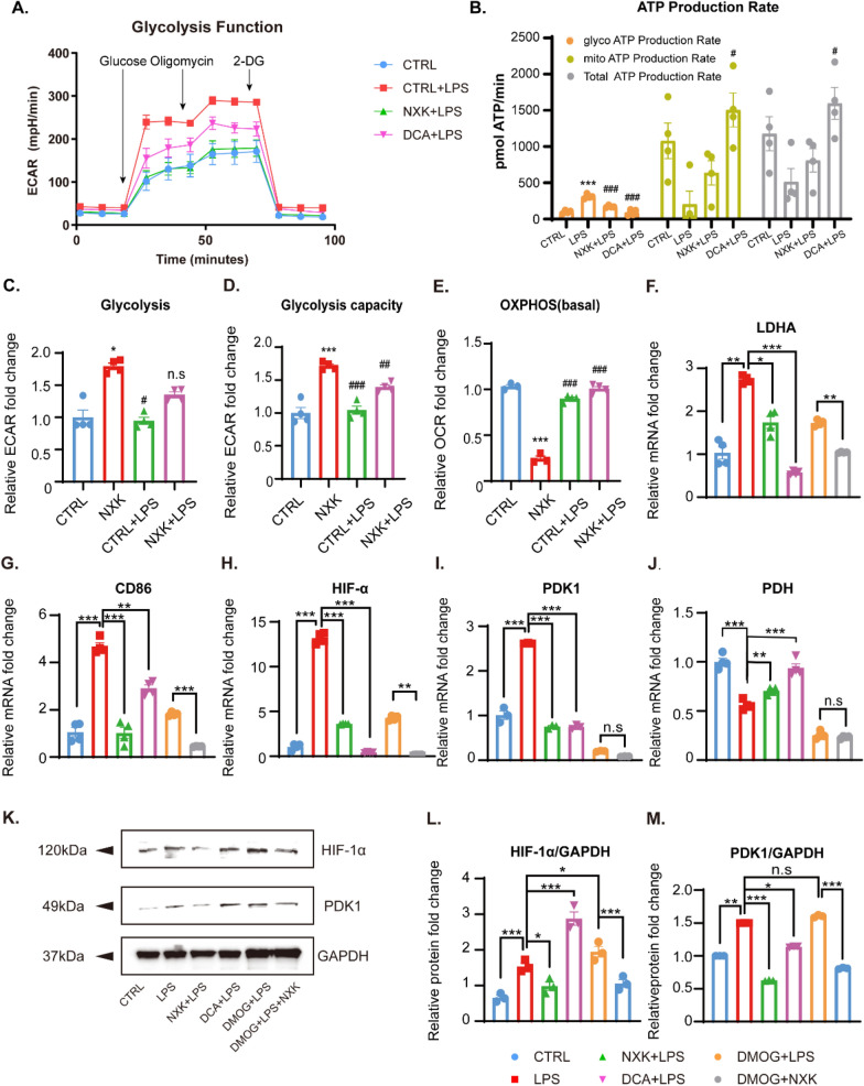
Results: In vivo, MI mice exhibited a decline in cardiac function, adverse ventricular remodeling, and an increase in glycolysis, coupled with M1-dominant polarization mediated by the HIF-1α/PDK1 axis. Notably, robust responses were evident with high-dose NXK treatment (1.65 g/kg/day), leading to a significant enhancement in cardiac function, inhibition of cardiac remodeling, and partial suppression of macrophage glycolysis and the inflammatory phenotype in MI mice. This effect was achieved through the modulation of the HIF-1α/PDK1 axis. In vitro, elevated levels of mitochondrial ROS production and glycolysis were observed in LPS-induced macrophages. Conversely, treatment with NXK notably reduced the oxidative stress damage induced by LPS and enhanced oxidative phosphorylation (OXPHOS). Furthermore, NXK demonstrated the ability to modify the energy metabolism and inflammatory characteristics of macrophages by modulating the HIF-1α/PDK1 axis. The influence of NXK on this axis was partially counteracted by the HIF-1α agonist DMOG. And NXK downregulated PDK1 expression, curtailed glycolysis, and reversed LPS-induced M1 polarization in macrophages, similar to the PDK1 inhibitor DCA.

Conclusion: In conclusion, NXK protects against MI-induced cardiac remodeling by inducing metabolic reprogramming and phenotypic differentiation of macrophages, achieved through the modulation of the HIF-1α/PDK1 axis. This provides a novel and promising strategy for the treatment of MI.

Keywords: Energy metabolism; Macrophages polarization; Myocardial Infarction (MI); Nuanxinkang (NXK).


Conflict of interest statement

The authors declare that they have no competing interests.

Figures

Fig. 1
Fig. 1
NXK improves cardiac function in MI mice. A Representative M-mode echocardiographic images were shown from Sham, MI, MI + NXK groups 28 days post MI. BE LVEF, LVFS, SV, LVAWs, LVAWd, LVPWs, LVPWd, LVESV, and LVEDV were measured by echocardiography (n ≥ 6). Values were shown as mean ± SD, **P < 0.01, ***P < 0.001 vs the SHAM group, #P < 0.05, ##P < 0.01, ###P < 0.001 vs MI group. P < 0.05 vs the Perindopril group
Fig. 2
Fig. 2
NXK protects against the deterioration of cardiac structure after MI. A Representative micrographs depicting TTC staining in various groups. B Quantification of fibrosis area from different groups (n ≥ 3). C Representative photomicrographs of Masson’s trichrome staining respectively from different groups (upper bars, 1 mm; bottom bars, 200 μm). D RT-PCR quantification of COL1 and COL3 mRNA levels in mouse cardiac tissues (n = 6). E The concentration of LDH, cTn-I and CK-MB in the serum of mice (n ≥ 7). F Representative TUNEL staining (bars, 50 μm) and quantification of positive rate of apoptotic cardiomyocytes in peripheral tissue of infarction (n = 3). G Quantification of ANP and BNP mRNA expression (n = 5). F TUNEL staining revealed that NXK treatment alleviated cardiac apoptosis in mice. Values were shown as mean ± SD, *P < 0.05, **P < 0.01, ***P < 0.001 vs the SHAM group, #P < 0.05, ##P < 0.01 vs the MI group
Fig. 3
Fig. 3
NXK alleviates oxidative stress and inflammatory responses. A The representative figure of MitoSOX staining red positive cells in different groups of macrophages. Quantified percent of MitoSOX staining red positive cells (n ≥ 4), all the data were shown as fold change compared with CTRL. B Quantification of ROS production in mouse heart tissues (n = 6). C The levels of IL-1β and IL-6 antigens in mouse serum were determined by Elisa assays (n ≥ 5). D mRNA expression of IL-1β, TNF-α and iNOS were detected by qPCR (n ≥ 4). Values were shown as means ± SD, *P < 0.05, **P < 0.01, ***P < 0.001 vs the SHAM group, #P < 0.05, ##P < 0.01, ###P < 0.001 vs the MI group
Fig. 4
Fig. 4
NXK inhibited M1 polarization and promoted M2. A Representative flow diagrams of M1-like and M2-like macrophages in different groups. BD Quantified percent of total macrophages, neutrophils, M1-like and M2-like macrophages respectively (n ≥ 4). EH The mRNA expression of CD86, CD163, YM1, Rentla and TGF-β in vivo (n ≥ 4), were detected by qPCR. Values were shown as means ± SD, *P < 0.05, **P < 0.01, ***P < 0.001 vs the SHAM/CTRL group, #P < 0.05, ##P < 0.01, ###P < 0.001 vs the MI group/CTRL + LPS group
Fig. 5
Fig. 5
NXK coordinates macrophage polarization via energy metabolism reprogramming. A The mRNA expression of CD86 in vitro (n = 8), were detected by qPCR. B Glycolysis level were quantitatively analyzed (n ≥ 3). C Representative curves of cellular glycolysis function. D Representative curves of cellular mitochondrial respiration. EG Basal OXPHOS (%), spare respiratory capacity (%), maximal respiration, and ATP production based on mitochondrial aerobic respiration were analyzed quantitatively (n ≥ 3). I The real-time ATP production rate and total ATP production level of macrophages were shown (n = 4). Values were shown as means ± SD, *P < 0.05, **P < 0.01, ***P < 0.001 vs the CTRL group, #P < 0.05, ##P < 0.01, ###P < 0.001 vs the CTRL + LPS group, n.s, no significance
Fig. 6
Fig. 6
NXK modulates the HIF-1α/PDK1 axis. AC Representative WB images and qualified protein expression levels of cardiac HIF-1α and PDK1 (n ≥ 3). DG The mRNA expression level of cardiac LDHA, HIF-1α, PDK1, and PDH were detected by qPCR (n ≥ 4). H PDH activity was detected by an Elisa kit and analyzed following instructions (n = 3). IL The mRNA expression level of cellular LDHA, HIF-1α, PDK1, and PDH were detected by qPCR (n ≥ 3). Values showed as means ± SD, *P < 0.05, **P < 0.01, ***P < 0.001 vs the MI group/the CTRL group, #P < 0.05, ##P < 0.05, ###P < 0.001 vs the MI group/the CTRL + LPS group, n.s, no significance
Fig. 7
Fig. 7
NXK regulates the energy metabolism of macrophages via the HIF-1α/PDK1 axis. A Representative curves of glycolysis function. B The real-time different types of ATP production rate (n = 4). CE Glycolysis, glycolysis capacity, and basal OXPHOS were quantified (n ≥ 3). FJ The mRNA expression levels of cellular LDHA, CD86, HIF-1α, PDK1, PDH genes were quantified by qPCR (n ≥ 3). K Representative WB images of cellular HIF-1α and PDK1 in vitro. L-M Quantification of the cellular HIF-1α and PDK1 protein expression levels (n = 3). Values were shown as means ± SD, *P < 0.05, **P < 0.01, ***P < 0.001 vs the CTRL group, #P < 0.05, ##P < 0.01, ###P < 0.001 vs the CTRL + LPS group, n.s, no significance (Panel BE). P* < 0.05, P** < 0.01, P*** < 0.001, n.s, no significance (Panel FJ and LM)
Fig. 8
Fig. 8
NXK orchestrates macrophage polarization by reprogramming energy metabolism based on HIF-1α/PDK1 axis. In the early stages of MI, M1 macrophages are heavily recruited and localized in a relatively hypoxic state, which activates HIF-1α. HIF-1α can participate in macrophage migration and polarization through PDK1-induced active glycolysis, which reduces the flux of TCA circulation by inactivating PDH. These suggest NXK can participate in macrophage metabolic reprogramming by inhibiting the HIF-1α/PDK1 axis, thereby regulating macrophage polarization, affecting the ratio of M1/M2 during post-MI repair, and improves cardiac remodeling

References

    1. Boeing T, Da SL, Mariott M, et al. Diuretic and natriuretic effect of luteolin in normotensive and hypertensive rats: role of muscarinic acetylcholine receptors. Pharmacol Rep. 2017;69(6):1121–1124. doi: 10.1016/j.pharep.2017.05.010. - DOI - PubMed
    1. Chen M, Liu M, Luo Y, et al. Celastrol protects against cerebral ischemia/reperfusion injury in mice by inhibiting glycolysis through targeting HIF-1alpha/PDK1 axis. Oxid Med Cell Longev. 2022;2022:7420507. doi: 10.1155/2022/7420507. - DOI - PMC - PubMed
    1. Chen Z, Luo T, Zhang L, et al. A simplified herbal formula for the treatment of heart failure: efficacy, bioactive ingredients, and mechanisms. Pharmacol Res. 2019;147:104251. doi: 10.1016/j.phrs.2019.104251. - DOI - PubMed
    1. Fan Q, Tao R, Zhang H, et al. Dectin-1 contributes to myocardial ischemia/reperfusion injury by regulating macrophage polarization and neutrophil infiltration. Circulation. 2019;139(5):663–678. doi: 10.1161/CIRCULATIONAHA.118.036044. - DOI - PubMed
    1. Frantz S, Nahrendorf M. Cardiac macrophages and their role in ischaemic heart disease. Cardiovasc Res. 2014;102(2):240–248. doi: 10.1093/cvr/cvu025. - DOI - PMC - PubMed

LinkOut - more resources