Review
. 2024 May 30;11(1):33.
doi: 10.1186/s40779-024-00538-3.

Advancing skeletal health and disease research with single-cell RNA sequencing

Affiliations
Review

Advancing skeletal health and disease research with single-cell RNA sequencing

Peng Lin et al. Mil Med Res. .

Abstract

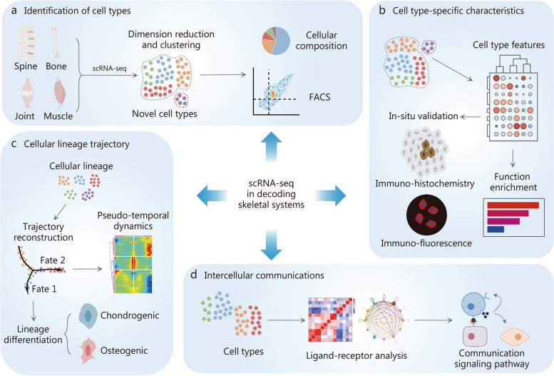
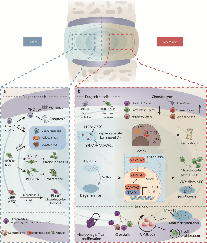
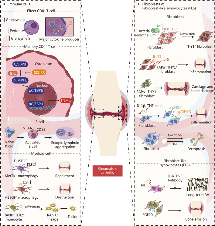
Orthopedic conditions have emerged as global health concerns, impacting approximately 1.7 billion individuals worldwide. However, the limited understanding of the underlying pathological processes at the cellular and molecular level has hindered the development of comprehensive treatment options for these disorders. The advent of single-cell RNA sequencing (scRNA-seq) technology has revolutionized biomedical research by enabling detailed examination of cellular and molecular diversity. Nevertheless, investigating mechanisms at the single-cell level in highly mineralized skeletal tissue poses technical challenges. In this comprehensive review, we present a streamlined approach to obtaining high-quality single cells from skeletal tissue and provide an overview of existing scRNA-seq technologies employed in skeletal studies along with practical bioinformatic analysis pipelines. By utilizing these methodologies, crucial insights into the developmental dynamics, maintenance of homeostasis, and pathological processes involved in spine, joint, bone, muscle, and tendon disorders have been uncovered. Specifically focusing on the joint diseases of degenerative disc disease, osteoarthritis, and rheumatoid arthritis using scRNA-seq has provided novel insights and a more nuanced comprehension. These findings have paved the way for discovering novel therapeutic targets that offer potential benefits to patients suffering from diverse skeletal disorders.

Keywords: Bioinformatic analysis; Cellular heterogeneity; Musculoskeletal system; Single cell suspension; Single-cell RNA sequencing (scRNA-seq); Skeletal disorders.


Conflict of interest statement

The authors declare that they have no competing interests.

Figures

Fig. 1
Fig. 1
Single-cell RNA sequencing (scRNA-seq) reveals the cellular heterogeneity in unprecedented resolution in skeletal research. a scRNA-seq can dissect the cellular composition of specific skeletal tissues in different conditions, providing strategies of prospective isolation for target cell populations using fluorescence-activated cell sorting (FACS). b Differential expression analysis of scRNA-seq data helps identify both classic and novel characteristics of cell clusters. c The fate of cells can be predicted using single-cell trajectory inference methods, which map the developmental pathways of cells based on their gene expression profiles. d The relationships and intercellular communications among different cell clusters can be predicted through scRNA-seq data, which is crucial for understanding tissue function and disease progression
Fig. 2
Fig. 2
Overview of single-cell acquisition technologies used to acquire high-quality single-cell suspensions from skeletal tissues. a Mechanical dissociation involves techniques such as microdissection, sectioning, and grinding to physically separate cells. b Enzymatic digestion is an effective method that utilizes enzymes like trypsin and collagenase to break down the extracellular matrix, facilitating cell separation. DNase is also employed to extract free DNA from cell clusters. c (i) Fluorescence-activated cell sorting is a pivotal and precise technology for isolating single cells through fluorescence markers. (ii) Magnetic bead sorting employs magnetic beads to acquire single-cell. (iii) Microfluidic technologies represent advanced methods for single-cell acquisition. These technologies are notable for their compact design, high throughput, and enhanced sensitivity, making them indispensable in modern cellular biology
Fig. 3
Fig. 3
Performance of different batch effect removal strategies in the integration of scRNA-seq datasets on human IVD cells. a Evaluation, applicable programming language and website of FastMNN, Seurat v3 (CCA), Harmony and scGen methods. b The dimensionality reduction plots of raw data, FastMNN, Seurat v3 (CCA), Harmony and scGen contain two rows. In the first row, cells are colored by different sites of intervertebral disc, and in the second by cell type. Seurat v3, Harmony are embedded in t-SNE, and FastMNN, scGen are embedded in UMAP. Each method can well eliminate the batch effect while FastMNN and scGen have better performance [23]. Copyright © 2021, Published by Springer Nature. AF annulus fibrosus, Chond chondrocyte, CEP cartilaginous endplate, FastMNN fast mutual nearest neighbors, NPPC nucleus pulposus progenitor cells, t-SNE t-distributed stochastic neighbor embedding, UMAP uniform manifold approximation and projection, PCA principal component analysis, CCA canonical correlation analysis, NP nucleus pulposus, IVD intervertebral disc, Noto notochord cell, EC endothelial cell
Fig. 4
Fig. 4
Single-cell RNA sequencing unveiled distinct cell types including progenitor cells and chondrocytes in healthy intervertebral discs (IVDs) and critical biological processes including matrix changes and immune activation during degenerative conditions. a In healthy IVDs, a variety of cells including progenitor cells, chondrocytes, and notochord cells play crucial roles in maintaining the IVD homeostasis. b In degenerated IVDs, there is a notable alteration in the phenotypes of progenitor cells and chondrocytes. The stiffening of the matrix activates YAP/TAZ signaling pathways, which in turn promotes chondrocyte proliferation and contributes to IVD fibrosis. Concurrently, macrophages and T cells not only proliferate but also engage in active crosstalk, influencing inflammatory responses. Additionally, the number of G-MDSCs increases, which plays a role in inhibiting matrix degeneration and suppressing T cell proliferation. AF annulus fibrosus, AFSC annulus fibrosus stem cell, Chond chondrocytes, NPC nucleus pulposus cell, NPPC nucleus pulposus progenitor cells, ProNP nucleus pulposus progenitors, TNC tenascin-C, TGF-β transforming growth factor-β, PDGFRA platelet-derived growth factor receptor alpha, FTL ferritin light chain, Fibro fibrogenic, MMP13 matrix metallopeptidase 13, YAP Yes-associated protein, TAZ tafazzin, TEAD1 TEA domain transcription factor 1, CCNB1 G2/mitotic-specific cyclin-B1, CTGF connective tissue growth factor, G-MDSCs granulocyte-like myeloid derived suppressor cells, UTS2R urotensin-2 receptor, PROCR protein C receptor, LEPR leptin receptor, SFMA/HAMA/FCI methacrylated SF/methacrylated HA/fibrochondrogenic inductive, HO-1 heme oxygenase-1
Fig. 5
Fig. 5
Single-cell RNA sequencing analysis elucidated critical alterations during the progression of osteoarthritis. a Different clusters of progenitor cells in distinguish stages of mice joints. b In osteoarthritis, chondrocytes undergo senescence and ferroptosis processes that contribute to cell aging and death. This cellular deterioration is associated with increased pain sensitivity in the affected joints. c Fibroblasts, and immune cells are triggered in osteoarthritis and improve inflammation, neuronal growth and chondrocyte senescence. OA osteoarthritis, ZEB1 Zinc finger E-box binding homeobox 1, FAP fibroblast activation protein, ECM extracellular matrix, TRPV1 transient receptor potential cation channel subfamily V member 1, GPX4 glutathione peroxidase 4, GSSG glutathione disulfide, GSH glutathione, EAAT solute carrier eamily 1 member 3, PIEZO2 piezo-type mechanosensitive ion channel component 2, Ntrk 1 high affinity nerve growth factor receptor, GDF5 growth/differentiation factor 5, CDKN2A cyclin-dependent kinase inhibitor 2 A, SERPINE1 plasminogen activator inhibitor 1, CHI3L1 chitinase-3-like protein 1, CD cluster of differentiation, RARRES2 retinoic acid receptor responder protein 2, LGALS1 galectin-1, Ly6e lymphocyte antigen 6E, Prg4 proteoglycan 4, Rspo2 r-spondin-2, IL interleukin, IGF2BP3 insulin-like growth factor 2 mRNA binding protein 3
Fig. 6
Fig. 6
Single-cell RNA analysis unveiled the intricate roles of immune cells, fibroblasts, and fibroblast-like synoviocyte subclusters in driving inflammation, bone erosion, and other pathological processes in rheumatoid arthritis. a Effect CD8+ T cells are the major cytokine producer in RA. IL1 activates C/EBPβ phosphorylation to promote TNF production in Memory CD4+ T cells while SIGIRR plays an inhibitory role. Ectopic lymphoid B cells are activated in RA synovium. Myeloid cells play an important role in joint repairment and destruction. b Fibroblasts promote joint inflammation in RA, and FLSs drives long-term RA and contribute to bone erosion. RA rheumatoid arthritis, pC/EBPβ phosphorylated CCAAT/enhancer binding protein β, DUSP1 dual specificity protein phosphatase 1, KLF2 krueppel-like factor 2, EGF epidermal growth factor, THY1 thy-1 membrane glycoprotein, TNF tumor necrosis factor, TGF-β transforming growth factor-β, FGF10 fibroblast growth factor 10, FLS fibroblast-like synoviocyte, IL interleukin, JAG1 protein jagged-1, NOTCH3 notch homolog protein 3, CD cluster of differentiation, NR4A1 nuclear receptor subfamily 4 immunity group A member 1, RANK receptor activator for nuclear factor-κB, TLR2 Toll-like receptor 2, FAP fibroblast activation protein, proNGF nerve growth factor precursor, p75NTR p75 neurotrophin receptor, SIGIRR single immunoglobulin IL-1R-related receptor

References

    1. Zhou M, Wang H, Zeng X, Yin P, Zhu J, Chen W, et al. Mortality, morbidity, and risk factors in China and its provinces, 1990–2017: a systematic analysis for the global burden of Disease Study 2017. Lancet. 2019;394(10204):1145–58. doi: 10.1016/S0140-6736(19)30427-1. - DOI - PMC - PubMed
    1. Knezevic NN, Candido KD, Vlaeyen JWS, Van Zundert J, Cohen SP. Low back pain. Lancet. 2021;398(10294):78–92. doi: 10.1016/S0140-6736(21)00733-9. - DOI - PubMed
    1. Hunter DJ, Bierma-Zeinstra S, Osteoarthritis Lancet. 2019;393(10182):1745–59. doi: 10.1016/S0140-6736(19)30417-9. - DOI - PubMed
    1. Smolen JS, Aletaha D, McInnes IB. Rheumatoid arthritis. Lancet. 2016;388(10055):2023–38. doi: 10.1016/S0140-6736(16)30173-8. - DOI - PubMed
    1. Compston JE, McClung MR, Leslie WD. Osteoporos Lancet. 2019;393(10169):364–76. doi: 10.1016/S0140-6736(18)32112-3. - DOI - PubMed

LinkOut - more resources