. 2024 Jul 18;5(3):100314.
doi: 10.1016/j.xhgg.2024.100314. Epub 2024 May 29.

Whole genome sequencing identifies elusive variants in genetically unsolved Italian inherited retinal disease patients

Affiliations

Whole genome sequencing identifies elusive variants in genetically unsolved Italian inherited retinal disease patients

Roberta Zeuli et al. HGG Adv. .

Abstract

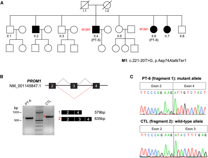
Inherited retinal diseases (IRDs) are a group of rare monogenic diseases with high genetic heterogeneity (pathogenic variants identified in over 280 causative genes). The genetic diagnostic rate for IRDs is around 60%, mainly thanks to the routine application of next-generation sequencing (NGS) approaches such as extensive gene panels or whole exome analyses. Whole-genome sequencing (WGS) has been reported to improve this diagnostic rate by revealing elusive variants, such as structural variants (SVs) and deep intronic variants (DIVs). We performed WGS on 33 unsolved cases with suspected autosomal recessive IRD, aiming to identify causative genetic variants in non-coding regions or to detect SVs that were unexplored in the initial screening. Most of the selected cases (30 of 33, 90.9%) carried monoallelic pathogenic variants in genes associated with their clinical presentation, hence we first analyzed the non-coding regions of these candidate genes. Whenever additional pathogenic variants were not identified with this approach, we extended the search for SVs and DIVs to all IRD-associated genes. Overall, we identified the missing causative variants in 11 patients (11 of 33, 33.3%). These included three DIVs in ABCA4, CEP290 and RPGRIP1; one non-canonical splice site (NCSS) variant in PROM1 and three SVs (large deletions) in EYS, PCDH15 and USH2A. For the previously unreported DIV in CEP290 and for the NCCS variant in PROM1, we confirmed the effect on splicing by reverse transcription (RT)-PCR on patient-derived RNA. This study demonstrates the power and clinical utility of WGS as an all-in-one test to identify disease-causing variants missed by standard NGS diagnostic methodologies.

Keywords: DIVs; IRD; SVs; WGS; deep intronic variants; inherited retinal diseases; structural variants; unsolved monoallelic cases.


Conflict of interest statement

Declaration of interests The authors declare no competing interests.

Figures

Figure 1
Figure 1
Clinical composition of the analyzed cohort
Figure 2
Figure 2
Pedigrees carrying biallelic CEP290 variants and effect on splicing of the deep-intronic variant c.6136-436A>G (A) Pedigrees of PT-9 and PT-10 showing the presumed autosomal recessive inheritance pattern of the RD and the segregation of the variants. (B) Graphical representation of the predicted effect of the DIV (c.6136-436A>G, p.Ile2046AspfsTer29) in CEP290, which interferes with the normal splicing (black dashed line) inducing the insertion of a 97-bp pseudo-exon (PE, red-framed square) between exons 44 and 45 (black squares) of CEP290. (C) Sanger sequencing of the RT-PCR products obtained from mRNA of patient (PT-10) and control (CTL) samples. The chromatogram shows the sequences of both the wild-type (exons 44–45) and mutant alleles (exons 44-PE-45) in PT-10, while only the wild-type sequence is detected in the CTL sample.
Figure 3
Figure 3
Effect on splicing of the non-canonical splice site variant in PROM1 (A) Pedigree of PT-5 and PT-6 showing the suspected autosomal recessive inheritance pattern of the RD. (B) Graphical representation of the predicted effect of the NCSS variant in PROM1 and agarose gel electrophoresis of the RT-PCR products obtained from mRNA of patient (PT-6) and control (CTL) samples. The c.221-20T>G, p.Asp74AlafsTer1 variant is predicted to induce skipping of exon 3 (red dashed line). RT-PCR on PT-6 yielded only one fragment with lower molecular weight (fragment 1, 579 bp) than the one obtained in the control sample (fragment 2, 635 bp), consistent with the skipping of exon 3 (56 bp). (C) Sanger sequencing of the RT-PCR products obtained from mRNA of PT-6 and control samples (CTL). PT-6 was homozygous for the c.221-20T>G variant, hence the chromatogram of the fragment 1 only shows the sequence of the mutant allele confirming skipping of exon 3.
Figure 4
Figure 4
Overview of the deletions detected in EYS, PCDH15, and USH2A Using the Manta Structural Variant caller, we identified three large deletions in EYS, PCDH15, and USH2A in three patients (PT-13, PT-21, PT-29) who carried monoallelic (likely) pathogenic variants in the respective gene. (A) Visualization of WGS data in the Integrative Genomics Viewer (IGV) revealed split reads (red colored reads) corresponding to the breakpoints of the deletions identified by Manta in all patients. In PT-13 a 380.9-kb deletion was detected, with breakpoints located in introns 22 and 13 of EYS, causing the deletion of exons 14 to 22. The deletion in PT-21 was spanning 179.2 kb, from intron 28 to intron 18 of PCDH15. In PT-29, a deletion of 77.4 kb encompassing exon 22 of USH2A was detected. (B) Agarose gel electrophoresis and schematic representation of the PCR products obtained using two different primer sets (Table S1), one amplifying the wild-type alleles (fragments 1, 3, and 5; red numbers) and the other spanning the breakpoint, thus amplifying the mutant alleles (fragments 2, 4, and 6). The fragments spanning the breakpoint were present in all patients (PT-13, PT-21, and PT-29) and in one carrier parent (father of PT-21) and were absent in control (CTL) samples. (C) The chromatograms obtained by Sanger sequencing of the wild-type (upper) and mutant (lower) alleles showed the breakpoints (vertical line) only in the fragments corresponding to the mutant alleles. The genomic coordinates of either breakpoint are reported below the chromatogram of each mutant allele. All genomic coordinates are referred to the Human Reference Genome build GRCh38/hg38.

References

    1. Berger W., Kloeckener-Gruissem B., Neidhardt J. The molecular basis of human retinal and vitreoretinal diseases. Prog. Retin. Eye Res. 2010;29:335–375. - PubMed
    1. Schneider N., Sundaresan Y., Gopalakrishnan P., Beryozkin A., Hanany M., Levanon E.Y., Banin E., Ben-Aroya S., Sharon D. Inherited retinal diseases: Linking genes, disease-causing variants, and relevant therapeutic modalities. Prog. Retin. Eye Res. 2022;89 - PubMed
    1. Cremers F.P.M., Lee W., Collin R.W.J., Allikmets R. Clinical spectrum, genetic complexity and therapeutic approaches for retinal disease caused by ABCA4 mutations. Prog. Retin. Eye Res. 2020;79 - PMC - PubMed
    1. Rivolta C., Sharon D., DeAngelis M.M., Dryja T.P. Retinitis pigmentosa and allied diseases: numerous diseases, genes, and inheritance patterns. Hum. Mol. Genet. 2002;11:1219–1227. - PubMed
    1. Eisenberger T., Neuhaus C., Khan A.O., Decker C., Preising M.N., Friedburg C., Bieg A., Gliem M., Charbel Issa P., Holz F.G., et al. Increasing the Yield in Targeted Next-Generation Sequencing by Implicating CNV Analysis, Non-Coding Exons and the Overall Variant Load: The Example of Retinal Dystrophies. Li T., editor. PLoS One. 2013;8 - PMC - PubMed

MeSH terms