. 2023 Dec;2(12):1159-1172.
doi: 10.1038/s44161-023-00375-y. Epub 2023 Dec 4.

Whole-genome sequencing uncovers two loci for coronary artery calcification and identifies ARSE as a regulator of vascular calcification

Paul S de Vries  1 Matthew P Conomos  2 Kuldeep Singh  3 Christopher J Nicholson  3 Deepti Jain  2 Natalie R Hasbani  1 Wanlin Jiang  3 Sujin Lee  3 Christian L Lino Cardenas  3 Sharon M Lutz  4   5 Doris Wong  6 Xiuqing Guo  7 Jie Yao  7 Erica P Young  8 Catherine Tcheandjieu  9   10 Austin T Hilliard  9   11 Joshua C Bis  12 Lawrence F Bielak  13 Michael R Brown  1 Shaila Musharoff  9   14 Shoa L Clarke  9   10 James G Terry  15 Nicholette D Palmer  16 Lisa R Yanek  17 Huichun Xu  18 Nancy Heard-Costa  19   20 Jennifer Wessel  21   22 Margaret Sunitha Selvaraj  23   24   25 Rebecca H Li  3 Xiao Sun  26   27 Adam W Turner  6 Adrienne M Stilp  2 Alyna Khan  2 Anne B Newman  28 Asif Rasheed  29 Barry I Freedman  30 Brian G Kral  31 Caitlin P McHugh  2 Chani Hodonsky  6 Danish Saleheen  29   32   33 David M Herrington  34 David R Jacobs Jr  35 Deborah A Nickerson  36   37 Eric Boerwinkle  1   38 Fei Fei Wang  2 Gerardo Heiss  39 Goo Jun  1 Greg L Kinney  40 Haakon H Sigurslid  3 HarshaVardhan Doddapaneni  38 Ira M Hall  41 Isabela M Bensenor  42 Jai Broome  2 James D Crapo  43 James G Wilson  44 Jennifer A Smith  13   45 John Blangero  46   47 Jose D Vargas  48 Jose Verdezoto Mosquera  6 Joshua D Smith  36   37 Karine A Viaud-Martinez  49 Kathleen A Ryan  18 Kendra A Young  40 Kent D Taylor  7 Leslie A Lange  50 Leslie S Emery  2 Marcio S Bittencourt  42 Matthew J Budoff  51 May E Montasser  18 Miao Yu  13 Michael C Mahaney  46   47 Mohammed S Mahamdeh  3 Myriam Fornage  1   52 Nora Franceschini  53 Paulo A Lotufo  42 Pradeep Natarajan  23   24   25 Quenna Wong  2 Rasika A Mathias  17   54 Richard A Gibbs  38   55 Ron Do  56   57 Roxana Mehran  58 Russell P Tracy  59 Ryan W Kim  60 Sarah C Nelson  2 Scott M Damrauer  61   62 Sharon L R Kardia  13 Stephen S Rich  6 Valentin Fuster  63   64 Valerio Napolioni  65 Wei Zhao  13 Wenjie Tian  3 Xianyong Yin  66 Yuan-I Min  67 Alisa K Manning  68   69 Gina Peloso  70 Tanika N Kelly  27 Christopher J O'Donnell  71   72 Alanna C Morrison  1 Joanne E Curran  46   47 Warren M Zapol  73 Donald W Bowden  16 Lewis C Becker  31 Adolfo Correa  67   74 Braxton D Mitchell  18   75 Bruce M Psaty  12   76   77 John Jeffrey Carr  15 Alexandre C Pereira  78   79 Themistocles L Assimes  9   10 Nathan O Stitziel  8   80   81 John E Hokanson  40 Cecelia A Laurie  2 Jerome I Rotter  7 Ramachandran S Vasan  20   82   83 Wendy S Post  31 Patricia A Peyser  13 Clint L Miller  6 Rajeev Malhotra  3
Affiliations

Whole-genome sequencing uncovers two loci for coronary artery calcification and identifies ARSE as a regulator of vascular calcification

Paul S de Vries et al. Nat Cardiovasc Res. 2023 Dec.

Abstract

Coronary artery calcification (CAC) is a measure of atherosclerosis and a well-established predictor of coronary artery disease (CAD) events. Here we describe a genome-wide association study (GWAS) of CAC in 22,400 participants from multiple ancestral groups. We confirmed associations with four known loci and identified two additional loci associated with CAC (ARSE and MMP16), with evidence of significant associations in replication analyses for both novel loci. Functional assays of ARSE and MMP16 in human vascular smooth muscle cells (VSMCs) demonstrate that ARSE is a promoter of VSMC calcification and VSMC phenotype switching from a contractile to a calcifying or osteogenic phenotype. Furthermore, we show that the association of variants near ARSE with reduced CAC is likely explained by reduced ARSE expression with the G allele of enhancer variant rs5982944. Our study highlights ARSE as an important contributor to atherosclerotic vascular calcification, and a potential drug target for vascular calcific disease.


Conflict of interest statement

Bruce M. Psaty serves on the Steering Committee of the Yale Open Data Access Project funded by Johnson & Johnson. Leslie S. Emery is now an employee of Celgene/Bristol Myers Squibb. Celgene/Bristol Myers Squibb had no role in the funding, design, conduct, or interpretation of this study. Nathan O. Stitziel has received research funding from Regeneron Pharmaceuticals, unrelated to this work. Karine A. Viaud-Martinez is an employee at Illumina Inc. Ryan W. Kim is an employee at Psomagen Inc. Roxona Mehran reports institutional research grants from Abbott Laboratories, Abiomed, Applied Therapeutics, AstraZeneca, Bayer, Beth Israel Deaconess, Bristol-Myers Squibb, CERC, Chiesi, Concept Medical, CSL Behring, DSI, Medtronic, Novartis Pharmaceuticals, OrbusNeich; consultant fees from Abbott Laboratories, Boston Scientific, CardiaWave, Chiesi, Janssen Scientific Affairs, Medscape/WebMD, Medtelligence (Janssen Scientific Affairs), Roivant Sciences, Sanofi, Siemens Medical Solutions; consultant fees paid to the institution from Abbott Laboratories, Bristol-Myers Squibb; advisory board, funding paid to the institution from Spectranetics/Philips/Volcano Corp; consultant (spouse) from Abiomed, The Medicines Company, Merck; Equity <1% from Claret Medical, Elixir Medical; DSMB Membership fees paid to the institution from Watermark Research Partners; consulting (no fee) from Idorsia Pharmaceuticals Ltd., Regeneron Pharmaceuticals; Associate Editor for ACC, AMA. Rajeev Malhotra is a consultant for MyoKardia (now owned by BMS), Epizon Pharma, Renovacor, and Third Pole, a co-founder of Patch and Angea Biotherapeutics, and has received research funding from Angea Biotherapeutics, Bayer Pharmaceuticals, and Amgen. Adrienne M. Stilp receives funding from Seven Bridges Genomics to develop tools for the NHLBI BioData Catalyst consortium. Pradeep Natarajan reports grants from Amgen, Apple, Boston Scientific, AstraZeneca, Allelica, Novartis, and Genentech, consulting income from GV, Blackstone Life Sciences, Foresite Labs, Apple, AstraZeneca, Allelica, Novartis, HeartFlow, and Genentech, is a scientific advisor to Esperion Therapeutics, Preciseli, and TenSixteen Bio, is a scientific co-founder of TenSixteen Bio, and spousal employment at Vertex, all unrelated to the present work. Clint L. Miller received a research grant from AstraZeneca for an unrelated project. The remaining authors declare no competing interests.

Figures

Extended Data Fig. 1:
Extended Data Fig. 1:. Coronary artery calcification levels across ARSE index variant genotypes suggest a recessive mode of inheritance.
Mean log (coronary artery calcification + 1) across genotypes for index variants at ARSE. Error bars indicate the 95% confidence interval for the mean (n=22,400).
Extended Data Fig. 2:
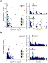
Extended Data Fig. 2:. Co-localization of genetic associations with coronary artery calcification and genetic associations with ARSE expression in a) cultured fibroblasts and b) aorta.
These plots were created with Locuscomparer (https://github.com/boxiangliu/locuscomparer) using the African 1000G ph3 reference population to calculate the linkage disequilibrium r. Unadjusted two-sided P values are provided. The posterior probability for causal variant sharing was 99.9% in cultured fibroblasts and 8.9% in aorta.
Extended Data Fig. 3:
Extended Data Fig. 3:. Co-localization of genetic associations with coronary artery calcification and genetic associations with MMP16 expression in aorta.
This plot was created with Locuscomparer (https://github.com/boxiangliu/locuscomparer) using the European 1000G ph3 reference population to calculate the linkage disequilibrium r. Unadjusted two-sided P values are provided. The posterior probability for causal variant sharing was 81.9%.
Extended Data Fig. 4:
Extended Data Fig. 4:. Cell type-specific gene expression of ARSE and MMP16 in an integrated human atherosclerosis reference dataset.
(a-b) Uniform Manifold Approximation and Projection (UMAP) embeddings from an integrated human carotid and coronary artery atherosclerosis single-cell RNA-seq reference dataset (Methods), showing (a) ARSE and (b) MMP16, normalized gene expression depicted by the heatmap from SCTransform normalized read counts. Individual sequencing libraries across four studies were harmonized after QC and batch correction with reciprocal PCA (rPCA). Clusters were annotated with level 1 cell type labels using transfer learning with cell labels from the Tabula Sapiens vasculature subset. Level 2 cell type label for endothelial-mesenchymal transition (EndoMT) endothelial cells expressing ARSE and MMP16 are also highlighted. (c-d) Scatter plots showing the normalized expression level of (c) ARSE and (d) MMP16, across the level 1 cell types. EC: Endothelial cells; SMC: Smooth muscle cells; T/NK: T cells and Natural Killer cells; pDC: plasmacytoid dendritic cells.
Extended Data Fig. 5:
Extended Data Fig. 5:. Silencing MMP16 expression has no effect on osteogenic phenotypic switching in human coronary artery vascular smooth muscle cells.
a) Treatment of human coronary artery vascular smooth muscle cells (n = 6 biologically independent samples in each group) with osteogenic media decreased MMP16 mRNA expression by ~74% (left panel). Treatment of cells grown in osteogenic media with siMMP16 (resulting in >90% knockdown of MMP16 mRNA) had no effect on RUNX2 (middle panel) or CNN1 (right panel) mRNA levels. Statistical comparisons were made using a two-tailed one-way ANOVA with Sidak’s post-hoc comparison testing. The mean ± SEM is depicted in plots. b) Treatment of human coronary artery vascular smooth muscle cells grown in osteogenic media with siMMP16 had no effect on calcification, as evidenced by decreased Alizarin Red S staining.
Extended Data Fig. 6:
Extended Data Fig. 6:. Silencing ARSE expression increases contractile gene expression in human aortic vascular smooth muscle cells.
Silencing ARSE in cells (n = 12 biologically independent samples in each group) grown in normal media increased a) ACTA2 and b) TAGLN mRNA levels by ~ 56% and 35%, respectively. Statistical comparisons were made using a two-tailed Student t test. The mean is depicted in plots, with the error bars representing the standard error of the mean.
Extended Data Fig. 7:
Extended Data Fig. 7:. Human aortic vascular smooth muscle cell calcification, bone and contractile marker expression, and contractility are affected by changes in ARSE expression.
a) Treatment of human aortic vascular smooth muscle cells (n = 12 biologically independent samples in each group) with osteogenic media increased ARSE mRNA expression > 2-fold. b) Treatment of cells grown in osteogenic media with siARSE (resulting in >90% knockdown of ARSE mRNA) decreased RUNX2 (left panel), and BGLAP (middle panel) mRNA levels by ~20% and ~43% respectively, and increased CNN1 mRNA levels by > 150% (right panel). Silencing ARSE in cells grown in normal media increased CNN1 mRNA levels by > 2.5-fold. c) Treatment of cells grown in osteogenic media with siARSE reduced calcification, as evidenced by decreased Alizarin Red S staining (n=5 biologically independent samples in each group). d) Reduced ARSE expression in cells grown in collagen discs (left panel) resulted in a >3-fold increase in contraction (right panel, n=6 biologically independent samples in each group). e) Protein expression of ARSE, RUNX2 and CNN1 were confirmed by Western blot using antibodies directed against ARSE, RUNX2, CNN1 and GAPDH (for a loading control). Adenoviral expression of the 70-kDa isoform of ARSE in human aortic vascular smooth muscle cells was associated with a >15-fold increase in RUNX2 protein levels and an approximately 34% decrease in CNN1 protein levels, when cells were harvested 5 days after viral transduction (n=3 biologically independent samples in each group). f) As shown by Alizarin Red staining, increased ARSE expression resulted in augmented calcification in human aortic vascular smooth muscle cells (n=3 biologically independent samples in each group). g) Increased ARSE expression also caused a >70% decrease (right panel, n=6 biologically independent samples in each group) in contraction of human aortic vascular smooth muscle cells grown in collagen discs (left panel). Statistical comparisons were made using either a two-tailed one-way ANOVA with Sidak’s post-hoc comparison testing or a two-tailed Student t test. The mean is depicted in plots, with the error bars representing the standard error of the mean.
Extended Data Fig. 8:
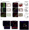
Extended Data Fig. 8:. ARSE expression and calcification in normal and ischemic human coronary arteries.
a) Cross sections of human coronary arteries from control subjects and patients with ischemic coronary artery disease (n=3 individuals in each group with 2 sections stained for each individual) were stained for ARSE (red), α-smooth muscle actin (green) and DNA (blue, DAPI). Immunofluorescence analysis shows a higher expression of ARSE in diseased arteries. Alizarin red staining for calcification was high in the coronary arteries of diseased patients with no significant stain observed in the control group. Scale bars, 200 µm for each immunofluorescence image; 500 µm for each Alizarin red staining image. Statistical comparisons were made using a two-tailed Student’s t test. The mean is depicted in plots, with the error bars representing the standard error of the mean. b) Cross sections of human coronary arteries (n=1 each for control and ischemic patient) were stained for ARSE (red), RUNX2 (green, VSMC calcification marker) and DNA (blue, DAPI). Immunofluorescence analysis shows a higher expression of ARSE in calcified diseased arteries that colocalized with increased RUNX2 expression. Scale bar 500 µm.
Extended Data Fig. 9:
Extended Data Fig. 9:. Luciferase reporter assay to analyze the functional impact of SNP rs5982944 (A/G).
The rs5982944-A allele and rs5982944-G allele firefly luciferase constructs were co-transfected with renilla luciferase plasmid into human coronary smooth muscle cells (HCSMCs), human aortic smooth muscle cells (HASMCs) and HEK-293 cells (n=6 biologically independent samples for HCSMCs and HASMCs and n=5 biologically independent samples for HEK-293). Firefly luciferase activity and renilla luciferase activity (internal control reporter vector) were measured sequentially in cell lysates. Firefly luciferase activity in cell lysates transfected with either the rs5982944-A construct or the rs5982944-G construct was normalized to renilla luciferase activity. The mean is depicted in plots, with the error bars representing the standard error of the mean. Statistical comparisons were made using the two-tailed unpaired t-test.
Extended Data Fig. 10:
Extended Data Fig. 10:. Model of ARSE-induced phenotype switch from contractile to osteogenic vascular smooth muscle cells.
Atherosclerotic vascular calcification is characterized by the phenotype switch of vascular smooth muscle cells (VSMCs) from a contractile phenotype to a proliferative, osteogenic phenotype. The osteogenic phenotype of VSMCs is characterized by decreased expression of contractile proteins such as calponin (CNN1), but increased expression of Runt-related transcription factor 2 (RUNX2), a master regulator of the phenotype switch, in addition to other markers of calcification such as bone gamma-carboxyglutamate protein (BGLAP) and alkaline phosphatase (ALPL). We identified ARSE as a major regulator of the phenotype switch.
Fig. 1:
Fig. 1:. Manhattan plot for the genome-wide association study of log(CAC+1).
In the Manhattan plot, each genetic variant is inluded as a dot, with the position on the x axis corresponding to their genomic position and the position on the y axis corresponding to the significance the association, denoted by –log10 transformed two-sided P-values. The plot shows 6 genetic loci (including 2 novel and 4 known) associated with coronary artery calcification score at a significance level of P <5×10 (dotted line) in the pooled analysis of 22,400 individuals from 10 studies.
Fig. 2:
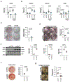
Fig. 2:. Silencing ARSE expression inhibits osteogenic phenotype switch in human coronary artery vascular smooth muscle cells.
a) Treatment of human coronary artery vascular smooth muscle cells (n = 6 biologically independent samples in each group) with osteogenic media for 3 days increased ARSE mRNA expression approximately 5-fold. Treatment of cells grown in osteogenic media with siARSE resulted in >90% knockdown of ARSE mRNA. b) Protein expression of ARSE was measured by immunoblot (left panel) using antibodies directed against ARSE and GAPDH (for a loading control). Treatment of cells with osteogenic media increased ARSE protein levels by 1.5-fold (right panel). Treatment of cells grown in osteogenic media with siARSE resulted in >70% reduction of ARSE protein (n=4 biologically independent samples in each group). c) Treatment of cells grown in osteogenic media with siARSE ameliorated osteogenic phenotype switch as evidenced by decreased RUNX2, BGLAP, and ALPL mRNA levels, and increased CNN1 mRNA levels ~2-fold. Of note, silencing ARSE in cells grown in normal media increased CNN1 mRNA levels by > 5-fold (n=6 biologically independent samples in each group except n=5 for siARSE CNN1 data). d) Reduced ARSE expression was also associated with an approximately 50% decrease in RUNX2 and >30% increase in CNN1 protein levels assessed by immunoblot using antibodies directed against RUNX2, CNN1 and VCL (for a loading control) (n=6 biologically independent samples in each group). e) Treatment of cells grown in osteogenic media with siARSE reduced calcification by approximately 60% (right panel, n = 3 biologically independent samples in each group), as evidenced by decreased Alizarin Red staining (left panel). f) Reduced ARSE expression with siARSE treatment in human coronary artery vascular smooth muscle cells grown in collagen discs (left panel) resulted in a >3-fold increase in contraction (right panel, n=6 biologically independent samples in each group). Statistical comparisons were made using either a two-tailed one-way ANOVA with Sidak’s post-hoc comparison testing (for more than two groups) or a two-tailed Student t test (for two groups). The mean is depicted in plots, with the error bars representing the mean ± the standard error of the mean.
Fig. 3:
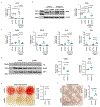
Fig. 3:. Overexpression of ARSE induces calcification in human coronary artery vascular smooth muscle cells.
a) Adenoviral expression of ARSE in human coronary artery vascular smooth muscle cells was associated with an 8-fold increase in RUNX2 protein levels and an approximately 70% decrease in CNN1 protein levels, when cells were harvested 5 days after viral transduction (n=3 biologically independent samples in each group). Protein expression was determined by immunoblot (left panel) using antibodies directed against ARSE, RUNX2, CNN1 and GAPDH (for a loading control) with quantification shown in the right panel. b) As shown by Alizarin Red staining (left panel), increased ARSE expression resulted in augmented calcification in human coronary artery vascular smooth muscle cells (right panel, n = 3 biologically independent samples in each group). Two independent experiments were performed with representative images shown. c) Increased ARSE expression also caused a >70% decrease (right panel, n=6 biologically independent samples in each group) in contraction of human coronary artery vascular smooth muscle cells grown in collagen discs (left panel). Statistical comparisons were made using a two-tailed Student t test. The mean is depicted in plots, with the error bars representing the mean ± the standard error of the mean.

References

    1. Kavousi M et al. Evaluation of newer risk markers for coronary heart disease risk classification: a cohort study. Ann Intern Med 156, 438–44 (2012). - PubMed
    1. Budoff MJ et al. Ten-year association of coronary artery calcium with atherosclerotic cardiovascular disease (ASCVD) events: the multi-ethnic study of atherosclerosis (MESA). Eur Heart J 39, 2401–2408 (2018). - PMC - PubMed
    1. Greenland P, Blaha MJ, Budoff MJ, Erbel R & Watson KE Coronary Calcium Score and Cardiovascular Risk. J Am Coll Cardiol 72, 434–447 (2018). - PMC - PubMed
    1. Polonsky TS et al. Coronary artery calcium score and risk classification for coronary heart disease prediction. JAMA 303, 1610–6 (2010). - PMC - PubMed
    1. Shi X et al. Calcification in Atherosclerotic Plaque Vulnerability: Friend or Foe? Front Physiol 11, 56 (2020). - PMC - PubMed

METHODS-ONLY REFERENCES

    1. Fang H et al. Harmonizing Genetic Ancestry and Self-identified Race/Ethnicity in Genome-wide Association Studies. Am J Hum Genet 105, 763–772 (2019). - PMC - PubMed
    1. Taliun D et al. Sequencing of 53,831 diverse genomes from the NHLBI TOPMed Program. bioRxiv (2019). - PMC - PubMed
    1. Agatston AS et al. Quantification of coronary artery calcium using ultrafast computed tomography. J Am Coll Cardiol 15, 827–32 (1990). - PubMed
    1. Carr JJ et al. Calcified coronary artery plaque measurement with cardiac CT in population-based studies: standardized protocol of Multi-Ethnic Study of Atherosclerosis (MESA) and Coronary Artery Risk Development in Young Adults (CARDIA) study. Radiology 234, 35–43 (2005). - PubMed
    1. Stilp AM et al. A system for phenotype harmonization in the NHLBI Trans-Omics for Precision Medicine (TOPMed) Program. bioRxiv (2020). - PMC - PubMed