

Computational studies on rep and capsid proteins of CRESS DNA viruses
- PMID: 38817400
- PMCID: PMC11133267
- DOI: 10.1007/s13337-024-00858-x
Computational studies on rep and capsid proteins of CRESS DNA viruses
Abstract
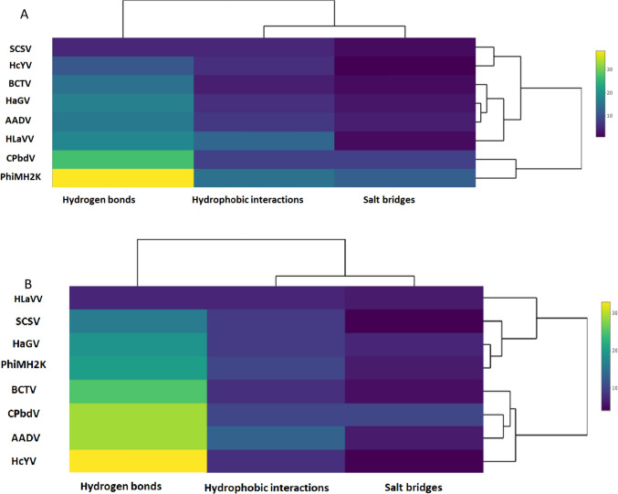
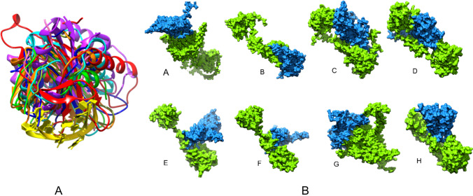
The circular rep-encoding single-stranded DNA viruses (CRESS DNA viruses) are among the smallest, with 2-6 kb ssDNA genomes that encode for a coat protein (C) and a replication protein (R). To comprehend the complexity and divergence of the C and R proteins, we have created predictive structural models of representative viruses infecting unique hosts from each family using the neural network-based method AlphaFold2 and carried out molecular dynamic simulations to assess their stability. The structural characteristics indicate that differences in loops and amino-terminus may play a significant role in facilitating adaptations to multiple hosts and vectors. In comparison to the C, the Rs show a high degree of conservation and structural mimicry of the nuclease-helicase domains of plasmids. A phylogenetic analysis based on the structures and sequences of the C and R proteins reveals evolutionary variances. Our study also highlights the conservation of structural components involved in the interaction of R with the conserved intergenic region of the genome. Further, we envisage that the adaptability of R's central linker may be crucial for establishing interactions with multiple protein partners, including C.
Supplementary information: The online version contains supplementary material available at 10.1007/s13337-024-00858-x.
Keywords: Capsid and rep protein; Interaction; MD simulation; Phylogenetic analysis; Single-stranded DNA; Virus.
© The Author(s), under exclusive licence to Indian Virological Society 2024. Springer Nature or its licensor (e.g. a society or other partner) holds exclusive rights to this article under a publishing agreement with the author(s) or other rightsholder(s); author self-archiving of the accepted manuscript version of this article is solely governed by the terms of such publishing agreement and applicable law.
Conflict of interest statement
Conflict of interestNone.
Figures


Similar articles
-
Pervasive Chimerism in the Replication-Associated Proteins of Uncultured Single-Stranded DNA Viruses.Viruses. 2018 Apr 10;10(4):187. doi: 10.3390/v10040187. Viruses. 2018. PMID: 29642587 Free PMC article.
-
Unveiling Crucivirus Diversity by Mining Metagenomic Data.mBio. 2020 Sep 1;11(5):e01410-20. doi: 10.1128/mBio.01410-20. mBio. 2020. PMID: 32873755 Free PMC article.
-
Genetic Diversity and Characterization of Circular Replication (Rep)-Encoding Single-Stranded (CRESS) DNA Viruses.Microbiol Spectr. 2022 Dec 21;10(6):e0105722. doi: 10.1128/spectrum.01057-22. Epub 2022 Nov 8. Microbiol Spectr. 2022. PMID: 36346238 Free PMC article.
-
Eukaryotic Circular Rep-Encoding Single-Stranded DNA (CRESS DNA) Viruses: Ubiquitous Viruses With Small Genomes and a Diverse Host Range.Adv Virus Res. 2019;103:71-133. doi: 10.1016/bs.aivir.2018.10.001. Epub 2018 Dec 5. Adv Virus Res. 2019. PMID: 30635078 Review.
-
Diversity of small, single-stranded DNA viruses of invertebrates and their chaotic evolutionary past.J Invertebr Pathol. 2016 Oct;140:83-96. doi: 10.1016/j.jip.2016.09.005. Epub 2016 Sep 20. J Invertebr Pathol. 2016. PMID: 27663091 Review.
Cited by
-
Twenty years of advances in prediction of nucleic acid-binding residues in protein sequences.Brief Bioinform. 2024 Nov 22;26(1):bbaf016. doi: 10.1093/bib/bbaf016. Brief Bioinform. 2024. PMID: 39833102 Free PMC article. Review.
References
-
- Bennett A, Agbandje-McKenna M. Geminivirus structure and assembly. Cambridge: Academic press; 2020. pp. 1–32. - PubMed
-
- Chattopadhyay A, Mandal B. Applied plant virology. Netherlans: Elsevier; 2020. Hypotheses of virus origin and evolutionary patterns of plant viruses; pp. 779–796.
LinkOut - more resources
Full Text Sources
