Unveiling Nasal Glial Heterotopia: A Pathological Perspective
- PMID: 38817464
- PMCID: PMC11137777
- DOI: 10.7759/cureus.59341
Unveiling Nasal Glial Heterotopia: A Pathological Perspective
Abstract
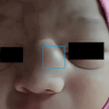
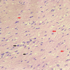
The uncommon, non-hereditary congenital abnormalities known as nasal glial heterotopias (NGH) are composed of heterotopic neuroglial tissue. Typically, NGH manifests in infancy, but occasionally it can also be seen in older children and adults. To rule out intracranial extension, magnetic resonance imaging (MRI) and computed tomography (CT) scans should be performed. Numerous cases have been documented where NGH was mistakenly identified as encephaloceles, teratomas, dermoid cysts, capillary haemangiomas, and even desmoids. A proper clinical, sonological, and even CT and MRI evaluation can lead to a near-final diagnosis; nonetheless, surgical excision and histological confirmation are the gold standards. We report a rare case of a firm, subcutaneous, non-tender, non-reducible midline 2 x 2 x 1 cm swelling with bluish-red skin near the root of the nose that was not affected by posture or pressure. Encephalocele, NGH, and dermoid were the differential diagnoses made based on the oedema found on CT and MRI scans. Histopathology provided a conclusive NGH diagnosis. The instance illustrates the significance of histology as the gold standard for NGH diagnosis.
Keywords: encephalocele; extranasal; intranasal; nasal glial heterotopia; nasal glioma.
Copyright © 2024, Deotale et al.
Conflict of interest statement
The authors have declared that no competing interests exist.
Figures


Similar articles
-
Congenital nasal neuroglial heterotopia and encephaloceles: An update on current evaluation and management.Laryngoscope. 2016 Sep;126(9):2161-7. doi: 10.1002/lary.25864. Epub 2016 Jan 13. Laryngoscope. 2016. PMID: 26763579
-
Histopathologic features of nasal glial heterotopia (nasal glioma).Childs Nerv Syst. 2022 Jan;38(1):63-75. doi: 10.1007/s00381-021-05369-4. Epub 2021 Sep 25. Childs Nerv Syst. 2022. PMID: 34562130
-
Pediatric subcutaneous nasal glial heterotopia.Surg Neurol Int. 2025 Jan 3;16:1. doi: 10.25259/SNI_93_2024. eCollection 2025. Surg Neurol Int. 2025. PMID: 39926455 Free PMC article.
-
[Nasal glial heterotopia: embryological and clinical approaches].Rev Stomatol Chir Maxillofac. 2006 Feb;107(1):44-9. doi: 10.1016/s0035-1768(06)76981-9. Rev Stomatol Chir Maxillofac. 2006. PMID: 16523177 Review. French.
-
Nasal glial heterotopia: a clinicopathologic and immunophenotypic analysis of 10 cases with a review of the literature.Ann Diagn Pathol. 2003 Dec;7(6):354-9. doi: 10.1016/j.anndiagpath.2003.09.010. Ann Diagn Pathol. 2003. PMID: 15018118 Review.
Cited by
-
A case of large oropharyngeal glial heterotopia causing airway obstruction in a newborn: A case report and brief review of literature.Int J Surg Case Rep. 2025 Sep;134:111809. doi: 10.1016/j.ijscr.2025.111809. Epub 2025 Aug 14. Int J Surg Case Rep. 2025. PMID: 40818395 Free PMC article.
References
-
- Management of the congenital midline nasal mass: a review. Hughes GB, Sharpino G, Hunt W, Tucker HM. Head Neck Surg. 1980;2:222–233. - PubMed
-
- Neuroglial heterotopia causing neonatal airway obstruction: presentation, management, and literature review. Husein OF, Collins M, Kang DR. https://doi.org/10.1007/s00431-008-0810-2. Eur J Pediatr. 2008;167:1351–1355. - PubMed
-
- A clinicopathological study of 15 patients with neuroglial heterotopias and encephaloceles of the middle ear and mastoid region. Gyure KA, Thompson LD, Morrison AL. Laryngoscope. 2000;110:1731–1735. - PubMed
Publication types
LinkOut - more resources
Full Text Sources
