. 2024 Apr 25;15(21):7982-7991.
doi: 10.1039/d4sc01755b. eCollection 2024 May 29.

Structural elucidation of HIV-1 G-quadruplexes in a cellular environment and their ligand binding using responsive 19F-labeled nucleoside probes

Affiliations

Structural elucidation of HIV-1 G-quadruplexes in a cellular environment and their ligand binding using responsive 19F-labeled nucleoside probes

Sarupa Roy et al. Chem Sci. .

Abstract

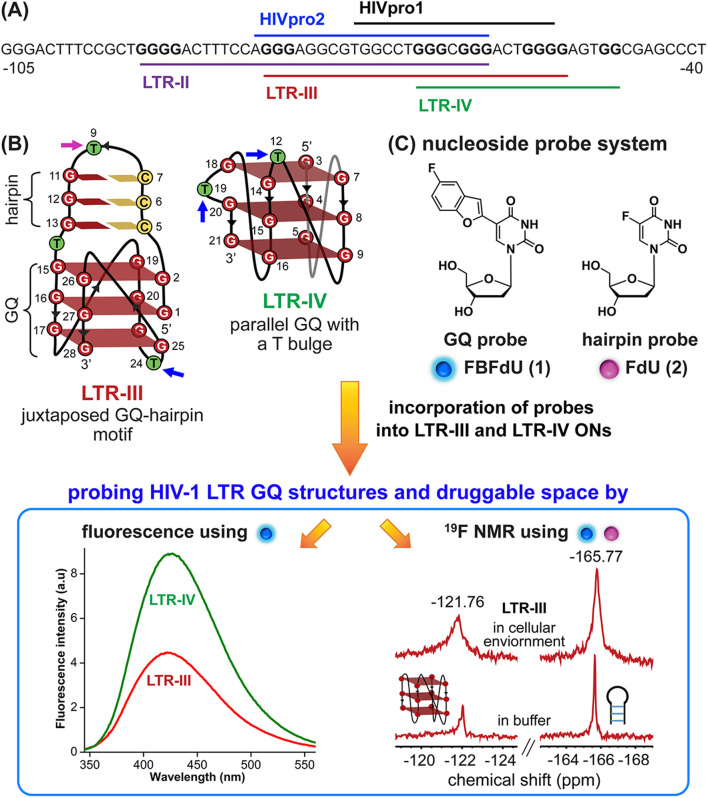
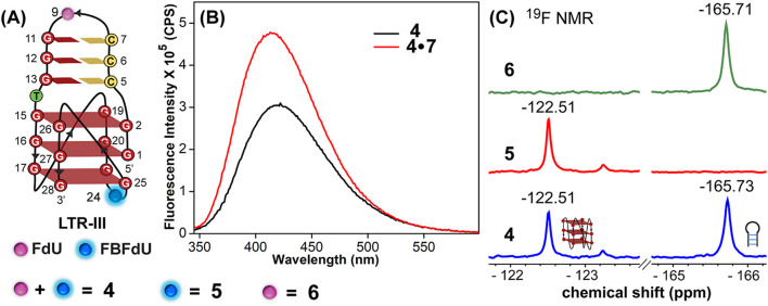
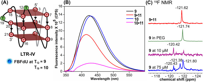
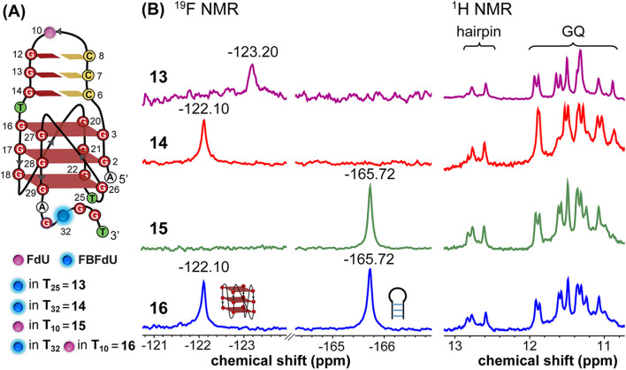
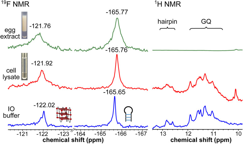
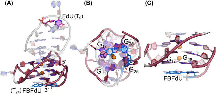
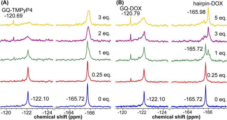
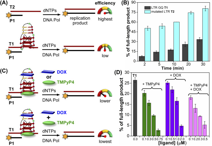
Understanding the structure and recognition of highly conserved regulatory segments of the integrated viral DNA genome that forms unique topologies can greatly aid in devising novel therapeutic strategies to counter chronic infections. In this study, we configured a probe system using highly environment-sensitive nucleoside analogs, 5-fluoro-2'-deoxyuridine (FdU) and 5-fluorobenzofuran-2'-deoxyuridine (FBFdU), to investigate the structural polymorphism of HIV-1 long terminal repeat (LTR) G-quadruplexes (GQs) by fluorescence and 19F NMR. FdU and FBFdU, serving as hairpin and GQ sensors, produced distinct spectral signatures for different GQ topologies adopted by LTR G-rich oligonucleotides. Importantly, systematic 19F NMR analysis in Xenopus laevis oocytes gave unprecedented information on the structure adopted by the LTR G-rich region in the cellular environment. The results indicate that it forms a unique GQ-hairpin hybrid architecture, a potent hotspot for selective targeting. Furthermore, structural models generated using MD simulations provided insights on how the probe system senses different GQs. Using the responsiveness of the probes and Taq DNA polymerase stop assay, we monitored GQ- and hairpin-specific ligand interactions and their synergistic inhibitory effect on the replication process. Our findings suggest that targeting GQ and hairpin motifs simultaneously using bimodal ligands could be a new strategy to selectively block the viral replication.


Conflict of interest statement

There are no conflicts to declare.

Figures

Fig. 1
Fig. 1. (A) Schematic representation of the HIV-1 LTR G-rich region. (B) Secondary structures of LTR-III and LTR-IV GQs are depicted using the respective NMR structures (PDB: 6H1K and 2N4Y). (C) Environment-sensitive nucleoside system designed to probe the structural polymorphism and druggable space of LTR GQs by fluorescence and 19F NMR techniques in cell-free and cellular environments. Potential sites for incorporation of the GQ probe (FBFdU 1) and hairpin probe (FdU 2) into the loop region of LTR-III and LTR-IV G-rich sequences are shown with blue and magenta arrows, respectively.
Fig. 2
Fig. 2. (A) Schematic representation of the juxtaposed GQ-hairpin structure of LTR-III. ON 4 is modified with FdU at T9 and FBFdU at T24 positions. ON 5 contains FBFdU at the T24 position. ON 6 contains FdU at the T9 position. (B) Fluorescence spectra of ON 4 (GQ) and its duplex 4·7. The samples were excited at 330 nm with excitation and emission slit widths of 7 nm and 9 nm, respectively. (C) 19F NMR spectra of ONs 4–6.
Fig. 3
Fig. 3. FBFdU reports the formation of LTR-IV GQs. (A) Schematic representation of the parallel GQ structure of LTR-IV ON. ON 9 contains FBFdU at the T12 position. ON 10 contains FBFdU at the T19 position. 9·11 and 10·11 are corresponding duplexes. (B) Fluorescence spectra (1 μM) of ONs 9 and 10 (GQ) and their duplexes. The samples were excited at 330 nm with excitation and emission slit widths of 6 nm and 7 nm, respectively. (C) 19F NMR spectra of ON 9 under different conditions.
Fig. 4
Fig. 4. (A) Schematic representation of the juxtaposed GQ-hairpin structure of the LTR-III + IV region. (B) 19F NMR and partial 1H NMR spectra of ONs 13–16.
Fig. 5
Fig. 5. LTR region forms a GQ-hairpin structure in a cellular environment as detected using FBFdU and FdU. 19F and 1H NMR spectra of ON 16 (100 μM) in IO buffer, frog egg lysate and extract (ex vivo cell model).
Fig. 6
Fig. 6. Representative images of major clusters of LTR-III ON 4. (A) Overall structure with FdU and FBFdU in ON 4. (B) Axial view showing the stacking of FBFdU over the bottom quartet. (C) Zoomed-in image showing the perpendicular orientation of FBFdU stacked with G17 and G28. GQ bases are represented in maroon, FdU in magenta and FBFdU in blue. K+ ions are represented as orange spheres. The clusters have been obtained from the 500 ns MD simulation. Details of MD simulations are provided in the ESI.
Fig. 7
Fig. 7. FBFdU and FdU report structure-specific ligand binding to the LTR GQ-hairpin structure. (A) and (B) 19F NMR spectra of ON 16 as a function of increasing TMPyP4 and DOX concentration, respectively.
Fig. 8
Fig. 8. Schematic representation of primer extension reactions using Taq DNA polymerase (A) with non-GQ forming template T2 and GQ forming template T1, (C) in the presence of ligands TMPyP4 or DOX and TMPyP4 + DOX. (B) and (D) Percentage of the full-length product obtained from Taq DNA polymerase reactions. (B) Reactions performed using templates T1 and T2 at different time intervals. (D) Reactions performed using T1 with increasing concentrations of ligands TMPyP4, DOX and TMPyP4 + DOX at 20 min. For gel images, see Fig. S28 and S29. Values are denoted as mean ± s.d for 2 independent experiments.

Similar articles

References

    1. Peng Y. Zong Y. Wang D. Chen J. Chen Z.-S. Peng F. Liu Z. Front. Pharmacol. 2023;26:1294966. doi: 10.3389/fphar.2023.1294966. - DOI - PMC - PubMed
    1. Pomerantz R. J. Clin. Infect. Dis. 2002;34:91–97. doi: 10.1086/338256. - DOI - PubMed
    1. Gupta R. K. Gregson J. Parkin N. Haile-Selassie H. Tanuri A. Andrade F. L. Kaleebu P. Watera C. Aghokeng A. Mutenda N. Dzangare J. Hone S. Hang Z. Z. Garcia J. Garcia Z. Marchorro P. Beteta E. Giron A. Hamers R. Inzaule S. Frenkel L. M. Chung M. H. de Oliveira T. Pillay D. Naidoo K. Kharsany A. Kugathasan R. Cutino T. Hunt G. Rios S. A. Doherty M. Jordan M. R. Bertagnolio S. Lancet Infect. Dis. 2018;18:346–355. doi: 10.1016/S1473-3099(17)30702-8. - DOI - PMC - PubMed
    1. Archin A. Liberty L. Kashuba A. D. Choudhary S. K. Kuruc J. D. Crooks A. M. Parker D. C. Anderson E. M. Kearney M. F. Strain M. C. Richman D. D. Hudgens M. G. Bosch R. J. Coffin J. M. J Eron J. Hazuda D. J. Margolis D. M. Nature. 2012;487:482–485. doi: 10.1038/nature11286. - DOI - PMC - PubMed
    1. Nguyen W. Jacobson J. Jarman K. E. Jousset Sabroux H. Harty L. McMahon J. Lewin S. R. Purcell D. F. Sleebs B. E. J. Med. Chem. 2019;62:5148–5175. doi: 10.1021/acs.jmedchem.9b00462. - DOI - PubMed